The small bowel mesentery is a large fan-shaped fold of the peritoneum that suspends the small intestines and attaches them to the posterior wall of the abdomen. The small bowel mesentery can be represented schematically as a wagon wheel. The rim of the wheel is the small bowel. The spokes of the wheel are two peritoneal reflections that contain blood vessels, lymph nodes, lymphatic channels, and abundant fat. The center is the root of the small bowel mesentery that encompasses the superior mesenteric artery and the superior mesenteric vein and extends diagonally from the ligament of Treitz down to the ileocecal region.
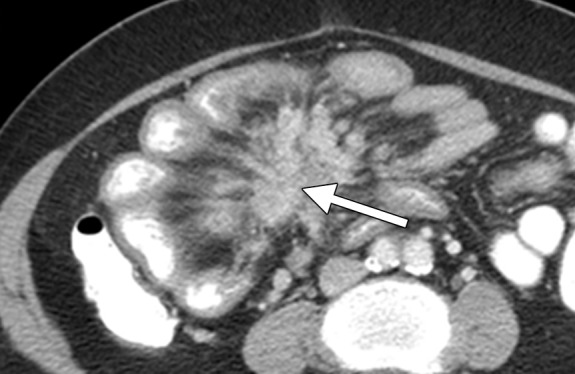
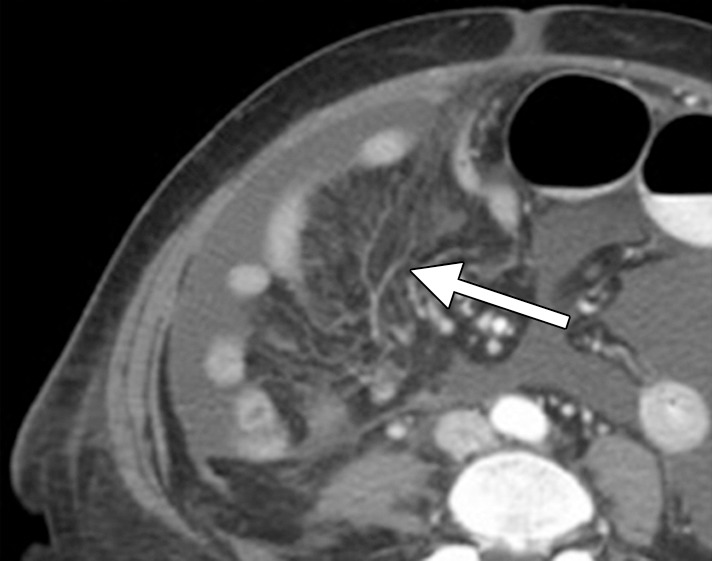
Primary mesenteric neoplasms are uncommon. Most of these lesions originate from the mesenchyme, and the majority are benign. In contrast, secondary involvement of the small bowel mesentery is frequent, and most of the masses are malignant at histopathologic examination. Tumors that arise elsewhere in the abdomen can reach and spread through “spokes of the mesenteric wheel” by direct tumor extension, dissemination via lymphatic channels, embolic hematogenous spread, intraperitoneal seeding, or a combination of these routes (Figs 1, 2).
Figure 1.

Carcinoid tumor metastatic to the mesentery in a 36-year-old woman with abdominal pain. Axial contrast material–enhanced computed tomographic (CT) image shows radiating nodular soft-tissue strands in the mesentery (arrow), as well as tethering, angulation, and retraction of the small bowel loops producing the characteristic spoke-wheel appearance due to tumor-induced fibrosis and desmoplastic reaction. Most carcinoids originate in the small intestines and secondarily involve the mesentery via direct tumor extension and/or lymphatic spread. Primary intestinal tumor is often difficult to detect at imaging because of its small size.
Figure 2.

Advanced-stage high-grade serous ovarian carcinoma in a 49-year-old woman with abdominal pain and abdominal distention. Axial contrast-enhanced CT image shows ill-defined infiltration of the small bowel mesentery (arrow) consistent with seeding of the mesentery by tumor via intraperitoneal dissemination.
In this online presentation, a framework is presented for viewers to remember various primary and secondary mesenteric masses by organizing them according to lesion composition and growth pattern (for primary mesenteric lesions) and type of tumor spread (for secondary mesenteric neoplasms). Characteristic imaging appearances of mesenteric tumors are emphasized, and key imaging pearls and pitfalls at CT and magnetic resonance imaging are highlighted. Major complications that may be caused by mesenteric tumors and should be recognized by the radiologist are illustrated, including (a) bowel-related complication such as mechanical obstruction, perforation, or fistula formation; (b) vascular complication such as vascular compression, erosion, invasion, or thrombosis; and (c) intratumoral hemorrhage. Finally, various surgical and medical therapies that may be appropriate for patients with certain primary and secondary mesenteric lesions are listed, and the role of imaging as a road map for predicting resectability on the basis of tumor location and extent is emphasized.
Supplementary Material
Download:
(19 Mb) |
(38 Mb)
Footnotes
All authors have disclosed no relevant relationships.
Funding: This work was supported in part by the National Institutes of Health and the National Cancer Institute (grant no. P30 CA008748).
Suggested Readings
- Forstner R, Sala E, Kinkel K, Spencer JA; European Society of Urogenital Radiology. ESUR guidelines: ovarian cancer staging and follow-up. Eur Radiol 2010;20(12):2773–2780. [DOI] [PubMed] [Google Scholar]
- Nougaret S, Addley HC, Colombo PE, et al. Ovarian carcinomatosis: how the radiologist can help plan the surgical approach. RadioGraphics 2012;32(6):1775–1800. [DOI] [PubMed] [Google Scholar]
- Sheth S, Horton KM, Garland MR, Fishman EK. Mesenteric neoplasms: CT appearances of primary and secondary tumors and differential diagnosis. RadioGraphics 2003;23(2):457–473. [DOI] [PubMed] [Google Scholar]


