

Abstract
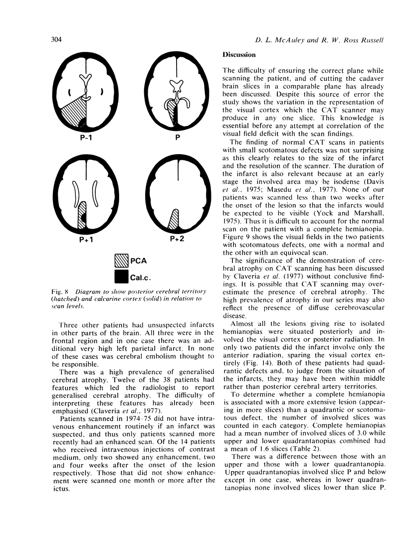
Thirty-nine patients with various types of isolated homonymous hemianopias resulting from ischaemic lesions in the posterior parts of the cerebral hemisphere was examined by CAT scanning. Most had localised low density lesions withing the distribution of the posterior cerebral artery. The location of the lesion (deduced from a separate anatomical study of postmortem brain cut in the plane of the CAT scanner) was correlated with visual field defects. Lesions giving rise to quadrantic defects were smaller than those causing total hemianopias; lower quadrantic defects tended to occur in superior cuts and vice versa. Macular sparing was associated with survival of the occipital pole in some instances. Bilateral cases had a higher prevalence of associated defects.
Full text
PDF


Images in this article


Selected References
These references are in PubMed. This may not be the complete list of references from this article.
- Davis K. R., Taveras J. M., New P. F., Schnur J. A., Roberson G. H. Cerebral infarction diagnosis by computerized tomography. Analysis and evaluation of findings. Am J Roentgenol Radium Ther Nucl Med. 1975 Aug;124(4):643–660. doi: 10.2214/ajr.124.4.643. [DOI] [PubMed] [Google Scholar]
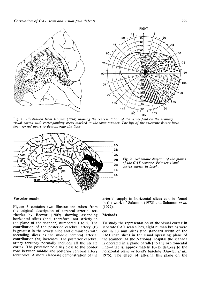
- Gawler J., Bull J. D., Du Boulay G. H., Marshall J. Computerized axial tomography: the normal EMI scan. J Neurol Neurosurg Psychiatry. 1975 Oct;38(10):935–947. doi: 10.1136/jnnp.38.10.935. [DOI] [PMC free article] [PubMed] [Google Scholar]
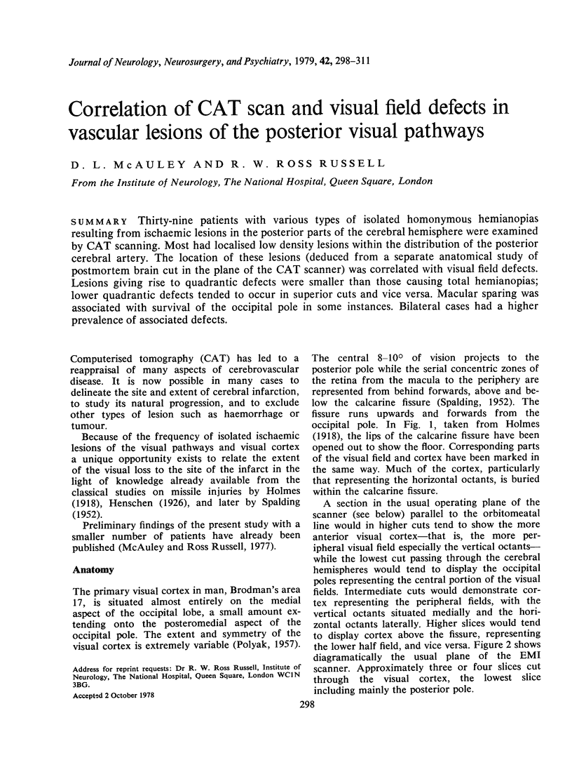
- Holmes G. DISTURBANCES OF VISION BY CEREBRAL LESIONS. Br J Ophthalmol. 1918 Jul;2(7):353–384. doi: 10.1136/bjo.2.7.353. [DOI] [PMC free article] [PubMed] [Google Scholar]
- Masdeu J. C., Azar-Kia B., Rubino F. A. Evaluation of recent cerebral infarction by computerized tomography. Arch Neurol. 1977 Jul;34(7):417–421. doi: 10.1001/archneur.1977.00500190051007. [DOI] [PubMed] [Google Scholar]
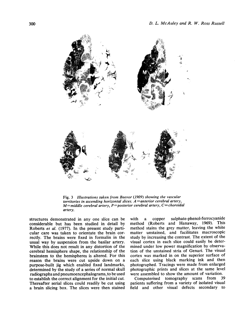
- Roberts M., Hanaway J. Preparation of brain slices for macroscopic study by the copper sulfate-phenol-ferrocyanide technique. Stain Technol. 1969 May;44(3):143–146. doi: 10.3109/10520296909063340. [DOI] [PubMed] [Google Scholar]
- Yock D. H., Jr, Marshall W. H., Jr Recent ischemic brain infarcts at computed tomography: appearances pre- and postcontrast infusion. Radiology. 1975 Dec;117(3 Pt 1):599–608. doi: 10.1148/117.3.599. [DOI] [PubMed] [Google Scholar]