Abstract
Loxosceles reclusa (brown recluse spider) bites often cause local envenomation reactions, however acute hemolysis from systemic loxoscelism is rare. To highlight this important diagnostic consideration for unexplained hemolysis in areas endemic for brown recluse spiders, we report six adolescents with acute hemolytic anemia from presumed L. reclusa bites.
Keywords: hemolytic anemia, loxoscelism, brown recluse envenomation
Loxosceles reclusa (brown recluse spider) bites can cause a variety of local and systemic effects related to envenomation. Most patients remain asymptomatic or develop a mild skin reaction at the site of envenomation, although substantial dermonecrosis can occur 1,2. Loxoscelism, a term denoting systemic effects resulting from L. reclusa venom, is less common but can result in significant morbidity from hemolytic anemia, disseminated intravascular coagulation, and acute renal failure 3. Occasional reports of multiorgan failure and even death have been described, and may be more common among children 4. Although dermatologic manifestations following L. reclusa envenomation have been reported extensively, hematologic manifestations of loxoscelism are less recognized, especially among children. In western Tennessee, an endemic area for brown recluse spiders, many patients seek medical attention for dermatologic complications following a bite. Additionally, some pediatric patients have evidence of hemolysis following envenomation. With the approval of the Institutional Review Board, we performed a retrospective chart review and identified six adolescents with symptomatic acute hemolytic anemia requiring hospital admission and hematology consultation; each had history and physical findings consistent with recent L. reclusa toxin exposure.
CASE REPORTS
The six described patients are unrelated, previously healthy adolescents who presented to a major urban children’s hospital for medical care during 2006 and 2007 with an acute systemic illness featuring hematological manifestations (Table). The primary symptoms at initial presentation in all six patients included fever, pallor, and diffuse rash; they also had fatigue (4), jaundice (4), and dark/red urine (2). After specific inquiry, three children recalled a spider bite within the previous week and could identify its location, but the other three had no recollection of a bite even after the characteristic dermonecrotic wound was identified on physical examination. None of the patients had palpable hepatosplenomegaly or lymphadenopathy.
Table.
Laboratory and clinical course of 6 patients with acute hemolytic anemia from presumed L. reclusa toxin exposure. All laboratory values are at clinical presentation except for the hemoglobin nadir (Lowest Hb), peak reticulocyte count, and the maximum total bilirubin value (Peak T Bili).
| Patient 1 17 y/o AAF |
Patient 2 16 y/o AAM |
Patient 3 13 y/o AAM |
Patient 4 12 y/o AAF |
Patient 5 13 y/o AAF |
Patient 6 14 y/o AAF |
|
|---|---|---|---|---|---|---|
| Initial Hb (gm/dL) | 4.8 | 12.9 | 8.8 | 6.5 | 4.9 | 13.7 |
| Lowest Hb (gm/dL) | 4.8 | 7.1 | 8.7 | 5.6 | 4.5 | 5.9 |
| Initial Reticulocytes × 109/L (%) |
25 (1.6%) | 41 (1.1%) | 181 (6.3%) | 177 (8.3%) | 207 (14.6%) | 125 (4.2%) |
| Peak Reticulocytes × 109/L (%) |
255 (8.3%) | 229 (8.8%) | 226 (7.4%) | 222 (11.7%) | 364 (15.1%) | 155 (3.8%) |
| Platelets (× 109/L) | 260 | 182 | 535 | 383 | 344 | 222 |
| WBC (× 109/L) | 37.4 | 16.6 | 17.9 | 18.8 | 35.5 | 19 |
| ANC (× 109/L) | 34.4 | 11.0 | 9.7 | 11.8 | 20.6 | 14.8 |
| Peak T Bili (mg/dL) | 13.8 | 7.2 | 3.2 | 3.2 | 2.7 | 15.1 |
| LDH (U/L) | Not available | Not available | Not available | 4656 | 483 | 2611 |
| DAT | Positive | Positive | Positive | Positive | Positive | Positive |
| Surface IgG | Trace | Positive | − | − | − | Positive |
| Surface C3 | 3+ | Positive | 2+ | 1+ | 3+ | Positive |
| Hospital admission | ICU | Inpatient | Inpatient | Inpatient | ICU | ICU |
| BP Support | IV fluids, Dopamine |
Not required | Not required | Not required | IV fluids, Dopamine |
IV fluids |
| PRBC Transfusion | 4 units | 2 units | Not required | Not required | 1 unit | 5 units |
| Surgical wound care | I&D | Bedside Debride | Not required | Not required | Not required | Not required |
All six patients developed substantial anemia during their hospital stay (median Hb 7.1 gm/dL). The direct antiglobulin test (DAT) was positive for surface complement component C3 in all six patients, and surface IgG additionally was detected in three patients (Table). All patients developed reticulocytosis and hyperbilirubinemia. Other causes of anemia (e.g., nutritional deficiency, blood loss, marrow failure) were excluded by thorough history, physical examination, laboratory testing and review of peripheral blood smears.
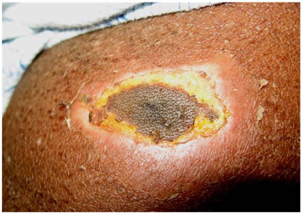
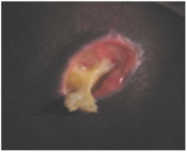
All patients were hospitalized for medical management, including 3 patients who required admission to the intensive care unit to receive fluid resuscitation, RBC transfusion, and dopamine infusion for hypotension. Four patients received packed red blood cell transfusions (range 1-5 units), and 2 patients with stable vital signs, physical examination and laboratory values were carefully observed without transfusion. In all patients, the bite progressed with local dermonecrosis of varying severity with evolution of surrounding desquamation. Wound care varied; two patients had surgical intervention with an open incision and drainage at the bite location (Figure). All 6 patients had full resolution of anemia within several weeks and have had no recurrence of hemolysis during the follow-up period of almost two years.
Figure.
Sequential progression of L. reclusa spider bite in Patient 1 who presented with acute hemolytic anemia requiring transfusion of 4 units of packed RBCs and ICU admission. A, (Day 2) Visible bite mark with demarcation around the 1cm lesion, which featured tenderness and induration, with early signs of dermonecrosis and a papular rash. B, (Day 5) Illustrates evolving central necrosis with circumferential desquamation of approximately 4 cm; there was continued tenderness and induration. C, (Day 14) Shows complete central necrosis with inspissated pus, requiring I&D to promote healing; pain had subsided.
DISCUSSION
Loxoscelism, defined as systemic manifestations resulting from envenomation by the spider genus Loxosceles, is notfamiliar to most pediatric hematologists. Few members of the Loxosceles family of spiders are endemic to the United States, and their distribution is geographically limited to the midsouthern and southwestern regions of the country (http://spiders.ucr.edu/images/colorloxmap.gif). Loxosceles reclusa (brown recluse) is the most prevalent and medically significant Loxosceles spider in the US, and the most common cause of spider venom-associated morbidity. Loxosceles venom is highly toxic, but the dose of venom injected, sex of the spider, amount of sphingomyelinase activity, and host factors may account for clinical variability 5-8. Although the majority of persons bitten by a brown recluse spider probably will not seek medical attention, approximately 16% develop systemic loxoscelism 1, with children having more severe systemic symptoms. Children presenting for medical care may be more likely to exhibit hematologic complications than are adults7.
Our retrospective review describes cases coming to medical attention at a tertiary medical center and requiring hematology consultation for anemia. All six patients had signs and symptoms of systemic illness including fever, pallor, and rash, along with acute anemia featuring both intravascular and extravascular hemolysis. The DAT was positive for C3 on the RBC surface in all 6 patients, and positive surface IgG in 3 patients, supporting the pathophysiology of immune-mediated hemolysis. Severe anemia developed in all 6 patients, and the peripheral blood smear contained fragmented erythrocytes with microspherocytes (Figure). Three patients could recall a recent spider bite but in the others, a bite was identified only after thorough examination. In Case 1, the initially small bite was hidden but soon evolved to become a painful local wound requiring surgical debridement (Figure).
A positive DAT result has been reported only rarely in patients with loxoscelism 5,7. Previous reports have described positive C3 on the RBC surface, although at least one described IgG positivity surface 7. All 6 of our patients in this time period had positive C3 with variable IgG on the RBC surface. The pathophysiology of venom-associated hemolysis is incompletely understood, but likely involves erythrocyte lysis induced by sphyngomyelinase venom toxins. Two purified sphingomyelinase toxins in the related Loxosceles intermedia induce complement-dependent erythrocyte lysis9. The alternative complement pathway is activated by a cascade with endogenous metalloproteases inducing cleavage of surface RBC glycophorins. Additionally, disruption of cell membrane asymmetry results in phosphatidylserine exposure 10. Our cases suggest that anemia in loxoscelism results from direct toxin-related erythrocyte damage and complement-mediated immune destruction, featuring both intravascular and extravascular hemolysis. We cannot exclude the possibility that concomitant G6PD deficiency played a role in the etiology of hemolysis in these patients. Despite the beneficial role of corticosteroids in the treatment of children with idiopathic warm-reactive autoimmune hemolytic anemia, use of corticosteroid therapy is unproven in the setting of loxoscelism. Supportive care measures including hemodynamic support and blood product transfusion remain the standard of care in the United States.
Healthcare providers in Loxosceles endemic areas should consider this unusual diagnosis in the appropriate clinical setting of acute unexplained hemolytic anemia. Detailed history and careful search for a spider bite can help elucidate this etiology; acute management may require emergency intervention but the anemia is typically self- limited with a good long-term prognosis. It is important to remember that these spiders favor uninhabited areas and are non-aggressive by nature. No confirmed cases have been documented outside endemic areas, and given the reclusive nature of these spiders, this diagnosis should only be considered after excluding other causes of acute hemolytic anemia. Medical providers practicing in areas inhabited by Loxosceles spiders should be familiar with the possible hematologic consequences of envenomation.
Footnotes
Publisher's Disclaimer: This is a PDF file of an unedited manuscript that has been accepted for publication. As a service to our customers we are providing this early version of the manuscript. The manuscript will undergo copyediting, typesetting, and review of the resulting proof before it is published in its final citable form. Please note that during the production process errors may be discovered which could affect the content, and all legal disclaimers that apply to the journal pertain.
The authors declare no conflicts of interest.
REFERENCES
- 1.Swanson DL, Vetter RS. Loxoscelism. Clin Dermatol. 2006;24:213–221. doi: 10.1016/j.clindermatol.2005.11.006. [DOI] [PubMed] [Google Scholar]
- 2.da Silva PH, da Silveira RB, Appel MH, Mangili OC, Gremski W, Veiga SS. Brown spiders and loxoscelism. Toxicon. 2004;44:693–709. doi: 10.1016/j.toxicon.2004.07.012. [DOI] [PubMed] [Google Scholar]
- 3.de Souza AL, Malaque CM, Sztajnbok J, Romano CC, Duarte AJ, Seguro AC. Loxosceles venom-induced cytokine activation, hemolysis, and acute kidney injury. Toxicon. 2008;51:151–156. doi: 10.1016/j.toxicon.2007.08.011. [DOI] [PubMed] [Google Scholar]
- 4.Futrell JM. Loxoscelism. Am J Med Sci. 1992;304:261–267. doi: 10.1097/00000441-199210000-00008. [DOI] [PubMed] [Google Scholar]
- 5.Lane DR, Youse JS. Coombs-positive hemolytic anemia secondary to brown recluse spider bite: a review of the literature and discussion of treatment. Cutis. 2004;74:341–347. [PubMed] [Google Scholar]
- 6.Hostetler MA, Dribben W, Wilson DB, Grossman WJ. Sudden unexplained hemolysis occurring in an infant due to presumed loxosceles envenomation. J Emerg Med. 2003;25:277–282. doi: 10.1016/s0736-4679(03)00202-6. [DOI] [PubMed] [Google Scholar]
- 7.Elbahlawan LM, Stidham GL, Bugnitz MC, Storgion SA, Quasney MW. Severe systemic reaction to loxosceles recluse spider bites in a pediatric population. Pediatr Emerg Care. 2005;21:177–180. [PubMed] [Google Scholar]
- 8.de Oliveira KC, Goncalves de Andrade RM, Piazza RMF, Ferreira JM, Jr, van den Berg CW, Tambourgi DV. Variations in loxosceles spider venom composition and toxicity contribute to the severity of envenomation. Toxicon. 2005;45:421–429. doi: 10.1016/j.toxicon.2004.08.022. [DOI] [PubMed] [Google Scholar]
- 9.Tambourgi DV, Magnoli FC, Von Eickstedt VRD, Benedetti ZC, Petricevich VL, Da Silva WD. Incorporation of a 35-kilodalton purified protein from loxosceles intermedia spider venom transforms human erythrocytes into activators of autologous complement alternative pathway. J Immunol. 1995;155:4459–4466. [PubMed] [Google Scholar]
- 10.Tambourgi DV, De Sousa Da Silva M, Billington SJ, Goncalves De Andrade RM, Magnoli FC, Songer JG, et al. Mechanism of induction of complement susceptibility of erythrocytes by spider and bacterial sphingomyelinases. Immunology. 2002;107:93–101. doi: 10.1046/j.1365-2567.2002.01483.x. [DOI] [PMC free article] [PubMed] [Google Scholar]


