

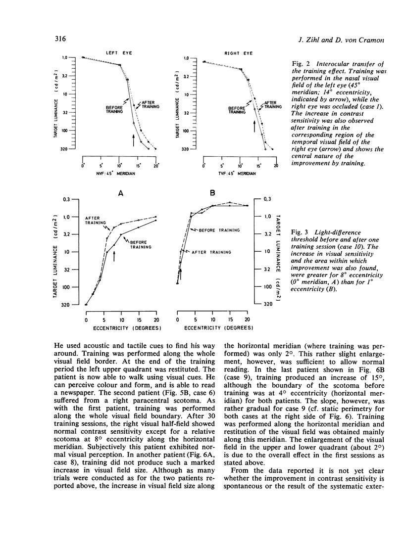
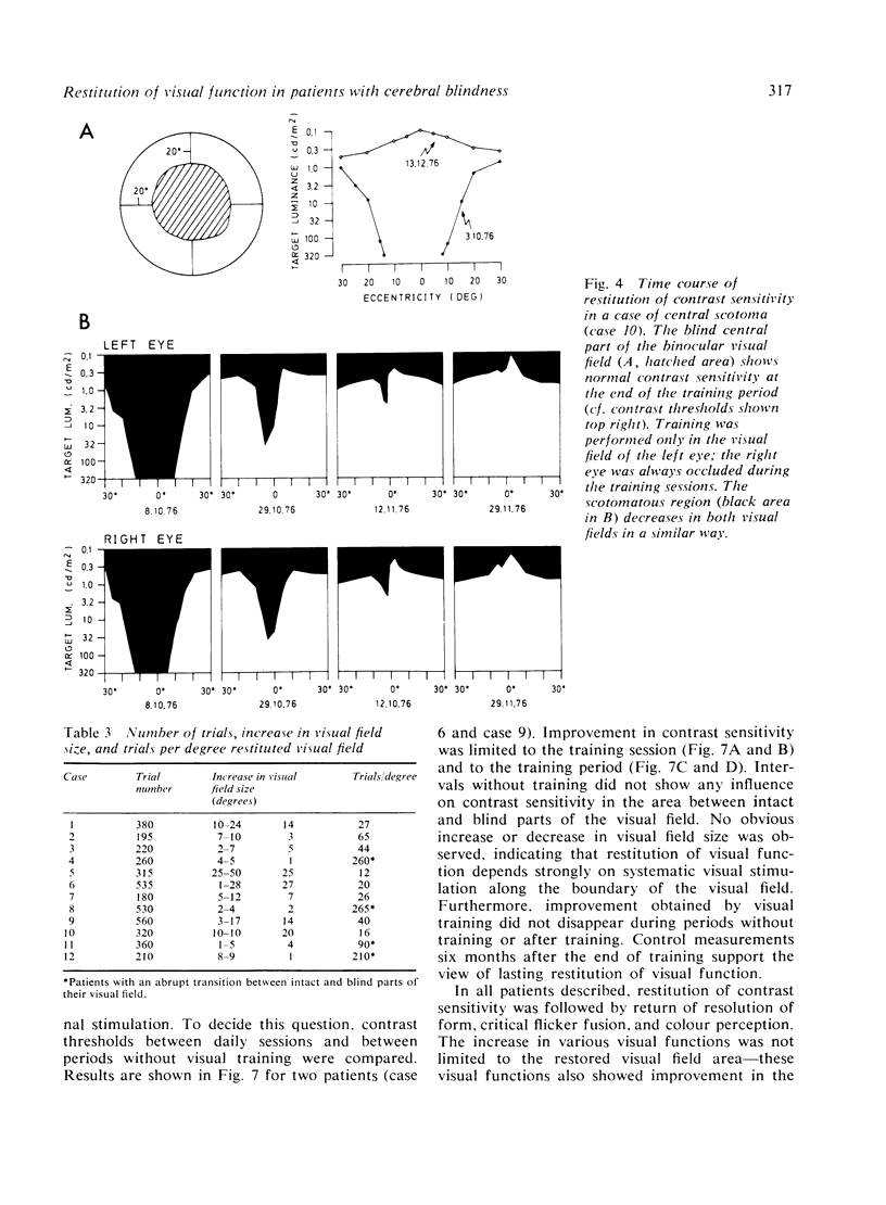
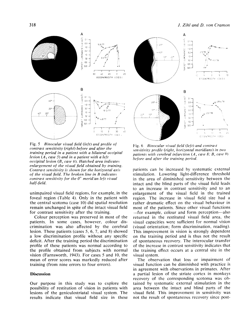
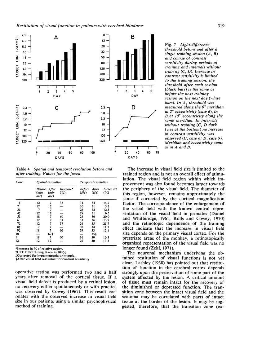
Abstract
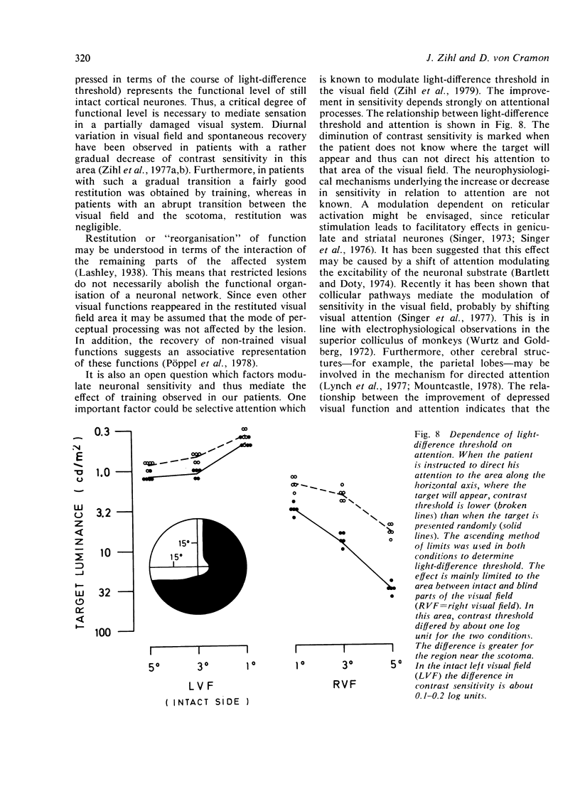
Patients with postchiasmatic visual field defects were trained at the border of their visual field. Using a psychophysical method, light-difference thresholds were determined repeatedly in this visual field area. Improvement in contrast sensitivity and increase in size of the visual field could be obtained by this training procedure. The improvement was confined to the trained visual field area and showed interocular transfer indicating its central nature. Althoughh only contrast sensitivity was trained, the observed improvement was not limited to this visual function. Visual acutity, critical flicker fusion, and colour perception also showed and improvement suggesting an association of these functions. The improvement was restricted to the training period-no spontaneous recovery was observed between or after the periods of training. It is suggested that a lesion in the central visual system does not always result in a complete and permanent loss of function. The critical level of function that normally has to be reached for sufficient neuronal sensitivity may be obtained by systematic visual stimulation in the area between the intact and blind parts of the visual field. This increase in neuronal sensitivity leads to an improvement in visual performance.
Full text
PDF
Selected References
These references are in PubMed. This may not be the complete list of references from this article.
- Bartlett J. R., Doty R. W., Sr Influence of mesencephalic stimulation on unit activity in striate cortex of squirrel monkeys. J Neurophysiol. 1974 Jul;37(4):642–652. doi: 10.1152/jn.1974.37.4.642. [DOI] [PubMed] [Google Scholar]
- Cowey A. Perimetric study of field defects in monkeys after cortical and retinal ablations. Q J Exp Psychol. 1967 Aug;19(3):232–245. doi: 10.1080/14640746708400098. [DOI] [PubMed] [Google Scholar]
- DANIEL P. M., WHITTERIDGE D. The representation of the visual field on the cerebral cortex in monkeys. J Physiol. 1961 Dec;159:203–221. doi: 10.1113/jphysiol.1961.sp006803. [DOI] [PMC free article] [PubMed] [Google Scholar]
- Koerner F., Teuber H. L. Visual field defects after missile injuries to the geniculo-striate pathway in man. Exp Brain Res. 1973 Aug 31;18(1):88–113. doi: 10.1007/BF00236558. [DOI] [PubMed] [Google Scholar]
- Lashley K. S. MASS ACTION IN CEREBRAL FUNCTION. Science. 1931 Mar 6;73(1888):245–254. doi: 10.1126/science.73.1888.245. [DOI] [PubMed] [Google Scholar]
- Lynch J. C., Mountcastle V. B., Talbot W. H., Yin T. C. Parietal lobe mechanisms for directed visual attention. J Neurophysiol. 1977 Mar;40(2):362–389. doi: 10.1152/jn.1977.40.2.362. [DOI] [PubMed] [Google Scholar]
- Mountcastle V. B. Brain mechanisms for directed attention. J R Soc Med. 1978 Jan;71(1):14–28. doi: 10.1177/014107687807100105. [DOI] [PMC free article] [PubMed] [Google Scholar]
- Poppel E., Held R., Frost D. Leter: Residual visual function after brain wounds involving the central visual pathways in man. Nature. 1973 Jun 1;243(5405):295–296. doi: 10.1038/243295a0. [DOI] [PubMed] [Google Scholar]
- Pöppel E., Brinkmann R., von Cramon D., Singer W. Association and dissociation of visual functions in a case of bilateral occipital lobe infarction. Arch Psychiatr Nervenkr (1970) 1978 Mar 7;225(1):1–21. doi: 10.1007/BF00367348. [DOI] [PubMed] [Google Scholar]
- Rolls E. T., Cowey A. Topography of the retina and striate cortex and its relationship to visual acuity in rhesus monkeys and squirrel monkeys. Exp Brain Res. 1970;10(3):298–310. doi: 10.1007/BF00235053. [DOI] [PubMed] [Google Scholar]
- SYMONDS C., MACKENZIE I. Bilateral loss of vision from cerebral infarction. Brain. 1957 Dec;80(4):415–455. doi: 10.1093/brain/80.4.415. [DOI] [PubMed] [Google Scholar]
- Singer W. The effect of mesencephalic reticular stimulation on intracellular potentials of cat lateral geniculate neurons. Brain Res. 1973 Oct 26;61:35–54. doi: 10.1016/0006-8993(73)90514-3. [DOI] [PubMed] [Google Scholar]
- Singer W., Tretter F., Cynader M. The effect of reticular stimulation on spontaneous and evoked activity in the cat visual cortex. Brain Res. 1976 Jan 30;102(1):71–90. doi: 10.1016/0006-8993(76)90576-x. [DOI] [PubMed] [Google Scholar]
- Singer W., Zihl J., Pöppel E. Subcortical control of visual thresholds in humans: evidence for modality specific and retinotopically organized mechanisms of selective attention. Exp Brain Res. 1977 Aug 31;29(2):173–190. doi: 10.1007/BF00237040. [DOI] [PubMed] [Google Scholar]
- Sloan L. L. The Tubinger perimeter of Harms and Aulhorn. Recommended procedures and supplementary equipment. Arch Ophthalmol. 1971 Dec;86(6):612–622. doi: 10.1001/archopht.1971.01000010614002. [DOI] [PubMed] [Google Scholar]
- Teuber H. L. Recovery of function after brain injury in man. Ciba Found Symp. 1975;(34):159–190. doi: 10.1002/9780470720165.ch10. [DOI] [PubMed] [Google Scholar]
- Wurtz R. H., Goldberg M. E. The primate superior colliculus and the shift of visual attention. Invest Ophthalmol. 1972 Jun;11(6):441–450. [PubMed] [Google Scholar]
- Zeki S. M. Cortical projections from two prestriate areas in the monkey. Brain Res. 1971 Nov;34(1):19–35. doi: 10.1016/0006-8993(71)90348-9. [DOI] [PubMed] [Google Scholar]
- Zihl J., Pöppel E., von Cramon D. Diurnal variation of visual field size in patients with postretinal lesions. Exp Brain Res. 1977 Mar 30;27(3-4):245–249. doi: 10.1007/BF00235501. [DOI] [PubMed] [Google Scholar]
- Zihl J., von Cramon D., Brinkmann R., Backmund H. Verlaufskontrolle und Prognose bei Gesichtsfeldausfällen von Patienten mit cerebrovaskulären Störungen. Nervenarzt. 1977 Apr;48(4):219–224. [PubMed] [Google Scholar]
- Zihl J., von Cramon D., Pöppel E. Sensorische Rehabilitation bei Patienten mit postchiasmatischen Sehstörungen. Nervenarzt. 1978 Feb;49(2):101–111. [PubMed] [Google Scholar]