Description
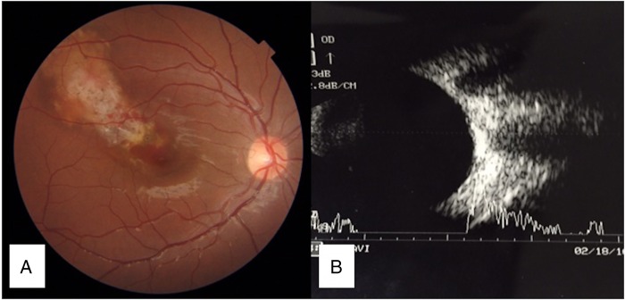
A 16-year-old boy presented with best-corrected visual acuity of 6/18 OD. OS was within normal limits, as was anterior segment OD. A focal whitish-yellow plaque lesion with overlying areas of depigmentation, and retinal atrophy in the central and superior lateral macula were noted in OD. A choroidal neovascular membrane (CNVM) with subretinal haemorrhage was seen at the lesion's inferior medial edge, involving the fovea (figure 1A). Therefore, diagnosis of choroidal osteoma1 (CO) was made and confirmed on sonography, with a highly acoustic elevated lesion causing a corresponding shadow (figure 1B).
Figure 1.
(A) Fundus photograph of the OD showing focal whitish-yellow plaque lesion with overlying area of depigmentation and atrophy. Medial edge of osteoma near fovea shows subretinal haemorrhage with choroidal neovascular membrane. (B) Sonography of the OD showing slightly elevated lesion with high acoustic reflectivity with corresponding shadow giving a false appearance of double optic nerve.
Fluorescein angiography (FA) confirmed an active CNVM at the medial edge of the osteoma (figure 2A). A lattice-like reflective pattern corresponding to the bony trabeculae2 of the tumour in deeper choroid, with overlying area of retinal atrophy and type 1 CNVM was seen on optical coherence tomography (OCT) (figure 2B). Swept-source OCT angiography (SS-OCTA) additionally revealed a superficial lacy network of interconnected new vessels seen in the region of the CNVM, which were thick and dilated (figure 2C). Also, a deeper layer of thin vessels arranged in a ‘Medusa head appearance’ was seen in the region of the tumour (figure 2C, D). A connection between these groups of vessels could also be deciphered at two junctions (figure 2C). Multiple feeder vessels make such CNVMs poor candidates for traditional laser therapy. Findings can be masked in FA3 due to mottled hyperfluorescence related to the CO. SS-OCTA is more informative than standard spectral-domain (SD)-OCTA due to superior image quality.
Figure 2.
(A) Fluorescein angiography of the OD showing patchy choroidal hyperfluorescence underlying the tumour, with a neovascular membrane at the medial edge of the osteoma. (B) Optical coherence tomography of the OD showing type 1 choroidal neovascular membrane (CNVM). Lattice-work reflectivity is seen corresponding to bone trabeculae (white arrow). (C) Swept-source optical coherence tomography angiograph showing high-resolution superficial network of dilated vessels (white arrowhead) corresponding at the level of CNVM. Tumour vessels in a Medusa head appearance with two visible connections, possibly feeders for CNVM, are also seen (white arrow). (D) Swept-source optical coherence tomography angiograph showing a deeper network of fine thin vessels at the level of the tumour (white arrow).
Learning points.
Swept-source optical coherence tomography angiography (SS-OCTA) additionally reveals high-resolution choroidal vasculature characteristics, which can otherwise get masked due to high tumour density on fluorescein angiography.
There are multiple connections between the tumour vessels and choroidal neovascular membrane in a case of choroidal osteoma.
Footnotes
Competing interests: None declared.
Patient consent: Obtained.
Provenance and peer review: Not commissioned; externally peer reviewed.
References
- 1.Mansour AM, Arevalo JF, Al Kahtani E et al. . Role of intravitreal antivascular endothelial growth factor injections for choroidal neovascularization due to choroidal osteoma. J Ophthalmol 2014;2014:210458 doi:10.1155/2014/210458 [DOI] [PMC free article] [PubMed] [Google Scholar]
- 2.Gass JD, Guerry RK, Jack RL et al. . Choroidal osteoma. Arch Ophthalmol 1978;96:428–35. [DOI] [PubMed] [Google Scholar]
- 3.Szelog JT, Bonini Filho MA, Lally DR et al. . Optical coherence tomography angiography for detecting choroidal neovascularization secondary to choroidal osteoma. Ophthalmic Surg Lasers Imaging Retina 2016;47:69–72. doi:10.3928/23258160-20151214-10 [DOI] [PubMed] [Google Scholar]


