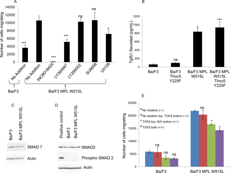
Figure 4. A role for TGFβ-1 in the MPL W515L induced chemotaxis.
A. The effect of inhibitor treatment on chemokinesis in MPL W515L was assessed. Cells were pre-incubated with inhibitors for 2 hours before undertaking the motility assay. 105 cells were used and the number of cells migrating into the bottom well counted after 6 hours incubation. Results are the mean ± SEM of at least three experiments. Inhibitors used were 50 μM INCB018424 (JAK2 inhibitor), 5μM LY364947 (TGFβ inhibitor), 10μM LY294002 (PI3K inhibitor), 20μM SU6656 (Src inhibitor) and 10μM U0126 (MEK 1/2 inhibitor). Cell viability was greater than 90% prior to and post the migration assay. B. The levels of TGFβ-1 were measured in culture supernatants from parental Ba/F3, MPL W515L, THOC5 Y225F and MPL W515L co-transfected with THOC5 Y225F expressing cells using the Quantikine ELISA from R&D systems. Results are displayed as pg/ml of cell culture supernatant +/−SEM (n = 3). The levels of SMAD7 C., SMAD2 and phospho S465/467 SMAD2 D. expression in Ba/F3 and MPL W515L expressing cells was assessed by western blot. E. Chemokinesis was measured using Boyden chamber assays in parental Ba/F3 cells and MPL W515L expressing cells with 5ng/ml TGFβ added to either the top well (+/−) bottom well (−/+) or both wells (+/+). Results shown are the number of cells migrating (mean ± SEM, n = 3). The results of a t-test against MPL W515L no addition (A) Ba/F3 (B) or no addition (E) are shown and represented by; * < 0.05, ** < 0.01, *** < 0.001.

