
Figure 1. Expression ofHNF4α in clinical gastric tissues and its role in regulating gastric cell proliferation.
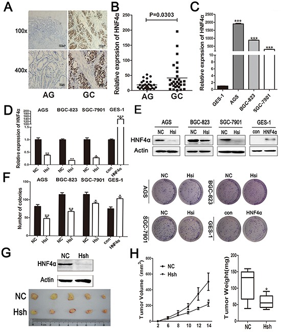
A. IHC staining of HNF4α in atrophic gastritis and gastric cancer samples. Representative images are shown here (magnification 100x,400x; Scale bars:200μm,50μm). B. The mRNA levels of HNF4α in 30 atrophic gastritis samples and 30 gastric cancers were measured by real-time PCR. The horizontal bars indicate the mean value of each sample group. Mann-Whitney U-test was used to calculate the P value. C. HNF4α mRNA expression levels in indicated gastric cancer cell lines and immortalized gastric cells GES-1. ***p<0.001 by Student's t-test. D. and E. HNF4α mRNA and protein levels in indicated cells transfected with HNF4α siRNA or HNF4α over-expression plasmid. *p<0.05, **p<0.01, ***p<0.001 by Student's t-test. F. Foci formation after cells transfected HNF4α siRNA or HNF4α over-expression plasmid. *p<0.05, **p<0.01 by Student's t-test. Representative images are shown here from three independent biological replicates. G. Western blot shows loss of HNF4α in BGC-823 cells transfected with lenti-HNF4α shRNA; Tumors formed in nude mice induced by BGC-823 cells transfected with lenti-negative control group(NC)and lenti-HNF4α shRNA group(Hsh). H.Tumor growth curve and tumor weight of NC group and Hsh group. *p<0.05 by Student's t-test.