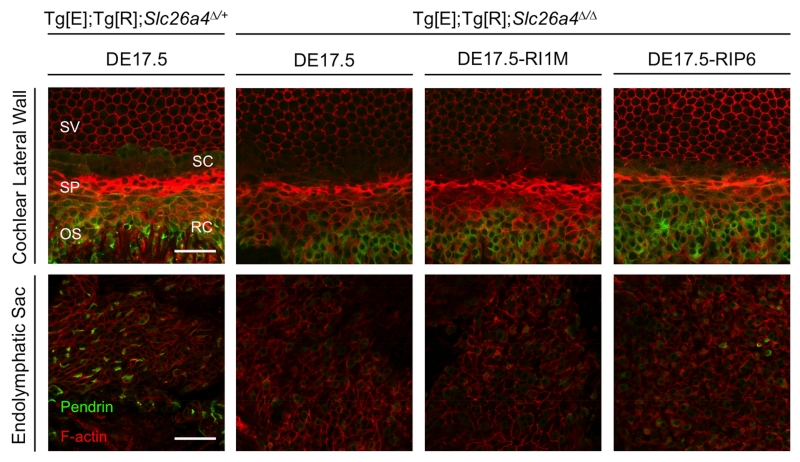
Figure 4.
Pendrin Expression.
Representative immunostaining is shown for pendrin (green) and F-actin (red) in whole-mounted lateral wall of the middle turn of the cochlea and endolymphatic sac of Tg[E];Tg[R];Slc26a4Δ/+ DE17.5 ears (n = 8 and 6, respectively), Tg[E];Tg[R];Slc26a4Δ/Δ DE17.5 ears (n = 17 each), Tg[E];Tg[R];Slc26a4Δ/Δ DE17.5-RI1M ears (n = 12 each), and Tg[E];Tg[R];Slc26a4Δ/Δ DE17.5-RIP6 ears (n = 10 and 9, respectively). Pendrin expression in spindle-shaped cells was reduced in Tg[E];Tg[R];Slc26a4Δ/Δ ears in comparison to Tg[E];Tg[R];Slc26a4Δ/+ ears. Scale bars: 50 μm. SV, stria vascularis; SP, spiral prominence; OS, outer sulcus; SC, spindle-shaped cell; RC, root cell.

