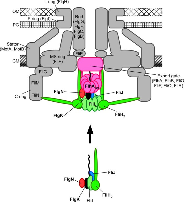
Figure 7.

Schematic diagram of the flagellar type III protein export apparatus. The export gate is composed of six transmembrane proteins, FlhA, FlhB, FliO, FliP, FliQ, and FliR and is located within the MS ring. The C‐terminal cytoplasmic domain of FlhA (FlhAC) forms part of the docking platform for FliH, FliI ATPase, FliJ, flagellar type III secretion chaperones, and export substrates. FliI forms a hetero‐trimer with the FliH dimer in the cytoplasm. The FliH2–FliI complex acts as a dynamic carrier to deliver FliJ and the FlgN–FlgK chaperone‐export substrate complex to the FlhAC ring complex (FlhAC 9). Upon formation of the FliI6FliJ ring complex on the FlhAC 9 ring, the FliI6FliJ ring complex hydrolyses ATP and activates the export gate through an interaction between FliJ and FlhA, allowing the gate to translocate FlgK into the central channel of the growing flagellar structure. OM, outer membrane; PG, peptidoglycan layer; CM, cytoplasmic membrane.
