Abstract
Classical swine fever (CSF) is a highly contagious swine disease caused by classical swine fever virus (CSFV). Previously, we demonstrated that rAdV-SFV-E2, an adenovirus-delivered, Semliki Forest virus replicon-vectored marker vaccine against CSF, is able to protect pigs against lethal CSFV challenge. From an economical point of view, it will be beneficial to reduce the minimum effective dose of the vaccine. This study was designed to test the adjuvant effects of Salmonella enteritidis-derived bacterial ghosts (BG) to enhance the protective immunity of rAdV-SFV-E2 in pigs. Groups of 5-week-old pigs (n = 4) were immunized intramuscularly twice with 105 median tissue culture infective doses (TCID50) rAdV-SFV-E2 combined with 1010 colony forming units (CFU) BG, 106 or 105 TCID50 rAdV-SFV-E2 alone or 1010 CFU BG alone at an interval of 3 weeks, and challenged with the highly virulent CSFV Shimen strain at 1 week post-booster immunization. The results show that the pigs inoculated with 105 TCID50 rAdV-SFV-E2 plus BG or 106 TCID50 rAdV-SFV-E2 alone were completely protected from lethal CSFV challenge, in contrast with the pigs vaccinated with 105 TCID50 rAdV-SFV-E2 or BG alone, which displayed partial or no protection following virulent challenge. The data indicate that BG are a promising adjuvant to enhance the efficacy of rAdV-SFV-E2 and possibly other vaccines.
Introduction
Classical swine fever (CSF) is one of the most contagious diseases and characterized by high fever and high mortality, resulting in huge economic losses to the pig industry [1]. Classical swine fever virus (CSFV), one of the members of the Pestivirus genus in the Flaviviridae family, is the causative pathogen of CSF. The genome of CSFV is a single-stranded, positive-sense RNA of about 12.3 kb, which encodes a polyprotein that is processed co- and posttranslationally into 12 proteins (Npro-C-Erns-E1-E2-p7-NS2-NS3-NS4A-NS4B-NS5A-NS5B) [2, 3].
Currently, immunization with modified live vaccines (MLV, e.g. C-strain) is a major strategy to control CSF in many countries [4]. However, the European Union has banned vaccination using traditional CSF MLV against CSF since 1990, as antibodies induced by MLV or field CSFV strains cannot be distinguished serologically [5]. Therefore, developing a safe and effective marker vaccine allowing differentiation of infected from vaccinated animals (DIVA) is very important. To address this issue, we developed a marker CSF vaccine rAdV-SFV-E2 based on human adenovirus type 5 (HAdV-5)/alphavirus replicon chimeric vector. We demonstrate that rAdV-SFV-E2 can elicit strong cellular and humoral responses in pigs and provide sterile immunity and complete protection against lethal CSFV challenge comparable to the C-strain [6, 7]. From an economic point of view, it is necessary to reduce the minimum effective dose (MED) of the vaccine.
Co-administration of adjuvants, such as aluminum and mineral oil, is an effective method to improve the efficacy of a suboptimal vaccine. Adjuvants can help antigens in activating pathways significantly in the induction of innate immunity, predominantly targeting antigen-presenting cells (APC) and consequently influencing the adaptive immune response [8]. Well-characterized bacterial ghosts (BG)-based adjuvants have unique advantages. BG are nonliving cell envelope preparations from Gram-negative bacteria, devoid of cytoplasmic contents, while their cellular morphology and native surface antigenic structures remain preserved. So they are potentially powerful adjuvants due to the presence of bacterial membrane components such as lipopolysaccharides, peptidoglycans and monophosphoryl lipid A (MPL) [9]. MPL interacts with toll-like receptor 4 [10], induces the production and release of cytokines [11] and increases the migration and maturation of dendritic cells [12]. Owing to the particulate nature of BG and the fact that they contain many well-known immune-stimulating compounds, BG have the potential to enhance immune responses to various antigens [13]. Therefore, we hypothesize that rAdV-SFV-E2 with BG can provide a better protection against CSF in pigs.
The present study was aimed at evaluating the adjuvant effects of BG to enhance the protective immunity of rAdV-SFV-E2 in pigs.
Materials and methods
Bacterial ghost adjuvant, vaccines and viruses
The Salmonella enteritidis-derived BG adjuvant was produced by controlled expression of the modified lysis gene E (mE) from bacteriophage ΦX174 [14]. Briefly, S. enteritidis DH091 harboring the recombinant bacteriolytic plasmid pBV-mE expressing the mE that is able to lyse the bacteria when induced at 42 °C, was cultured to an OD600nm of 1.0 at 37 °C. Then the culturing temperature was raised to 42 °C for mE expression, resulting in lysis of the bacteria. After 1 h, when the lysis curve started to decline, 10 μL of the cell suspension was spread onto LB plates containing ampicillin, followed by a 12-h incubation at 37 °C. Viable colonies were determined as colony forming units (CFU)/mL. The OD600nm was measured every 15 min till no further decline in OD600nm. After lysis, the BG were harvested by centrifugation (4000 × g for 10 min), washed with PBS (pH 7.2), suspended in 20 mL of sterile distilled water, lyophilized and stored at −20 °C. rAdV-SFV-E2 is an adenovirus-delivered, alphavirus replicon-vectored vaccine encoding the E2 glycoprotein of CSFV [6]. The highly virulent CSFV Shimen strain [7] maintained at Harbin Veterinary Research Institute (HVRI) was used for challenge.
Animals
Twenty 5-week-old cross-bred weanling piglets, free of CSFV-specific antibodies and antigens, were raised in the animal facility at HVRI. All experimental procedures involving animals were approved by the Experimental Animal Ethics Committee of HVRI.
Immunization-challenge experiment
The piglets were randomly divided into five groups of four animals each. Groups A and C were respectively vaccinated with 106 TCID50 and 105 TCID50 rAdV-SFV-E2 alone; Group B were co-immunized intramuscularly with 105 TCID50 rAdV-SFV-E2 and 1010 CFU BG; Groups D and E were injected intramuscularly with 1010 CFU BG and DMEM (2 mL), respectively, serving as controls. Three weeks later, all the pigs were given a booster immunization with the same vaccine, dose and route of administration. All the pigs were challenged intramuscularly with 106 TCID50 CSFV Shimen strain 1 week post-booster immunization. Following challenge, the rectal temperature and clinical signs were recorded every day. All the pigs were euthanized at 15 days post-challenge (dpc). The tissues from all the pigs were subjected to pathological examinations as described previously [15].
Serological assays
Serum samples were collected at different time points post-immunization. The presence of the E2-specific antibodies in samples were tested using the IDEXX HerdChek* CSFV antibody test kit (IDEXX Laboratories, Shiphol-Rijk, The Netherlands).
To test the level of CSFV-specific neutralizing antibodies (NAbs), a serum-virus neutralization test (SVNT) was carried out in 96-well flat-bottom microtiter plates (Coring, USA) as described previously [16].
Real-time RT-PCR
The total RNA was extracted from EDTA-treated blood samples collected at different days after challenge, and detection of CSFV RNA was performed by real-time RT-PCR with a CSFV-specific probe (5′-FAM-AGG ACT AGC AAA CGG AGG GAC TAG CCG-TAMRA-3′) and a primer pair (5′-GAA CTG GGC TAG CCA TG-3′ and 5′-ACT GTC CTG TAC TCA GGA C-3′) [17].
Pathology
All surviving pigs were euthanized at 15 dpc. Various organs (spleen, kidney, tonsils, lymph nodes and bladder) were collected and subjected to pathological and histopathological examinations as described previously [6, 18].
Statistical analysis
Statistical analysis was conducted using the SPSS 14.0 software. One-way ANOVA followed by Duncan’s multiple-range tests were used to compare the parameters among the different groups.
Results
Antibody production
E2-specific antibodies and NAbs were tested by blocking ELISA and SVNT following vaccination and challenge. The E2-specific antibodies were first detected in all the pigs in Group B (105 TCID50 rAdV-SFV-E2 plus 1010 CFU BG) at 1 week post-booster immunization, with the mean antibody blocking rate of 59.91%. Most pigs (3/4) in Group A (106 TCID50 rAdV-SFV-E2 alone) seroconverted at 1 week post-booster immunization. After challenge, the anti-E2 antibodies in Groups A and B decreased transiently at 0–3 dpc and then increased sharply after 3 dpc, and the antibody titers peaked at 9 dpc, with the mean antibody blocking rates of about 80%. The E2-specific antibodies were undetectable in Group C (105 TCID50 rAdV-SFV-E2 alone) prior to challenge and detected at 9 dpc, with the mean antibody blocking rates of 40.96%. As expected, no E2-specific antibody was detected in Groups D (1010 CFU BG alone) and E (DMEM) throughout the experiment. There was a significant difference at 0 and 3 dpc (P < 0.05), a very significant difference at 6 dpc (P < 0.001) between Groups B and C, and no significant difference in antibody titers between Groups A and B during the experiment (P > 0.05) (Figure 1).
Figure 1.

Detection of serum antibodies in immunized pigs by blocking ELISA. Five groups of pigs (n = 4) were immunized and challenged as described in the “Materials and methods” section. Serum samples were taken at 21 days post-immunization and 0 (7 days post-booster immunization), 3, 6, 9 and 12 days post-challenge and tested for CSFV-specific NAbs. Standard deviations were shown as error bars. *Significant difference between Groups B and C (P < 0.05); **very significant difference between Groups B and C (P < 0.001); CFU: colony forming units.
Based on SVNT, anti-CSFV NAbs were also tested. At 21 days post-vaccination (dpv), anti-CSFV NAbs were detected in Group B and the mean neutralization titers were 1:14. The neutralization titers rose to a peak at 28 dpv (7 days post-booster immunization), with 1:122 in Group A, 1:164 in Group B. Following virulent challenge, anti-CSFV NAbs levels increased remarkably after a transient slight decrease in Group A or B, and at 9 dpc, with mean neutralization titers of 1:841 for Group A, 1:612 for Group B. In Group C, anti-CSFV NAbs were detected at 9 dpc, with mean neutralization titers of 1:194. No neutralizing titers were detectable in Groups D and E. There was a significant difference in anti-CSFV NAbs levels between Groups B and C at 9 dpc (P < 0.05) (Table 1).
Table 1.
CSFV-specific neutralizing antibodies in pigs following lethal CSFV challenge
| Groups | Days post-immunization (days post-challenge) | |||||
|---|---|---|---|---|---|---|
| 0 | 21 | 28 (0) | 31 (3) | 34 (6) | 37 (9) | |
| A: rAdV-SFV-E2 (106 TCID50) | <10 | <10 | 122 ± 52 | 62 ± 9 | 699 ± 88 | 841 ± 258 |
| B: rAdV-SFV-E2 (105 TCID50) + BG (1010 CFU) | <10 | 14 ± 2 | 164 ± 39 | 147 ± 65 | 484 ± 66 | 612 ± 89* |
| C: rAdV-SFV-E2 (105 TCID50) | <10 | <10 | <10 | <10 | <10 | 194 ± 83 |
| D: BG (1010 CFU) | <10 | <10 | <10 | <10 | <10 | <10 |
| E: DMEM | <10 | <10 | <10 | <10 | <10 | <10 |
Five groups of pigs (n = 4) were immunized and challenged as described in the “Materials and methods” section. Serum samples were collected at different times following immunization and challenge and subjected to detection of anti-CSFV neutralizing antibody titers by serum–virus neutralization test. The diluted sera were mixed with equal volume of 200 TCID50 CSFV Shimen strain and incubated for 60 min at 37 °C. The serum–virus mixtures were inoculated to confluent PK-15 cells cultured in 96-well plates and incubated for 60 min at 37 °C. The inoculated cells were then incubated for 72 h at 37 °C. IFA was performed as described previously [6]. The cells were examined under a fluorescence microscope, and the titers of CSFV-specific neutralizing antibodies were determined and expressed as the reciprocal of the highest dilution at which infection of the PK-15 cells was inhibited in 50% of the culture wells.
* Significant difference between groups B and C (P < 0.05).
CFU: colony forming units.
Clinical protection of vaccinated pigs from virulent CSFV challenge
No adverse reactions were observed in any pigs following immunization. Following virulent challenge, no clinical symptoms were observed in Group A or B. Three out of four pigs in Group C exhibited a short-term fever (ranging from 40.5 to 42 °C), and all returned to normal 2–5 days later and survived at 15 dpc. One pig in Group C and all the pigs in Groups D and E showed typical CSF clinical signs, such as fever, inappetence, depression, chill, constipation, prostration and incoordination, followed by diarrhea, locomotorataxia and posterior paresis from 3 dpc to the end of the experiment. The fever frequencies in Groups D (27/46) and E (27/44) were the highest, followed by Group C (14/50) (Table 2).
Table 2.
Clinical outcome of the immunized pigs following virulent challenge
| Groups | Days to fever onset | Fever ratea | Fever frequencyb | Survival rate |
|---|---|---|---|---|
| A: rAdV-SFV-E2 (106 TCID50) | – | 0/4 | 0/60 | 4/4 |
| B: rAdV-SFV-E2 (105 TCID50) + BG (1010 CFU) | – | 0/4 | 0/60 | 4/4 |
| C: rAdV-SFV-E2 (105 TCID50) | 3 | 3/4 | 14/50 | 3/4 |
| D: BG (1010 CFU) | 3 | 4/4 | 27/46 | 0/4 |
| E: DMEM | 3 | 4/4 | 27/44 | 0/4 |
Five groups of pigs (n = 4) were immunized and challenged as described in the “Materials and methods” section. Following challenge, the clinical signs and rectal temperatures were recorded daily. Fever is defined as rectal temperature ≥40.5 °C.
– no fever, CFU: colony forming units.
aNumbers of pigs showing fever/total numbers of pigs in each group.
bTotal days with any pig showing fever/total days of monitored for all the pigs in a group following virulent challenge.
Virological protection of vaccinated pigs from virulent CSFV challenge
Viral RNA was undetectable in Group A or B. Low-level viral RNA (about 103 copies/μL) was detected in some pigs (2/4) in Group C at 6, 9 and 12 dpc. In Groups D and E, viral RNA loads higher than 103 copies/μL were detected from 6 dpc to death (Table 3).
Table 3.
Detection of viral RNA in whole blood samples from immunized pigs after virulent challenge by real-time RT-PCR
| Groups | Pig No. | Days post-challenge | ||||
|---|---|---|---|---|---|---|
| 0 | 3 | 6 | 9 | 12 | ||
| A: rAdV-SFV-E2 (106 TCID50) | A1 | – | – | – | – | – |
| A2 | – | – | – | – | – | |
| A3 | – | – | – | – | – | |
| A4 | – | – | – | – | – | |
| B: rAdV-SFV-E2 (105 TCID50) + BG (1010 CFU) | B1 | – | – | – | – | – |
| B2 | – | – | – | – | – | |
| B3 | – | – | – | – | – | |
| B4 | – | – | – | – | – | |
| C: rAdV-SFV-E2 (105 TCID50) | C1 | – | – | – | – | – |
| C2 | – | – | 1.49 × 10⁴ | 3.70 × 10³ | / | |
| C3 | – | – | – | – | – | |
| C4 | – | – | 2.70 × 10³ | 4.40 × 10³ | 1.97 × 10² | |
| D: BG (1010 CFU) | D1 | – | – | 2.35 × 10⁴ | 5.04 × 10⁴ | / |
| D2 | – | – | 4.86 × 10⁴ | 2.43 × 10⁴ | 2.55 × 10⁵ | |
| D3 | – | – | 5.04 × 10⁵ | 6.96 × 10⁵ | / | |
| D4 | – | – | 3.44 × 10⁴ | 6.81 × 10³ | 1.68 × 10⁴ | |
| E: DMEM | E1 | – | – | 1.31 × 10⁴ | 2.67 × 10⁴ | / |
| E2 | – | – | 4.21 × 10⁵ | 4.70 × 10⁴ | 9.08 × 10⁴ | |
| E3 | – | – | 9.76 × 10⁴ | 6.55 × 10⁵ | / | |
| E4 | – | – | 3.06 × 10⁵ | 3.92 × 10³ | / | |
Five groups of pigs (n = 4) were immunized and challenged as described in the “Materials and methods”. Whole blood samples were collected at days 0, 3, 6, 9 and 12 post-challenge. CSFV RNA was extracted and quantified by a real-time RT-PCR described previously [17].
– not detectable, / died, CFU: colony forming units.
Pathological protection of vaccinated pigs from lethal CSFV challenge
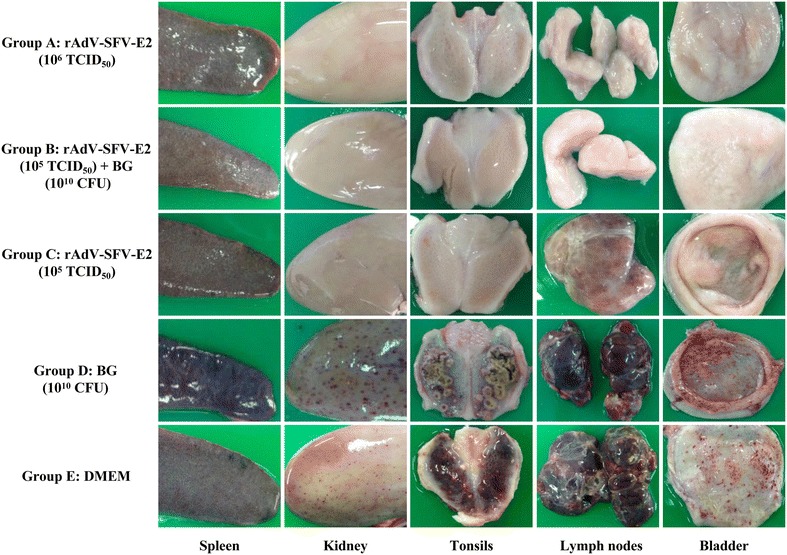
At 15 dpc, all surviving pigs were euthanized and subjected to pathological and histopathological examinations. All the pigs in Groups A and B did not show any pathological changes. Most pigs (3/4) in Group C showed mild lesions (including infarcts in the spleen, slight hemorrhages in the lymph nodes and necrotic foci in the tonsils). Similar severe pathological changes were observed in one pig of Group C and all the pigs of Groups D and E, including infarcts in the spleen, massive petechiae in the kidney and bladder, hemorrhages with necrotic foci in the tonsils, enlargement and hemorrhage of the lymph nodes and button-like ulcers in the ileocecal valve (Figure 2).
Figure 2.

Representative pathological changes of immunized pigs challenged with CSFV Shimen strain. Five groups of pigs (n = 4) were immunized and challenged as described in the “Materials and methods” section. At 15 days post-challenge (dpc), all surviving pigs were euthanized and different tissues (spleen, kidney, tonsils, lymph nodes and bladder) were collected and pathological examinations performed. CFU: colony forming units.
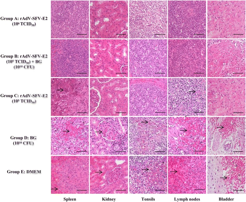
No histopathological changes were observed for pigs in Groups A and B. The pigs in Group C displayed slight to moderate histopathological changes in some tissues, including focal necrosis in the splenic parenchyma and depletion of lymphocytes in the white pulp in the spleen and hemorrhages in the lymph nodes. The pigs in Groups D and E displayed similar severe histopathological changes in most tissues, including depleted lymphoid follicles and hemorrhages in the lymph nodes and tonsils, diffuse hemorrhages throughout the splenic parenchyma and depletion of lymphocytes in the white pulp in the spleen, hemorrhages in the interstitial spaces in the kidney and diffuse hemorrhages in the bladder (Figure 3).
Figure 3.

Representative histopathological changes in pigs challenged with CSFV Shimen strain. Five groups of pigs (n = 4) were immunized and challenged as described in the “Materials and methods” section. At 15 days post-challenge (dpc), various tissues (spleen, kidney, tonsils, lymph nodes and bladder) were collected from the challenged animals, fixed with buffered 4% formalin and subsequently embedded in paraffin wax. Tissue sections (around 4-μm thick) were prepared and stained with hematoxylin and eosin for histopathological examinations. In Groups D and E, severe hemorrhages in the lymph nodes, tonsils, spleen, kidney and bladder were indicated as arrows; in Group C, slight to moderate histopathological changes were found in some tissues, such as focal necrosis in the splenic parenchyma (arrow) and depletion of lymphocytes (arrow) in the white pulp in the spleen and hemorrhages (arrow) in the lymph nodes. All the pigs in Group A or B did not show any histopathological changes. CFU: colony forming units. Bars 50 μm.
Discussion
In this study, the efficacy of rAdV-SFV-E2 in combination with BG was evaluated. The results show that pigs injected with 105 TCID50 rAdV-SFV-E2 plus 1010 CFU BG provided complete protection against lethal CSFV challenge and the efficacy was comparable to 106 TCID50 rAdV-SFV-E2, which indicates that BG can decrease the effective immunization dose of rAdV-SFV-E2 by at least 10-fold.
Adjuvants, such as microbial proteins or carbohydrates may activate APC and induce specific immune responses [19]. BG have the same potential because of their adjuvant components [20, 21]. A recent study has demonstrated that internalization of BG by porcine APC leads to enhanced expression of antigen-presenting molecules on the surface of APC and significantly increases the antigen-presenting capacity of APC [20]. Therefore, more memory B cells were generated following immunization with rAdV-SFV-E2 plus BG than rAdV-SFV-E2 alone, since more antigens were presented to B cells by activated APC. Another possible mechanism might be the effective delivery of rAdV-SFV-E2 by BG, since BG can function as carriers of protein antigens, drugs and a high loading capacity for DNA [9, 21]. Thus, immunization with BG-adjuvanted rAdV-SFV-E2 induced higher E2-specific antibodies and NAbs than rAdV-SFV-E2 alone after booster immunization (Figure 1; Table 1).
No significant difference in serum interferon γ (IFN-γ) and interleukin 4 (IL-4) was found between Groups B (105 TCID50 rAdV-SFV-E2 plus 1010 CFU BG) and C (105 TCID50 rAdV-SFV-E2) (data not shown). Therefore, it is necessary to evaluate cellular immune responses in details in future work, especially the CSFV-specific CD8+ cytotoxic T lymphocytes, which represent an important defense mechanism in the elimination of cells infected by CSFV [5, 22, 23]. In this study, we also found that some pigs immunized with BG displayed transient allergic reactions. In the following experiments, different amounts of the BG will be evaluated and compared with several commonly used adjuvants regarding the enhancement of the protective immunity of rAdV-SFV-E2.
In conclusion, the BG adjuvant can significantly enhance the protective immunity induced by the chimeric vector-based vaccine rAdV-SFV-E2 against CSF in pigs and it may be a promising adjuvant for other vaccines.
Competing interests
The authors declare that they have no competing interests.
Authors’ contributions
S-LX carried out most of the experiments and drafted the manuscript. J-LL, MD, YW, XC and G-TX carried out most of the experiments. L-FL carried out the real‑time PCR test. SL and ED contributed to the study design. SY prepared the Salmonella bacterial ghost adjuvant. H-JQ and YS designed the study, supervised the work and revised the manuscript. All authors read and approved the final manuscript.
Acknowledgements
This study was supported by the grants from the National Natural Science Foundation of China (No. 31200690), National High Technology Research and Development Program of China (2011AA10A210) and Science and Technology Innovation Project from Harbin (2014RFQYJ126).
Footnotes
Shui‑Li Xia and Jian‑Lin Lei are contributed equally to this work
Contributor Information
Shui-Li Xia, Email: xiashuili@126.com.
Jian-Lin Lei, Email: leijianlings@163.com.
Mingliang Du, Email: dumingliang1991@163.com.
Yimin Wang, Email: yiminwang1@hotmail.com.
Xin Cong, Email: 18346226592@163.com.
Guang-Tao Xiang, Email: xiangguangtao0723@163.com.
Lian-Feng Li, Email: lilianfeng124603@163.com.
Shenye Yu, Email: yushenye@163.com.
Enqi Du, Email: duenqi227@126.com.
Siguo Liu, Email: siguo_liu@hvri.ac.cn.
Yuan Sun, Email: sunyuan@hvri.ac.cn.
Hua-Ji Qiu, Email: huajiqiu@hvri.ac.cn.
References
- 1.Lindenbach BD, Murray CL, Thiel HJ. Flaviviridae. In: Knipe DM, Howley PM, Cohen JI, Griffin DE, Lamb RA, Martin MA, Racaniello VR, Roizman B, editors. Fields virology. 6. Philadelphia: Lippincott Williams & Wilkins; 2013. pp. 712–746. [Google Scholar]
- 2.Meyers G, Thiel HJ. Molecular characterization of pestiviruses. Adv Virus Res. 1996;47:53–118. doi: 10.1016/S0065-3527(08)60734-4. [DOI] [PubMed] [Google Scholar]
- 3.Rice CM. Flaviviridae: the viruses and their replication. In: Knipe DM, Howley PM, editors. Fundamental virology. 3. Philadelphia: Lippincott Raven; 1996. pp. 931–959. [Google Scholar]
- 4.Terpstra C. Hog cholera: an update of present knowledge. Br Vet J. 1991;147:397–406. doi: 10.1016/0007-1935(91)90081-W. [DOI] [PubMed] [Google Scholar]
- 5.Pauly T, Elbers K, König M, Lengsfeld T, Saalmüller A, Thiel HJ. Classical swine fever virus-specific cytotoxic T lymphocytes and identification of a T cell epitope. J Gen Virol. 1995;76:3039–3049. doi: 10.1099/0022-1317-76-12-3039. [DOI] [PubMed] [Google Scholar]
- 6.Sun Y, Li HY, Tian DY, Han QY, Zhang X, Li N, Qiu HJ. A novel alphavirus replicon-vectored vaccine delivered by adenovirus induces sterile immunity against classical swine fever. Vaccine. 2011;29:8364–8372. doi: 10.1016/j.vaccine.2011.08.085. [DOI] [PubMed] [Google Scholar]
- 7.Sun Y, Tian DY, Li S, Meng QL, Zhao BB, Li Y, Li D, Ling LJ, Liao YJ, Qiu HJ. Comprehensive evaluation of the adenovirus/alphavirus-replicon chimeric vector-based vaccine rAdV-SFV-E2 against classical swine fever. Vaccine. 2013;31:538–544. doi: 10.1016/j.vaccine.2012.11.013. [DOI] [PubMed] [Google Scholar]
- 8.Leroux-Roels G. Unmet needs in modern vaccinology: adjuvants to improve the immune response. Vaccine. 2010;28(Suppl 3):C25–C36. doi: 10.1016/j.vaccine.2010.07.021. [DOI] [PubMed] [Google Scholar]
- 9.Paukner S, Stiedl T, Kudela P, Bizik J, Al Laham F, Lubitz W. Bacterial ghosts as a novel advanced targeting system for drug and DNA delivery. Expert Opin Drug Deliv. 2006;3:11–22. doi: 10.1517/17425247.3.1.11. [DOI] [PubMed] [Google Scholar]
- 10.O’Hagan DT, MacKichan ML, Singh M. Recent developments in adjuvants for vaccines against infectious diseases. Biomol Eng. 2001;18:69–85. doi: 10.1016/S1389-0344(01)00101-0. [DOI] [PubMed] [Google Scholar]
- 11.Ulrich JT, Myers KR. Monophosphoryl lipid A as an adjuvant. Past experiences and new directions. Pharm Biotechnol. 1995;6:495–524. doi: 10.1007/978-1-4615-1823-5_21. [DOI] [PubMed] [Google Scholar]
- 12.De Becker G, Moulin V, Pajak B, Bruck C, Francotte M, Thiriart C, Urbain J, Moser M. The adjuvant monophosphoryl lipid A increases the function of antigen-presenting cells. Int Immunol. 2000;12:807–815. doi: 10.1093/intimm/12.6.807. [DOI] [PubMed] [Google Scholar]
- 13.Riedmann EM, Kyd JM, Cripps AW, Lubitz W. Bacterial ghosts as adjuvant particles. Expert Rev Vaccines. 2007;6:241–253. doi: 10.1586/14760584.6.2.241. [DOI] [PubMed] [Google Scholar]
- 14.Peng W, Si W, Yin L, Liu H, Yu S, Liu S, Wang C, Chang Y, Zhang Z, Hu S, Du Y. Salmonella enteritidis ghost vaccine induces effective protection against lethal challenge in specific-pathogen-free chicks. Immunobiology. 2011;216:558–565. doi: 10.1016/j.imbio.2010.10.001. [DOI] [PubMed] [Google Scholar]
- 15.Sun Y, Liu DF, Wang YF, Liang BB, Cheng D, Li N, Qi QF, Zhu QH, Qiu HJ. Generation and efficacy evaluation of a recombinant adenovirus expressing the E2 protein of classical swine fever virus. Res Vet Sci. 2010;88:77–82. doi: 10.1016/j.rvsc.2009.06.005. [DOI] [PubMed] [Google Scholar]
- 16.Buonavoglia C, Falcone E, Pestalozza S, Di Trani L, D’Amore E. A rapid serum neutralization test in microplates for the detection of antibodies to hog cholera virus. J Virol Methods. 1989;23:77–79. doi: 10.1016/0166-0934(89)90092-X. [DOI] [PubMed] [Google Scholar]
- 17.Zhao JJ, Cheng D, Li N, Sun Y, Shi Z, Zhu QH, Tu C, Tong GZ, Qiu HJ. Evaluation of a multiplex real-time RT-PCR for quantitative and differential detection of wild-type viruses and C-strain vaccine of classical swine fever. Vet Microbiol. 2008;126:1–10. doi: 10.1016/j.vetmic.2007.04.046. [DOI] [PubMed] [Google Scholar]
- 18.Wang Y, Xia SL, Lei JL, Cong X, Xiang GT, Luo Y, Sun Y, Qiu HJ. Dose-dependent pathogenicity of a pseudorabies virus variant in pigs inoculated via intranasal route. Vet Immunol Immunopathol. 2015;168:147–152. doi: 10.1016/j.vetimm.2015.10.011. [DOI] [PubMed] [Google Scholar]
- 19.Pashine A, Valiante NM, Ulmer JB. Targeting the innate immune response with improved vaccine adjuvants. Nat Med. 2005;11:S63–S68. doi: 10.1038/nm1210. [DOI] [PubMed] [Google Scholar]
- 20.Felnerova D, Kudela P, Bizik J, Haslberger A, Hensel A, Saalmuller A, Lubitz W. T cell-specific immune response induced by bacterial ghosts. Med Sci Monit. 2004;10:BR362–BR370. [PubMed] [Google Scholar]
- 21.Walcher P, Mayr UB, Azimpour-Tabrizi C, Eko FO, Jechlinger W, Mayrhofer P, Alefantis T, Mujer CV, DelVecchio VG, Lubitz W. Antigen discovery and delivery of subunit vaccines by nonliving bacterial ghost vectors. Expert Rev Vaccines. 2004;3:681–691. doi: 10.1586/14760584.3.6.681. [DOI] [PubMed] [Google Scholar]
- 22.Ceppi M, de Bruin MG, Seuberlich T, Balmelli C, Pascolo S, Ruggli N, Wienhold D, Tratschin JD, McCullough KC, Summerfield A. Identification of classical swine fever virus protein E2 as a target for cytotoxic T cells by using mRNA-transfected antigen-presenting cells. J Gen Virol. 2005;86:2525–2534. doi: 10.1099/vir.0.80907-0. [DOI] [PubMed] [Google Scholar]
- 23.Rau H, Revets H, Balmelli C, McCullough KC, Summerfield A. Immunological properties of recombinant classical swine fever virus NS3 protein in vitro and in vivo. Vet Res. 2006;37:155–168. doi: 10.1051/vetres:2005049. [DOI] [PubMed] [Google Scholar]


