Abstract
Introduction: The partial removal of dental caries aiming to maintain the integrity of the pulp has been considered the therapy of choice in the treatment of deep carious lesions, as long as certain principles of diagnosis are respected. Dentists are always looking for techniques to remove the decayed tissue with biosafety, what provides more comfort to the patient especially when it comes to children. Photodynamic therapy (PDT) is an antimicrobial treatment. PapacárieMblue is a modification of the regular Papacárie, with a photosensitizer added to it.
Case Report: PapacárieMblue was used in a patient who had deep carious lesions in a primary molar. After 5 minutes of application, the soft and infected tissues were removed from the side walls of the cavity and, after, PDT was held in the pulp wall with red laser (660 nm), energy of 30 J, output power of 100 mW and 5 minutes of exposure time. This caused a reduction in the amount of dental tissue removed, what favored the prognosis of the dental element. After a period of 3 months, a control of the case was done and we discovered that the tooth that received the PDT was not painful and the x-ray showed an absence of lesions in the furcation.
Conclusion: PDT with PapacárieMblue has been effective in the removal of a deep carious lesion that had a risk of pulp exposure.
Keywords: Dental caries, Photodynamic therapy, LLLT, Children, Primary teeth
Introduction
Dental caries remain highly prevalent throughout the world. The development of carious lesions results from a dynamic process involving acid produced by cariogenic bacteria, leading to the loss of minerals in the affected tooth.1
The principle of minimal intervention, along with the knowledge regarding the development of carious lesions, has led to transformations in the paradigm of restorative dental treatment, with the maximal preservation of sound dental tissue, capable of remineralization.2,3 The current concern is to maintain pulp integrity through the partial removal of carious tissue, which has become the treatment of choice for deep lesions, as long as basic principles are respected.3-5
PapacárieTM is a gel composed of papain and chloramine,3,6,7 the latter of which has disinfecting properties.8 Papain is an enzyme that is similar to human pepsin. It has bactericidal and bacteriostatic actions and serves as a debriding, anti-inflammatory agent that acts on carious tissue while causing no harm to sound dental tissue, thereby accelerating the healing process.8-10
Photodynamic therapy (PDT) has been studied for the treatment of dental caries.11 This method involves the use of a photosensitizer that is activated by light at a specific wavelength. There are different classes of chemical compounds that are used as photosensitizers, such as phenothiazines, phthalocyanines and porphyrins.12-14 In the presence of oxygen, the transfer of energy from the activated photosensitizer results in the formation of a kind of toxic oxygen species known as singlet oxygen and reactive oxygen species. These highly reactive chemical species cause harm to proteins, lipids, nucleic acid and other cell components in microorganisms.15,16 Photosensitizers have a pronounced cationic charge and bond quickly to bacterial cells. Thus, a photosensitizer presents a high degree of selectivity for killing microorganisms instead of host cells.17 Considering the properties of Papacárie and PDT, they have been combined as a treatment option that respects the principle of minimal intervention.
PapacárieMBlue is a modification of the regular Papacárie, with a photosensitizer added to it. The importance of this combination is the use of PapacarieMBlue on the lateral walls of a decayed tooth for the removal of the infected, irreversibly denatured dentin tissue, which is not capable of remineralization and affects the mechanical strength of the tooth, and PDT on the pulp wall to disinfect the affected region without the need for any invasive intervention.
The clinical case described here demonstrates the step-by-step administration of PapacarieMBlue combined with PDT for the treatment of a deep carious lesion in dentin of a primary tooth, as an example of a promising therapeutic option that does not damage the pulp.
Case Report
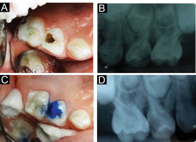
The patient underwent a previous selection, in which the oral health condition was initially assessed, a clinical examination was performed to detect carious lesions, x-rays were taken to analyze the depth of the lesion, risk of pulp involvement, and the furcation region. (Figure 1A and 1B). The research adopted regulations to studies in humans, according to Resolution 496/2012 of the CNS, with the approval of the Ethics Committee of the University Nove de Julho with the number 391.563.
Figure 1 .

A) Initial Photo of Carious Lesion. B) Initial X-Ray. C) Application of PapacárieMBlue. D) Control of the Case After 3 Months.
We selected one patient (male, 6 years old) to participate in the study and the informed consent form was presented to his legal guardian. The possible discomforts, risks, and benefits of this technique were explained to him before he signed the form. The selected tooth had a deep carious lesion, but without pulp involvement, and absence of symptoms.
A relative isolation was made (lip retractor, cotton roller and saliva sucking), followed by the application of PapacárieMBlue, which had an activation time of 5 minutes (Figure 1C). Then, the curettage of the carious lesions from the sidewall was done to remove the contaminated dentin, which had a soft aspect. After, PDT was performed in the pulp wall with red laser (660 nm) and active semiconductor (InGaA1P; Twin Flex Evolution®, MMOptics). The laser parameters were chosen to maintain total radiance energy of 30 J and with an output power of 100 mW. PapacárieMblue was used as the photosensitizer. The irradiation of the dental tissue for 5 minutes (single point) was performed with the use of safety glasses for the operator, assistant and the child.
We made a clinical evaluation through the inspection of the remaining dentin texture with a rounded tip explorer, cleaned the cavity with a cotton pellet and water and then with 2% chlorhexidine gluconate and restored the tooth with a conventional glass ionomer (Maxxion-FGM).
After a period of three months, a control of this case was carried out, when it was found that the tooth in which the PDT was performed presented a small tear in the glass ionomer (Maxxion-FGM), but there were no symptoms in this period, and absence of fistula.
The final x-ray showed no progression of carious lesions, and it was possible to detect that the furcation region remained whole, with the absence of any lesion (Figure 1D), indicating that PDT was effective. PapacárieMBlue promoted a proper removal of the infected dentin, as it was possible to avoid pulp exposure by preserving more dental structure.
Discussion
Infected or uninfected tissues which are capable of remineralization and more internal or affected reversibly denatured dentin should be preserved. The partial removal of carious tissue is performed, leaving a layer of affected dentin in the deepest portion of the cavity.3,18,19 A number of important aspects are involved in the indication of the procedures described here, such as the defense mechanism of the dentin-pulp complex in relation to carious tissues, the initial diagnosis, the differentiation of the types of dentin that compose the carious lesion and the materials indicated for the restorative process.20-22
The use of a photosensitiser with Papacárie potentiates the chemical and mechanical removal of carious tissue, reduces the amount of pathogenic bacteria and prevents the progression of caries, thereby preserving a maximal amount of sound dental tissue.
PDT has advantages such as: promoting a selective cell destruction; if it is necessary, the procedure can be repeated several times, since there is no cumulative toxicity; and the bacteria are eradicated of caries lesions, making the treatment more efficient.23,24
The photosensitizing action depends on the dye that is used, its concentration, fluency laser power intensity, contact time and the bacterial species involved.23,24 PapacarieMBlue has a maximum absorption in the 660 nm region (phototherapeutic window for PDT).23,24
PDT is a relevant and reverse alternative therapy for dental treatment including the treatment of dental cavities, particularly due to its antimicrobial activity, low cost, easy employment and good effectiveness. It is considered a conservative method, possibly able to eliminate the bacteria in the dentin.23 A study, which aimed to evaluate the antimicrobial effect of PDT with methylene blue and a halogen light source in carious lesions in vivo, concluded that this is a promising technique for eliminating bacteria in dental caries.25 A recent study, which used curcumin as a photosensitizing agent irradiated with a LED (L) in the blue wavelength as a light source, showed that PDT should be encouraged due to optimal results against cariogenic bacteria aiming to prevent or treat dental caries.26 Another recent study, which also used curcumin associated with a blue light, concluded that the therapy could efficiently inactivate planktonic cultures of Streptococcus mutans.27 Hakimiha et al compared two different photosensitizers and light sources and came to the conclusion that Streptococcus mutans colonies were susceptible to either 662 nm laser or LED light in the presence of Radachlorin and Toluidine blue, respectively.28 The main difference between these studies and the case presented in this study is the fact that, by using PapacárieMblue, we were able to remove the infected, soft tissue and disinfect the affected tissue through PDT with the same product, at the same time, combining the two treatments in order to improve the prognosis of the dental element in question.
Conclusion
This study concludes that PDT with low power laser using PapacárieMblue was effective in removing deep carious lesions, preventing the risk of pulpal exposure, and promoting microbial reduction.
Conflict of Interest
The authors declare that there is no conflict of interest regarding the publication of this paper.
Please cite this article as follows: da Mota AC, Leal CR, Olivan S, et al. Case report of photodynamic therapy in the treatment of dental caries on primary teeth. J Lasers Med Sci. 2016;7(2):131-133. doi:10.15171/jlms.2016.22.
References
- 1.Fejerskov O, Kidd E. Dental Caries: The disease and its clinical treatment 1st ed. Sao Paulo: Publisher Sa. ntos;2005:50–60. [Google Scholar]
- 2.Ericson D. Minimally invasive dentistry. Oral Health Prev Dent. 2003;1(2):91–22. [PubMed] [Google Scholar]
- 3.Bussadori SK, Guedes CC, Bachiega Bachiega, JC JC, Santis TO, Motta LJ. Clinical and radiographic study of chemical-mechanical removal of caries using Papacárie: 24month follow up. J Clin Pediatr Dent. 2011;35(3):251–254. doi: 10.17796/jcpd.35.3.75803m02524625h5. [DOI] [PubMed] [Google Scholar]
- 4.Frencken JE, Makoni E, Sithole WD. A traumatic restorative treatment and glass ionomer cement sealants in school oral health program in Zimbabwe Evaluation after 1 year. Caries Res. 2006;30(6):428–436. doi: 10.1159/000262355. [DOI] [PubMed] [Google Scholar]
- 5.Matsumoto SF, Motta LJ, Alfaya TA, Guedes CC, Fernandes KP, Bussadori SK. Assessment of chemomechanical removal of carious lesions using Papacárie Duo™: Randomized longitudinal clinical trial. Indian J Dent Res. 2013;24(4):488–492. doi: 10.4103/0970-9290.118393. [DOI] [PubMed] [Google Scholar]
- 6.Jawa D, Singh S, Somani R, Jaidka S, Sirkar K, Jaidka R. Comparative evaluation of the efficacy of chemomechanical caries removal agent (Papacárie) and conventional method of caries removal: an in vitro study. J Indian Soc Pedod Prev Dent. 2010;28(2):73–77. doi: 10.4103/0970-4388.66739. [DOI] [PubMed] [Google Scholar]
- 7.Bittencourt ST, Pereira JR, Rosa AW, Oliveira KS, Ghizoni JS, Oliveira MT. Mineral content removal after Papacarie application in primary teeth: a quantitative analysis. J Clin Pediatr Dent. 2010;34(3):229–231. doi: 10.4103/0970-4388.66739. [DOI] [PubMed] [Google Scholar]
- 8.Bussadori SK, Guedes CC, Hermida BM, RamD RamD. Chemo-mechanical removal of caries in an adolescent patient using a papain gel: case report. J Clin Pediatr Dent. 2008;32:177–180. doi: 10.17796/jcpd.32.3.1168770338617085. [DOI] [PubMed] [Google Scholar]
- 9.Beeley JA, Yip HK, Stevenson AG. Chemomechanical caries removal: a review of the techniques and latest developments. Br Dent. 2000;188(8):427–430. doi: 10.1038/sj.bdj.4800501. [DOI] [PubMed] [Google Scholar]
- 10.Tonami K, Araki K, Mataki S, Kurosaki N. Effects of cloramines and sodium hypoclorite on carious dentin. J Med Dent Sci. 2003;50(2):139–146. [PubMed] [Google Scholar]
- 11.Walsh LJ. Dental lasers: some basic principles. Postgrad Dent. 1994;4:26–29. [Google Scholar]
- 12. Pick RM, Miserendino LJ. Lasers in Dentistry. Chicago: Quintessence; 1995:17-25.
- 13.Goldman L, Goldman B, Van Lieu N. Current laser dentistry. Lasers Surg Med. 1987;6(6):559–562. doi: 10.1002/lsm.1900060616. [DOI] [PubMed] [Google Scholar]
- 14.Wilson M. Photolysis of oral bacteria and its potential use in the treatment of caries and periodontal disease. J Appl Bacteriol. 1993;75:299–306. doi: 10.1111/j.1365-2672.1993.tb02780.x. [DOI] [PubMed] [Google Scholar]
- 15.Wilson M. Lethal photosensitization of oral bacteria and its potential application in the photodynamic therapy or oral infections. J Photochem Photobiol. 2004;3:412–418. doi: 10.1039/b211266c. [DOI] [PubMed] [Google Scholar]
- 16.Pinheiro SL, Schenka AA, Neto AA, de Souza CP, Rodriguez HM, Ribeiro MC. Photodynamic therapy in endodontic treatment of deciduous teeth. Lasers Med Sci. 2009;24(4):521–526. doi: 10.1007/s10103-008-0562-2. [DOI] [PubMed] [Google Scholar]
- 17.Rios A, He J, Glickman GN, Spears R, Schneiderman ED, Honeyman AL. Evaluation of photodynamic therapy using a light emitting diode lamp against Enterococcus faecalis in extractic human teeth. J Endod. 2011;37(6):856–859. doi: 10.1016/j.joen.2011.03.014. [DOI] [PubMed] [Google Scholar]
- 18.El-Tekeya M, El-Habashy L, Mokhles N, El-Kimary E. Effectiveness of 2 chemomechanical caries removal methods on residual bacteria in dentin of primary teeth. Pediatr Dent. 2012;34(4):325–330. [PubMed] [Google Scholar]
- 19.Bijle MN, Patil S, Mumkekar SS, Arora N, Bhalla M, Murali KV. Awareness of dental surgeons in Pune and Mumbai, India, regarding chemomechanical caries removal system. J Contemp Dent Pract. 2013;1; 14(1):96–99. doi: 10.5005/jp-journals-10024-1278. [DOI] [PubMed] [Google Scholar]
- 20.Martins MD, Fernandes KP, Motta LJ, Santos EM, Pavesi VC, Bussadori SK. Biocompatibility analysis of chemomechanical caries removal material Papacarie on cultured fibroblasts and subcutaneous tissue. J Dent Child (Chic) 2009;76:123–129. [PubMed] [Google Scholar]
- 21.Bohari MR, Chunawalla YK, Ahmed BM. Clinical evaluation of caries removal in primary teeth using conventional, chemomechanical and laser technique: an in vivo study. J Contemp Dent Pract. 2012;13(1):40–47. doi: 10.5005/jp-journals-10024-1093. [DOI] [PubMed] [Google Scholar]
- 22.Kochhar GK, Srivastava N, Pandit IK, Gugnani N, Gupta M. An evaluation of differentcaries removal techniques in primary teeth: a comparitive clinical study. J Clin Pediatr Dent. 2011;36(1):5–9. doi: 10.17796/jcpd.36.1.u2421l4j68847215. [DOI] [PubMed] [Google Scholar]
- 23.Carneiro VS, Catão MH. Photodynamic therapy applications in dentistry. Rev Fac Odontol Lins. 2012;22(1):25–32. [Google Scholar]
- 24.Mesquita KS, Queiroz AM, Filho PN, Borsatto MC. Photodynamic therapy: a promising treatment in dentistry? Rev Fac Odontol Lins. 2013;23(2):45–52. [Google Scholar]
- 25.Araújo PV, Correia-Silva Jde F, Gomez RS, Massara Mde L, Cortes ME, Poletto LT. Antimicrobial effect of photodynamic therapy in carious lesions in vivo, using culture and realtime PCR methods. Photodiagnosis Photodyn Ther. 2015;12(3):401–7. doi: 10.1016/j.pdpdt.2015.06.003. [DOI] [PubMed] [Google Scholar]
- 26.Tonon CC, Paschoal MA, Correia M. Comparative effects of photodynamic therapy mediated by curcumin on standard and clinical isolate of Streptococcus mutans. J Contemp Dent Pract. 2015;16(1):1–6. doi: 10.5005/jp-journals-10024-1626. [DOI] [PubMed] [Google Scholar]
- 27.Manoil D, Filieri A, Gameiro C. et al. Flow cytometric assessment of Streptococcus mutans viability after exposure to blue light-activated curcumin. Photodiagnosis Photodyn Ther. 2014;11(3):372–9. doi: 10.1016/j.pdpdt.2014.06.003. [DOI] [PubMed] [Google Scholar]
- 28.Hakimiha N, Khoei F, Bahador A, Fekrazad R. The susceptibility of Streptococcus mutans to antibacterial photodynamic therapy: a comparison of two different photosensitizers and light sources. J Appl Oral Sci. 2014;22(2):80–4. doi: 10.1590/1678-775720130038. [DOI] [PMC free article] [PubMed] [Google Scholar]


