The Editor,
Sir,
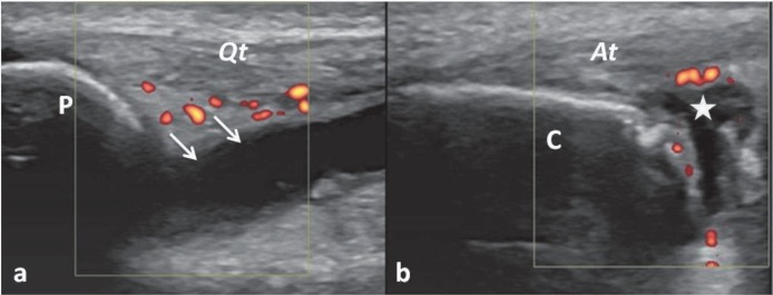
A 23-year old male patient who was previously diagnosed with peripheral spondyloarthropathy (SpA) presented to our clinic. During physical examination, pain and swelling were observed bilaterally in his knee and ankle joints. In this patient, musculoskeletal ultrasound imaging showed grade II synovitis [greater than grade 1 to < 50% of the intra-articular area filled with colour signals representing clear flow] (1) at both regions (Fig. 1A–1B).
Fig. 1. Ultrasound imaging (longitudinal view with power Doppler) for knee joint synovitis (a) and enthesitis of the Achilles tendon (b). The suprapatellar (arrows) and retrocalcaneal (star) bursae are distended with anechoic effusion. Peribursal synovitis is observed at both sites. P, patella; C, calcaneus; Qt, quadriceps tendon; At, Achilles tendon.
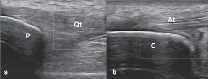
Since his current medical treatment (indomethacin 150 mg/day and sulfasalazine 400 mg/day) was considered insufficient, infliximab (5 mg/kg every 8 weeks) was commenced. On the 4th week of follow-up visit, the patient was found to be significantly improved both clinically and ultrasonographically (Fig. 2).
Fig. 2. A repeat imaging (4 weeks after treatment) reveals the disappearance of the effusions and the power Doppler signal at knee (a) and ankle (b). P, patella; C, calcaneus; Qt, quadriceps tendon; At, Achilles tendon.
Ultrasound can visualize a great spectrum of pathologies regarding peripheral SpA involvement [ie enthesitis, bone erosions, synovitis, bursitis and tenosynovitis] (2). Further, keeping in mind all of its advantages (handy, has high resolution, avoids radiation, provides dynamic imaging), ultrasound imaging of the joints and entheses can reasonably be incorporated as a complementary procedure into the overall assessment of disease activity and response to therapy (2–4). Apart from its great convenience for the clinician during patient follow-up (being as the ‘stethoscope’), the recent literature suggests that ultrasound images can be used as a visual biofeedback for the patients as well (5). Yet, it is not uncommon to have the patients comment and say, “It is not burning any more”, even while Doppler imaging is being performed.
REFERENCES
- 1.Backhaus M, Ohrndorf S, Kellner H, Strunk J, Backhaus TM, Hartung W, et al. Evaluation of a novel 7-joint ultrasound score in daily rheumatologic practice: a pilot project. Arthritis Rheum. 2009;61:1194–1201. doi: 10.1002/art.24646. [DOI] [PubMed] [Google Scholar]
- 2.Naredo E, Batlle-Gualda E, García-Vivar ML, García-Aparicio AM, Fernández-Sueiro JL, Fernández-Prada M, et al. Power Doppler ultrasonography assessment of entheses in spondyloarthropathies: response to therapy of entheseal abnormalities. J Rheumatol. 2010;37:2110–2117. doi: 10.3899/jrheum.100136. [DOI] [PubMed] [Google Scholar]
- 3.Özçakar L, Tok F, De Muynck M, Vanderstraeten G. Musculoskeletal ultrasonography in physical and rehabilitation medicine. J Rehabil Med. 2012;44:310–318. doi: 10.2340/16501977-0959. [DOI] [PubMed] [Google Scholar]
- 4.D’Agostino MA. Ultrasound imaging in spondyloarthropathies. Best Pract Res Clin Rheumatol. 2010;24:693–700. doi: 10.1016/j.berh.2010.05.003. [DOI] [PubMed] [Google Scholar]
- 5.Giggins MO, McCarthy U, Persson UM, Caulfield B. Biofeedback in rehabilitation. J Neuroeng Rehabil. 2013;10:60–60. doi: 10.1186/1743-0003-10-60. [DOI] [PMC free article] [PubMed] [Google Scholar]


