Abstract
Introduction
Alpha-fetoprotein (AFP) concentrations can be determined framing others from invasively acquired amnion fluid (AF-AFP). While the biological role of AFP remains unclear it is well known that AFP-levels can be altered in connection with specific clinical and/or genetic alterations of the fetus.
Materials and Method
here a retrospective study based on 3,119 singleton and 56 twin pregnancies is presented. The standard levels of amnion fluid derived alpha-fetoprotein level (AF-AFP) between 12th and 36th weeks of gestation were determined. Additionally, acetylcholinesterase (AChE) test results for 63 cases, ultrasonography results for 32 cases and abnormal karyotypic findings for 100 cases were available for selected cases.
Results and Discussion
according to the present data the AF-AFP test is reliable and provides expected test results in terms of population studies. However, individual AF-AFP test results can be subject to high individual variations. In this study AF-AFP multiple of medians (MoM) over 1.7 were indicative for neuronal tube defects and/or omphalocele in only 6.3% of the cases, while such AF-AFP values were hints on severe sonographic signs in 62% of the cases. Also, altered AF-AFP concentrations were present in 82% of cytogenetically abnormal cases. Overall, even though predicative value of the AF-AFP-test is matter of discussion it continues to be widely applied in invasive prenatal diagnostics. This study indicates that it only can be applied reliably in combination with other tests like banding cytogenetics, ultrasonography and all embedded in well-established genetic counseling.
Keywords: amnion fluid derived alpha-fetoprotein level (AF-AFP), acetylcholinesterase (AChE), chromosomal aberrations, ultrasonography
Introduction
In the last few decades medicine has advanced in many fields, including the screening tools available for prenatal diagnostics. Nowadays physicians are able to perform detailed diagnostics of unborn fetuses, a possibility being not more than pure science fiction only 50 years back in time. For George Lucas, producer of the first Star Wars film trilogy, it was out of his imagination at that time point, that it would be possible within the next than 25 years to predict anything about unborn children. Thus, in his story it was a surprise at birth (!) to Padmé Amidala that she was pregnant with twins (Leia and Luke Skywalker), instead with only a single child; this would be striking in a civilization being able to travel with more than light speed (1). Nowadays, here on earth, being far from laser swards and intergalactic traveling options, prenatal diagnostics is routine; it can be done invasively and non-invasively and went through many steps of developments, already (2). Apart from analyses of epidemiological data (like age, ethnicity or weight of mother), additional non-invasive and invasive tests can be offered to pregnant women. Routine invasive tests can be based on fetal blood, placenta/ chorion and amnion. To be mentioned as noninvasive options are ultrasonography, serum-screening and next generation screening for free placental DNA in maternal blood. According to the economic and ethno-cultural circumstances in which a baby is expected, different tests can be offered to the pregnant women and thus all mentioned tests are still applied and actual (2).
Alpha-fetoprotein (AFP) concentrations can be determined from maternal serum (S-AFP), as a part of non-invasive settings, or from invasively acquired amnion fluid (AF-AFP). Even though the biological role of AFP remains unclear (3), it was already described in 1977, that AFP-levels can be altered in connection with specific clinical and/or genetic alterations of the fetus (4). As summarized by Schneider et al. (5), the multiple of median (MoM) of AF-AFP is expected to be decreased in case of a trisomy 13, 18 or 21 and of a monosomy X, while it is increased in case of a triploidy.
An important hint for otherwise not detected hidden neural tube defects is expected by AF-AFP-test, too. In case of MoM >1.7 test for acetylcholinesterase (AChE) is recommended (6).
Here we re-evaluated the reliability of the AF-AFP-test retrospectively for 3,207 pregnancies, including 56 dizygotic twin pregnancies. We determined by that the MoM values for our lab between 12th and 36th week of gestation (w.o.g.). Ultrasonography results and abnormal karyotypic findings were correlated with AF-AFP-MoM values in selected cases and compared with the literature.
Material and Methods
AF-AFP-data was available for 3,175 pregnancies between 2000 and 2012. 3,119 were singletons, the remainder 56 dizygote twin pregnancies. AF-AFP was determined between 12th and 36th w.o.g. For 32 singletons with MoM >1.7 ultrasonographic data was available, as high resolution sonography was not necessarily done in the university clinic and thus data not available in retrospective for this study. For 100/3,119 (= 3.2%) singletons pregnancies abnormal karyotypic/ cytogenetic findings were reported. In 63 cases MoM was >1.7 and AChE test was performed in those. This value was chosen internally 0.3 points lower than the literature nowadays suggests (6) to achieve more security for the patients and not to possibly miss a neuronal tube defect.
Results
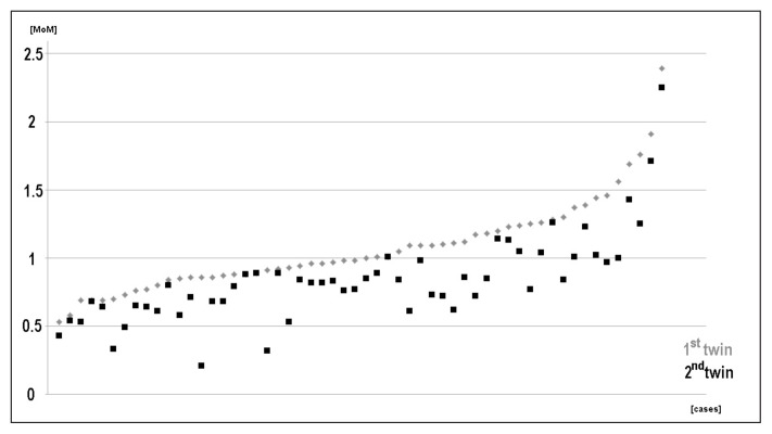
Based on the available data for the 3,119 singleton pregnancies a reference Table was established (Tab. 1). Less than 8 cases were available for w.o.g. 12, 25 and 27 to 36; thus, those values are shown in brackets. For twin pregnancies, data was available only between w.o.g. 14 and 23; more than 8 cases, each were obtainable for w.o.g. 15, 16 and 17, only (Tab. 1). In twin pregnancies for most w.o.g. there is a difference between the two twins of 0.48 to ~5.0 in MoM; one twin had an average MoM of 1.07 compared to 0.84 for the second twin (Fig. 1). Overall, the MoM in twin seems to be about the same as in singleton pregnancies (Tab. 1).
Table 1.
Multiple of median (MoM) of AFP as detected in 3,119 cases, sorted according to weeks of gestation (w.o.g.).
| w.o.g. | data from Habib (1977) median of AFP (μg/ml) | singletons | dizygote twins | ||||
|---|---|---|---|---|---|---|---|
| number of cases | median of AFP (μg/ml) | number of cases | median of AFP (μg/ml) | ||||
| 1st twin | 2nd twin | average | |||||
| 12 | 38 | 3 | (28.40) | - | - | - | - |
| 13 | 36 | 17 | 16.76 | - | - | - | - |
| 14 | 34 | 67 | 16.78 | 3 | (18.21) | (13.02) | (15.62) |
| 15 | 31 | 515 | 14.90 | 8 | 16.99 | 12.60 | 14.80 |
| 16 | 27 | 676 | 13.38 | 15 | 16.88 | 13.06 | 14.97 |
| 17 | 24 | 484 | 11.27 | 11 | 14.82 | 11.08 | 12.95 |
| 18 | 22 | 314 | 9.17 | 5 | (9.36) | (8.86) | (9.11) |
| 19 | 18 | 275 | 7.41 | 4 | (8.51) | (6.66) | (7.59) |
| 20 | 15 | 376 | 5.42 | 6 | (7.01) | (5.54) | (6.28) |
| 21 | 12 | 252 | 4.61 | 2 | (5.27) | (4.37) | (4.82) |
| 22 | 10 | 65 | 3.84 | 1 | (3.19) | (2.71) | (2.95) |
| 23 | 7 | 29 | 3.00 | 1 | (4.43) | (4.43) | (4.43) |
| 24 | 6 | 10 | 2.70 | - | - | - | - |
| 25 | 4 | 4 | (3.29) | - | - | - | - |
| 26 | 3.5 | 8 | 2.01 | - | - | - | - |
| 27 | 3 | 3 | (1.90) | - | - | - | - |
| 28 | 2.5 | 5 | (1.57) | - | - | - | - |
| 29 | 2 | - | - | - | - | - | - |
| 30 | 1.5 | 3 | (1.00) | - | - | - | - |
| 31 | 1 | 5 | (1.16) | - | - | - | - |
| 32 | 0.5 | 3 | (0.40) | - | - | - | - |
| 33 | 0.25 | 3 | (0.80) | - | - | - | - |
| 34 | 0 | - | - | - | - | - | - |
| 36 | 0 | 1 | (0.40) | - | - | - | - |
Figure 1.
Distribution of AF-AFP-MoM values in 56 twin pregnancies. The higher value was assigned to one twin, the lower to the other twin. They were denominated as 1st and 2nd twin for this chart and not as they were assigned due to sonographic data.
In 63 cases with AF-AFP MoM >1.7, AChE test was performed. Only in 4 of these cases (= 6.3%) AChE test was clearly indicative for a neuronal tube defect. In 50 cases (= 79.4%) the AChE test was negative. In the remainder 9 cases AChE levels were only slightly increased, giving no clear results (14.3%).
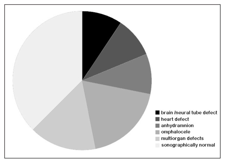
Thirty-two singletons with AF-AFP MoM >1.7 revealed that 38% of those did not show any sonographic signs, while the remainder had severe findings, but none of those had a neuronal tube defect (Fig. 2).
Figure 2.
Pie chart of 32 cases with AF-AFP-MoM >1.7 and the results of ultrasonography.
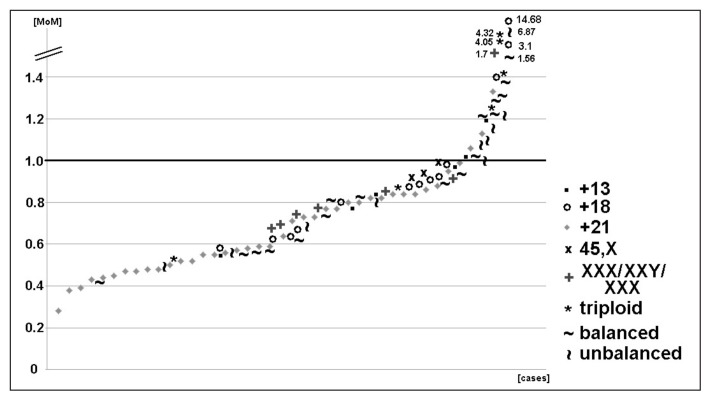
In Figure 3 AF-AFP MoM for 100 cases with different cytogenetic abnormalities are depicted. The abnormalities were summarized into 9 cytogenetic subgroups:
Figure 3.
Distribution of AF-AFP-MoM values in 100 cases with cytogenetic aberrations. The 100 cases are subdivided in 9 groups and depicted as 9 different symbols as indicated in the legend. For cases with MoM >1.4 the values are written beside the symbols.
- 40 cases with free trisomy 21 (average MoM of 0.68);
- 6 trisomy 13 cases (average MoM of 0.88);
- 13 trisomy 18 cases had an average MoM of 2.01 - if excluding the maximal value of 14.68 MoM, considering it as an outlier value, resulted in 1.0, only;
- 3 cases with karyotype 45,X (average MoM of 0.93);
- 6 cases with karyotypes 47,XXX, 47,XYY or 47,XXY (average MoM of 0.91);
- 6 triploidy cases (average of 2.07 MoM);
- 16 balanced chromosomal aberration cases (average MoM of 0.93);
- 10 unbalanced chromosomal aberration cases (average MoM of 1.43).
Details can be found in Table 2.
Table 2.
Cytogenetic result and previously determined MoM values are compared for the 100 cases with chromosomal abnormalities of the present study. The nine subgroups are subdivided, each for cases with decreased, normal and increased MoM.
| Karyotype | MoM decreased (<0.9) | MoM normal (0.9 to 1.1) | MoM increased (>1.1) |
|---|---|---|---|
| trisomy 13 | 50% | 33% | 17% |
| trisomy 18 | 54% | 23% | 23% |
| trisomy 21 | 87.5% | 7.5% | 5% |
| 45,X | 0% | 100% | 0% |
| 47,XXX/47,XXY/47,XYY | 83% | 17% | 0% |
| triploid | 33% | 0% | 67% |
| rearrangement balanced | 50% | 12.5% | 37.5% |
| details on the rearrangements | 46,XN,t(2;16);46,XY,t(6;11); 46,XY,t(3;18); 46,XN,t(8;14); 46,XY,t(9;13); 46,XX,t(18;22); 46,XY,t(2;17); 46,XX,t(9;22) 45,XX,t(14,21)(q10;q10); 45,XX,t(13,14)(q10;q10) 4 cases; 3 cases with heterochromatic extra marker chromosome |
||
| rearrangement unbalanced | 40% | 30% | 30% |
| details on the rearrangements | 46,XY,der(4)t(2;4); 46,XX,der(13)t(13;?) 2 cases; 46,XN,dup(1); 46,XN,del(22)(q11.2q11.2) 2 cases; 46,XN,del(4)(p16.3p16.3) 47,XY,+5[15]/46,XY[2] 1 cases with euchromatic extra marker chromosome 47,XX,+22 |
||
| Overall | 62% | 18% | 20% |
Individually, the MoM varied in these 100 cases between 0.28 and 14.68. Under careful evaluation considering values between 0.9 and 1.1 as normal, Table 2 shows the individual MoM results. Only in 50 to 87.5% of the cases the MoM was decreased in case of trisomy 13, 18 or 21. No decrease in MoM was detected in the three cases with karyotype 45,X, and only 2/3 of triploid cases showed an increased MoM, while in the remainder 33% MoM was even decreased. Decreased MoM was present in 83% of the cases with an additional sex-chromosome. Interestingly, 87.5% of cases with balanced and only 70% of such with unbalanced chromosomal aberrations showed an abnormal AF-AFP MoM.
Discussion
AF-AFP-test continues to be widely applied in invasive prenatal diagnostics. Still, its predicative value is matter of discussion (6, 7).
To the best of our knowledge there is only scarce data available on AF-AFP concentration in amnion fluid during the course of pregnancy. The data which is used up to present in textbooks and in internet (e.g. http://www.glowm.com/resources/glowm/cd/index.html) is based on a work from 1977 (8). The data published there is included in Table 1, and it is striking that AF-AFP concentrations there are always about double as high as in the data obtained in this study in >3,000 cases.
For twin pregnancies no data on AF-AFP-concentration to be expected per gestational week is available in the literature. Here we present a pilot study of 56 such cases. Overall the differences of twins AF-AFP concentrations compared to those of singleton AF-AFP values are modest (Tab. 1). However, differences of on average 0.23 MoM has to be expected here. Pijpers et al. (9) indicate that there might be also AF-AFP diffusion between the two amniotic sacs influencing the obtained data. Sharony et al. (10) could show in 2003 that there is a slight influence of gender in AFP-concentration in twin pregnancies, while the gender had no influence in singleton pregnancies (11); however, these results were also in question, recently (12).
AF-AFP-MoM of >1.7 is described as indication for additional AChE test. Still in the present study only 6.3% of such cases were confirmed to suffer from neuronal tube defects and 14.3% of the case gave no clear result after AChE test. This is in contrast to the literature which indicates much higher detection rates of AF-AFP combined with AChE test for neural tube or other defects including omphalocele (13, 14).
A severely enhanced MoM of >1.7 is also considered as strong hint on sonographically detectable fetal malformations. Robbin et al. (15) argue that combination of enhanced AF-AFP-levels and high resolution sonographic evaluation detect >99% of such fetal malformations. Even though true, there are also in the present study 38% of such pregnancies without any sonographic signs, indicating for a high false positive rate of the test.
The here for 100 cases aligned data concerning AF-AFP MoM and different cytogenetic abnormalities fits in general to the expected published data: the majority of the cases with trisomy 13, 18 and 21 show a decreased MoM. The fact that also subset of these cases can have enhanced MoM values was previously reported (16). The reported decrease of MoM in case of Turner syndrome was not observed in the three here studied cases, while a decrease was observed in 5/6 cases with gain of one sex chromosome. An enhanced MoM in triploidy was present in 67% of the cases, as expected (5). Overall 82% of cytogenetic aberrations were highlighted by altered AF-AFP-values. However, not only decreased but also enhanced AFP-concentrations can be a hint for a chromosomal abnormality.
Overall, the present study revealed that the AF-AFP test is reliable in terms of population studies. However, individual AF-AFP test results are rather subject to variations. While altered AF-AFP concentrations can be found in 82% of cytogenetically abnormal cases its value for detection of neuronal tube defects and/or severe sonographic signs could not be confirmed in 93.7 and 38% of cases with MoM >1.7. If the AF-AFP concentrations are also altered in case of submicroscopic changes like microdeletion- or microduplication- syndromes (17) remains to be determined. Overall, AF-AFP test seems to be obsolete in countries, where other tests like cytogenetics, molecular cytogenetics, array-comparative genomic hybridization, and/or even next generation sequencing can be offered as routine tests to the pregnant women. However, if after invasive procedure only banding cytogenetics can be routinely offered AF-AFP may be a valuable additional test providing hints for the ongoing pregnancy.
References
- 1.Lukas G. Star Wars: From the Adventures of Luke Sykwalker. The random publishing house; London: 1976. [Google Scholar]
- 2.Hixson L, Goel S, Schuber P, Faltas V, Lee J, Narayakkadan A, Leung H, Osborne J. An overview on prenatal screening for chromosomal aberrations. J Lab Autom. 2015 Oct;20(5):562–73. doi: 10.1177/2211068214564595. [DOI] [PubMed] [Google Scholar]
- 3.Mizejewski GJ. Biological roles of alpha-fetoprotein during pregnancy and perinatal development. Exp Biol Med (Maywood) 2004 Jun;229(6):439–63. doi: 10.1177/153537020422900602. [DOI] [PubMed] [Google Scholar]
- 4.Wald NJ, Cuckle H, Brock JH, Peto R, Polani PE, Woodford FP. Maternal serum-alpha-fetoprotein measurement in antenatal screening for anencephaly and spina bifida in early pregnancy. Report of U.K. collaborative study on alpha-fetoprotein in relation to neural-tube defects. Lancet. 1977 Jun 25;1(8026):1323–32. [PubMed] [Google Scholar]
- 5.Schneider H, Husslein P-W, Schneider KTM, editors. Die Geburtshilfe. Springer; Berlin, Heidelberg: 2011. p. 132. [Google Scholar]
- 6.Flick A, Krakow D, Martirosian A, Silverman N, Platt LD. Routine measurement of amniotic fluid alpha-fetoprotein and acetylcholinesterase: the need for a reevaluation. Am J Obstet Gynecol. 2014 Aug;211( 2):139.e1–6. doi: 10.1016/j.ajog.2014.02.005. [DOI] [PubMed] [Google Scholar]
- 7.Sepulveda W, Donaldson A, Johnson RD, Davies G, Fisk NM. Are routine alpha-fetoprotein and acetylcholinesterase determinations still necessary at second-trimester amniocentesis? Impact of high-resolution ultrasonography. Obstet Gynecol. 1995 Jan;85(1):107–12. doi: 10.1016/0029-7844(94)00325-8. [DOI] [PubMed] [Google Scholar]
- 8.Habib ZA. Maternal serum alpha-fetoprotein: its value in antenatal diagnosis of genetic disease and in obstetrical-gynaecological care. Acta Obstet Gynecol Scand Suppl. 1977;61:1–92. doi: 10.3109/00016347709156333. [DOI] [PubMed] [Google Scholar]
- 9.Pijpers L, Jahoda MG, Vosters RP, Niermeijer MF, Sachs ES. Genetic amniocentesis in twin pregnancies. Br J Obstet Gynaecol. 1988 Apr;95(4):323–6. doi: 10.1111/j.1471-0528.1988.tb06599.x. [DOI] [PubMed] [Google Scholar]
- 10.Sharony R, Drugan A, Amiel A, Grinshpun-Cohen J, Markov S, Fejgin MD. Are amniotic fluid alpha-fetoprotein levels influenced by the gender in twin pairs? Fetal Diagn Ther. 2003 Jul-Aug;18(4):281–3. doi: 10.1159/000071015. [DOI] [PubMed] [Google Scholar]
- 11.Drugan A, Yaron Y, Murphy J, Ebrahim SA, Kramer RL, Johnson MP, Evans MI. No effect of fetal sex on amniotic fluid alpha-fetoprotein. Fetal Diagn Ther. 1997 Sep-Oct;12(5):301–3. doi: 10.1159/000264491. [DOI] [PubMed] [Google Scholar]
- 12.Knippel AJ. Role of fetal sex in amniotic fluid alpha-fetoprotein screening. Prenat Diagn. 2002 Oct;22( 10):941–5. doi: 10.1002/pd.408. [DOI] [PubMed] [Google Scholar]
- 13.Tucker JM, Brumfield CG, Davis RO, Winkler CL, Boots LR, Krassikoff NE, Hauth JC. Prenatal differentiation of ventral abdominal wall defects. Are amniotic fluid markers useful adjuncts? J Reprod Med. 1992 May;37(5):445–8. [PubMed] [Google Scholar]
- 14.Crandall BF, Chua C. Risks for fetal abnormalities after very and moderately elevated AF-AFPs. Prenat Diagn. 1997 Sep;17(9):837–41. doi: 10.1002/(sici)1097-0223(199709)17:9<837::aid-pd157>3.0.co;2-o. [DOI] [PubMed] [Google Scholar]
- 15.Robbin M, Filly RA, Fell S, Goldstein RB, Callen PW, Goldberg JD, Golbus MS. Elevated levels of amniotic fluid alpha-fetoprotein: sonographic evaluation. Radiology. 1993 Jul;188(1):165–9. doi: 10.1148/radiology.188.1.7685529. [DOI] [PubMed] [Google Scholar]
- 16.Crandall BF, Matsumoto M. Risks associated with an elevated amniotic fluid alpha-fetoprotein level. Am J Med Genet. 1991 Apr 1;39(1):64–7. doi: 10.1002/ajmg.1320390114. [DOI] [PubMed] [Google Scholar]
- 17.Weise A, Mrasek K, Klein E, Mulatinho M, Llerena JC, Jr, Hardekopf D, Pekova S, Bhatt S, Kosyakova N, Liehr T. Microdeletion and microduplication syndromes. J Histochem Cytochem. 2012 May;60(5):346–58. doi: 10.1369/0022155412440001. [DOI] [PMC free article] [PubMed] [Google Scholar]


