Abstract
Intimate two-way interactions between the implantation-competent blastocyst and receptive uterus are prerequisite for successful embryo implantation. In humans, recurrent/repeated implantation failure (RIF) may occur due to altered uterine receptivity with aberrant gene expression in the endometrium as well as genetic defects in embryos. Several studies have been performed to understand dynamic changes of uterine transcriptome during menstrual cycles in humans. However, uterine transcriptome of the patients with RIF has not been clearly investigated yet. Here we show that several signaling pathways as well as many genes and microRNAs are dysregulated in the endometrium of patients with RIF (RIFE). Whereas unsupervised hierarchical clustering showed that overall mRNA and microRNA profiles of RIFE were similar to those of endometria of healthy women, many genes were significantly dysregulated in RIFE (cut off at 1.5 fold change). The majority (~75%) of differentially expressed genes in RIFE including S100 calcium binding protein P (S100P), Chemokine (C-X-C motif) ligand 13 (CXCL13) and SIX homeobox 1 (SIX1) were down-regulated, suggesting that reduced uterine expression of these genes is associated with RIF. Gene Set Enrichment analyses (GSEA) for mRNA microarrays revealed that various signaling pathways including Leukemia inhibitory factor (LIF) signaling and a P4 response were dysregulated in RIFE although expression levels of Estrogen receptor α (ERα) and Progesterone receptor (PR) were not significantly altered in RIFE. Furthermore, expression and phosphorylation of Signal transducer and activator of transcription 3 (STAT3) are reduced and a gene set associated with Janus kinase (JAK)-STAT signaling pathway is systemically down-regulated in these patients. Pairwise analyses of microRNA arrays with prediction of dysregulated microRNAs based on mRNA expression datasets demonstrated that 6 microRNAs are aberrantly regulated in RIFE. Collectively, we here suggest that dysregulation of several major signaling pathways and genes critical for uterine biology and embryo implantation may lead to uterine abnormalities in patients with RIF.
Introduction
Despite significant improvements in assisted reproductive technology (ART), a substantial numbers of patients undergoing ART fail to achieve successful pregnancy even after repeated attempts [1]. Failure to achieve a pregnancy following 2~6 In Vitro Fertilization (IVF) cycles with high-grade embryos transferred to the endometrium was defined as recurrent/repeated implantation failure (RIF) [2–4]. RIF still remains a major challenge for both clinicians and researchers to improve pregnancy outcomes in ART. Recently, many reports have suggested that local ‘endometrial injury’, such as endometrial biopsy or curettage prior to ART may improve the chance of embryo implantation in the endometrium of patients who suffer from RIF [3–5]. A recent meta-analysis reinforced improvement of clinical outcomes after endometrial injury prior to ART in these patients [6], although it is still controversial. In addition, intrauterine administration of human chorionic gonadotropin-treated autologous peripheral blood mononuclear cells (PBMCs) improves ART outcomes of women with RIF [7]. However, how these approaches may improve uterine environments with appropriate uterine receptivity in patients with RIF and what signaling pathways and/or genes are dysregulated in these patients remain largely unknown.
The endometrium reconstructs itself in each menstrual cycle to provide a favorable environment for blastocyst implantation [8–10]. Numerous factors including cytokines, growth factors, chemokines and adhesion molecules and their receptors have been suggested to participate in this complex process [10, 11]. Certain aspects of embryo implantation are considered similar between mice and humans, and thus, data obtained from diverse gene-manipulated mouse models have been used to extrapolate functional roles of these factors in humans [12–14]. However, the animal models still do have limitations to understand actual events of embryo implantation in humans. Thus, endometrial changes with uterine receptivity in humans have been persistently investigated even with strict ethical limitations. Several microarray experiments have been performed to obtain large-scale expression profiles of mRNAs and/or microRNAs in human endometrium during menstrual cycles [8, 9, 15–18]. They showed that expression patterns of many genes are dynamically changed during menstrual cycles. However, the lists of differentially expressed genes (DEGs) hardly overlap among these studies, suggesting that physiological changes of endometrium are far more complex than we assume [19]. Especially, molecular changes in the endometrium of patients with RIF (RIFE) which lead to implantation failure are largely unknown. Here we show that several signaling pathways including Leukemia inhibitory factor (LIF)-Janus kinase (JAK)-Signal transducer and activator of transcription 3 (STAT3) pathway as well as many genes and microRNAs are dysregulated in RIFE.
Materials and Methods
Patients and endometrial sampling
This study was approved by the Institution Review Board at CHA Bundang Medical Center, CHA University, before sample collection (IRB No 2011-01-001) and all women signed an informed consent form before participating in the study. The control group (n = 7) consisted of volunteer women under the age of 40 years with regular menstrual cycle, who had at least one normal pregnancy and delivery. Women who had a past record of infertility, those currently on oral contraceptive therapy and those with intrauterine contraceptive devices were excluded. The RIF group consisted of patients who had undergone at least three IVF cycles with good quality embryos, but failed to conceive (n = 15). All participants were recruited from Fertility Center of CHA Bundang Medical Center, CHA University (Seongnam, Gyeonggi, Korea). Uterine cavity of control women and patients with RIF was examined by transvaginal sonography and their endometrial thickness was measured. Endometrial biopsies were collected using Pipelle de Cornier® device (CCD Laboratories, Paris, France) on day 21 of the menstrual cycle (midluteal phase). Biopsied samples were immediately transferred to a research laboratory, and processed for snap frozen at −80°C for RNA extraction and/or to be embedded in paraffin for histological evaluation and immunohistochemistry.
RNA extraction and reverse transcription
Total RNA was extracted from each specimen using Trizol Reagent (Invitrogen life technologies, San Diego, CA, USA) according to the manufacturer’s protocols. The purity and concentration of all RNA samples were examined by using a microspectrophotometer (ND-1000, NanoDrop Technologies, Roackland, DE, USA). Five and three endometrial total RNA samples were randomly selected from each group to be used for mRNA and microRNA array experiments, respectively. Two μg of each RNA sample was reverse transcribed using random hexamer primers (Promega Corporation, Madison, WI, USA) and M-MLV reverse transcriptase (Promega Corporation, Madison, WI, USA) in a final volume of 40 μl. Subsequently, cDNA samples were used as the template for PCR using the specific primers (S1 Table) as designed for realtime RT- PCR.
Microarrays and data analyses with GSEA
Microarrays for mRNAs and microRNAs were initially performed with uterine total RNAs. Agilent Human genome 8 x 60 K arrays (Agilent Technologies, Santa Clara, CA, USA) and Affymetrix GeneChip®miRNA 3.0 arrays (Affymetrix, Santa Clara, CA, USA) were hybridized with appropriate cRNA probes at the core facility of GenoCheck (Ansan, Gyeonggi, Korea). The expression value and detection calls were computed from the raw data and Gene Set Enrichment Analyses (GSEA, version 3.7) was applied to interpret expression profiles from microarrays (Broad Institute, Cambridge, MA, USA). GSEA was originally developed to identify cohorts of genes whose functions are integrated into a certain biological process and/or signaling pathways [20]. Pathways were ranked according to the significance of enrichment, and the validation mode measure of significance was used to identify pathways of greatest enrichment.
Quantitative realtime RT-PCR
Realtime RT-PCR was performed using the iCycler (Bio-Rad, Hercules, CA, USA). QuantiTect SYBR Green PCR reagents (Qiagen, Disseldorf, Germany) were used for amplification and results were evaluated with the iQ5™ Optical system software. The gene expression level was calculated using the relative quantification approach based on the ΔΔCt method and this value was then normalized to the relative amounts of an internal control, rPL19 cDNA. All PCRs were performed in duplicate.
Immunohistochemistry
Immunohistochemistry for phosphorylated STAT3 (pSTAT3), estrogen receptor alpha (ERα) and progesterone receptor (PR) was performed on 5 μm sections of formalin-fixed, paraffin-embedded endometria as performed previously [21]. Paraffin sections were deparaffinized in xylene and hydrated in a series of graded ethanols. After a PBS rinse, the endogenous peroxidase activity was quenched on incubation for 10 min with 3% hydrogen peroxide. Sections were then incubated with blocking buffer (4% bovine serum albumin in PBS) containing 5% normal serum for 1 h at room temperature (RT), and incubated with primary antibody in the blocking buffer for 1 h at RT and overnight at 4°C. The primary antibodies were 1:400 anti-pSTAT3 (#MA5-11189, Thermo Fisher Scientific, Rockford, IL, USA), 1:50 ERα (#SC-542, Santa Cruz Biotechnology, Santa Cruz, CA, USA), and 1:200 PR (#PM-9102-S, Thermo Fisher Scientific, Rockford, IL, USA). After washing three times with PBS for 5 min, each section was incubated with 1:200 goat anti-rabbit IgG as the secondary antibody (Bio-Rad, Hercules, CA, USA) in the blocking buffer for 1 h at RT. Hematoxylin was used for nuclear counter-staining of the sections [22, 23]. Assessment of staining intensity and distribution of pSTAT3, ERα and PR was made using a modified semiquantitative analysis of HSCORE scoring system as described elsewhere [22, 23]. In all cases, 1000 cells/sample were evaluated by three independent observers.
Statistical analysis
All experiments were repeated at least three times. Quantitative variables are given as means ± standard deviation. The data were analyzed for statistical significance with the Student's t-test. p-values <0.05 were considered statistically significant.
Results
Characteristic and hormonal profiles of patients with RIF
The mean age, body mass index, and basal serum levels of hormones were similar between control healthy women and patients with RIF (S2 Table).
Overall mRNA expression profiles of RIFE are not distinctly different from those of healthy women
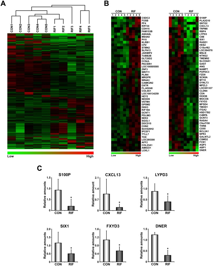
First we performed unsupervised hierarchical clustering for mRNA microarray data of endometria to determine whether overall endometrial transcriptome of patients with RIF is different from that of healthy women in midluteal phase. It shows that overall expression of RIFE is not distinctly different from that of healthy women (Fig 1A), suggesting that local signaling networks with genes critical for embryo implantation may be disturbed in RIFE. In fact, many genes are either up- or down-regulated (641 genes with 1.5 fold cut-off values) in RIFE (S3 Table). Of these, 164 genes (25%) were up-regulated and 477 genes (75%) were down-regulated, suggesting that DEGs in RIFE is mainly down-regulated. Fig 1B shows heatmaps of top 50 up- and down-regulated genes in RIFE. Realtime RT-PCR for S100 calcium binding protein P (S100P), Chemokine (C-X-C motif) ligand 13 (CXCL13), Ly6/PLAUR domain-containing protein 3 (LYPD3), SIX homeobox 1 (SIX1), FXYD domain containing ion transport regulator 3 (FXYD3) and Delta/Notch like EGF repeat containing (DNER) validated that these genes are significantly reduced in RIFE (Fig 1C).
Fig 1. Global expression profiles of the endometrium of patients with RIF are not distinctly different from those of healthy fertile women.
A) Unsupervised hierarchical clustering analysis for mRNA microarray data from endometria of healthy women and patients with RIF. B) Heatmaps for the 50 most increased and decreased genes in RIF. The color spectrum from green to red indicates low to high expression. C) Graphs of realtime RT-PCR results for genes whose expression is significantly reduced in RIF. CON and RIF represent endometrium of healthy fertile women and patients with RIF, respectively. *, p<0.05.
Identification of dysregulated signaling pathways in RIFE
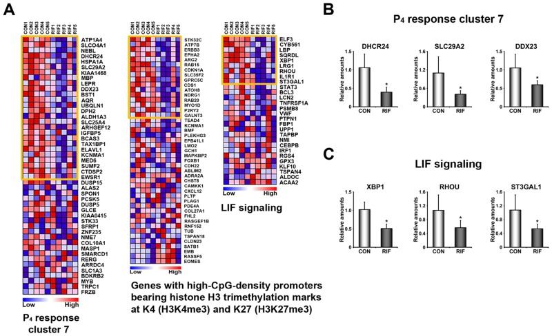
To gain insights into molecular causes leading to RIF in the endometrium, it is critical to identify significantly dysregulated signaling pathways and biological processes. GSEA, a supervised analysis program, was applied to provide insight into aberrantly regulated signaling pathways or biological processes in RIFE. The results suggested that various signaling pathways and biological processes are mainly reduced in RIFE (Tables 1 and 2). It is consistent with the result that 75% of DEGs are down-regulated in RIFE (S3 Table). Gene sets, such as epithelial_differentiation, metastasis and tumor_differentiated may be associated with poor differentiation of epithelial cells in RIFE. Furthermore, LIF signaling and a set of P4 response genes (Response_to_progesterone_cluster_7) are down-regulated in RIFE, suggesting that estrogen and progesterone actions may be impaired in these patients (Fig 2). Interestingly, a gene set for genes with high-CpG-density promoters bearing histone H3 trimethylation marks at K4 (H3K4me3) and K27 (H3K27me3) is dysregulated in the RIFE, suggesting that methylation profile may be also altered in these patients.
Table 1. The list of 22 down-regulated gene sets in the endometrium of patients with RIF.
| Name | Size | Nominal P |
|---|---|---|
| OVARIAN_CANCER | 51 | 0 |
| BREAST_CANCER | 44 | 0 |
| HGF_SIGNALING | 18 | 0 |
| SILENCED_BY_TUMOR_MICROENVIONMENT | 37 | 0 |
| EPITHELIAL_DIFFERENTIATION | 19 | 0 |
| CEBPA_TARGETS | 22 | 0 |
| METASTASIS | 87 | 0 |
| OXIDATIVE_STRESS | 17 | 0 |
| TUMOR_DIFFERENTIATED | 117 | 0 |
| H3K4ME3_AND_HEK27ME3 | 26 | 0.011 |
| ONCOGENIC_SIGNATURE | 92 | 0 |
| EGF_PERSISTENTLY | 17 | 0.010 |
| RESPONSE_TO_PROGESTERONE_CLUSTER_7 | 27 | 0.003 |
| ENDOCRINE_THERAPY_RESISTANCE_3 | 227 | 0 |
| IRF4_TARGETS_IN_MYELOMA | 36 | 0 |
| TARGETS_OF_SMAD2_OR_SMAD3 | 297 | 0 |
| PALILLARY_THYROID_CARCINOMA | 20 | 0.044 |
| NRG1_SIGNALING | 66 | 0.001 |
| LIF_SIGNALING | 18 | 0.011 |
| RESPONSE_TO GONADOTROP | 40 | 0.033 |
| ADIPOGENESIS | 28 | 0.049 |
| RESPONSE_TOSALIRASIB | 100 | 0.043 |
Table 2. The list of 3 up-regulated gene sets in the endometrium of patients with RIF.
| Name | Size | Nominal P |
|---|---|---|
| ENDOMETRIUIM_CANCER | 26 | 0.033 |
| HYPOXIA | 21 | 0.045 |
| BREAST_CANCER_CLUSTER_1 | 16 | 0.010 |
Fig 2. Dysregulated signaling pathways in the endometrium of patients with RIF.
A) Heatmaps of representative gene sets that are aberrantly regulated in the endometrium of patients with RIF listed in Table 1. Genes within the orange boxes are leading characters for building enrichment scores in CON. The color spectrum from blue to red indicates low to high expression. B-C) Validation of systemic alteration of genes in these gene sets with realtime RT-PCR. CON and RIF represent endometrium of healthy fertile women and patients with RIF, respectively. *, p<0.05.
LIF-STAT3 signaling pathway is systemically dysregulated in RIFE
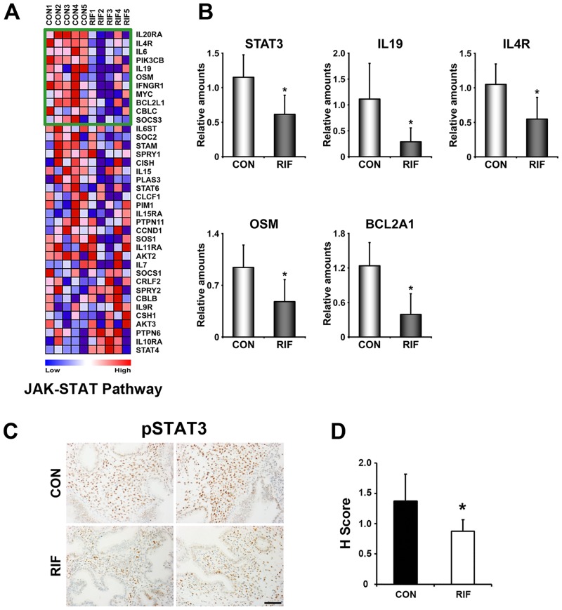
LIF is known as an essential factor of embryo implantation in mice and its expression in the human endometrium is also associated with uterine receptivity [24, 25]. Thus, dysregulation of LIF signaling in RIFE (Table 1) led us to examine whether STAT3, a major downstream effector of LIF signaling, is altered in these patients (Fig 3). Realtime RT-PCR results showed that STAT3 itself and a gene set associated with JAK-STAT signaling pathway including interleukin 19 (IL19), IL4 receptor (IL4R), Oncostatin M (OSM) and Bcl-2-related protein A1 (BCL2A1) are significantly reduced in RIFE (Fig 3A and 3B). Furthermore, immunohistochemistry of pSTAT3, an active form of STAT3, showed that STAT3 activity is notably lower in stromal cells of RIFE (Fig 3C). The HSCORE analysis for pSTAT3 reinforced a statistical significance in this comparison (Fig 3D).
Fig 3. JAK-STAT3 signaling pathway is significantly reduced in the endometrium of patients with RIF.
A) A heatmap of genes in the “JAK-STAT pathway” gene set. Genes within the green box are leading characters for building enrichment scores in CON. The color spectrum from blue to red indicates low to high expression. B) Realtime RT-PCR to confirm differential expression of genes within the green box of the gene set. C) Immunohistochemical analyses of phosphorylated signal transducer and activator of transcription 3 (pSTAT3) in RIF. Note that the number and intensity of STAT3 positive stromal cells were significantly reduced in RIF. D) HSCORE analysis for immunohistochemistry of pSTAT3 in endometrial stromal cells. CON and RIF represent endometrium of healthy fertile women and patients with RIF, respectively. *, p<0.05. Scale bar: 100 μm.
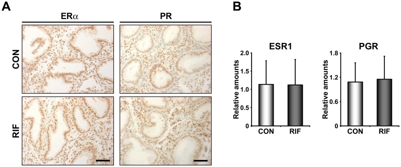
LIF is an established estrogen-responsive gene in the uterus [26, 27]. Since LIF signaling and a P4 response gene cluster are dysregulated in these patients (Fig 2), we examined whether ERα and PR are differentially regulated in RIFE. Realtime RT-PCR, immunohistochemistry for both ERα and PR, and HSCORE analyses for both receptors (Data not shown) collectively showed that there is no significant difference in their expression between healthy control endometrium and RIFE (Fig 4).
Fig 4. Expression levels of steroid hormone receptors are not altered in the endometrium of patients with RIF.
A) Immunohistochemistry of estrogen receptor α (ERα) and progesterone receptor (PR) in the endometria of fertile control women (CON) and patients with RIF (RIF). B) The graphs of realtime RT-PCR results to compare relative expression levels of ERα (ESR1) and PR (PGR) between CON and RIF. Scale bar: 100 μm.
Identification of dysregulated microRNAs in RIFE
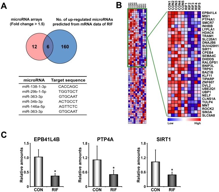
In addition to dysregulated signaling pathways and genes, microRNA arrays provided a list of microRNAs whose expression is either increased or decreased in RIFE (S4 Table). GSEA analyses with mRNA expression data sets (transcriptome) predicted that 166 microRNAs could be aberrantly up-regulated in RIFE. Pairwise analyses for transcriptome and microRNAome for RIFE showed that 6 up-regulated microRNAs (miR-138-1-3p, miR-29b-1-5p, miR-363-3p, miR-34b-3p, miR-146a-5p, and miR-363-3p) are overlapped by these two analyses (Fig 5A). Among these microRNAs, we have validated putative target genes of miR-138-1-3p in RIFE. Collective down-regulation of putative target genes of miR-138-1-3p suggests that reduced expression of these genes in RIFE may be affected by overexpression of these microRNAs (Fig 5B and 5C).
Fig 5. Dysregulated microRNAs in the endometrium of patients with RIF.
A) A Venn diagram to show the number of up-regulated microRNAs collected from microRNA arrays and mRNA arrays in the endometrium of patients with RIF. MicroRNA arrays provided a list of 18 differentially expressed microRNAs, and GSEA with total expression profiles of mRNAs predicted 166 putative microRNAs. Six microRNAs (as listed in a table at the bottom) are overlapped between these two microRNA lists. B) A heatmap of putative target genes of miR-138-1-3p analyzed by GSEA. Genes within the green box are leading characters for building enrichment scores in CON. The color spectrum from blue to red indicates low to high expression. C) The results of realtime RT-PCR to validate down-regulation of putative target genes of miR-138-1-3p which is one of significantly up-regulated microRNAs in RIF. CON and RIF represent endometrium of healthy fertile women and patients with RIF, respectively. *, p<0.05.
Discussion
Among the various potential causes of RIF, uterine factors (e.g., aberrant endometrial receptivity and immunological incompatibility) have become a recent focus of interest in ART [11, 28]. Endometrial receptivity could be modulated by a multitude of signaling molecules, including prostaglandins, growth factors, cytokines and chemokines [29, 30]. We showed that significantly down-regulated genes in RIFE including CXCL13 and S100P may be associated with poor embryo implantation in RIFE (Fig 1). It was suggested that CXCL13, a regulator of mucosal immunity, is abundantly secreted by human endometrial epithelium and specifically taken up by embryos with high implantation potential suggesting that it may be involved in embryo implantation [31]. S100P belongs to the EF-hand superfamily that mediates Ca2+-dependent signal transduction for cell growth, differentiation and metabolism. It is highly expressed during the implantation window in human endometrium [32, 33] and specifically induced in subluminal stromal cells surrounding the implanting blastocyst at the time of embryo implantation in mice [34]. Interestingly, S100P levels in natural cycle are higher than those in controlled ovarian hyperstimulation cycles [33], suggesting that it may be a marker for uterine receptivity in humans. The knockdown of S100A11, another S100 family member, in the uterus impaired embryo implantation in mice and it had adverse effects on the expression of factors related to endometrial receptivity and immune responses in human endometrial cells [35]. Furthermore, a recent proteomic analysis showed that S100A10 is up-regulated in midluteal phase and relatively down-regulated in the endometrium of infertile patients [36]. These reports collectively suggest that certain S100 family members may act as major players for establishing endometrial receptivity.
A variety of cytokines have been proposed to participate in sequential events of embryo implantation [37]. Especially, gene targeting experiments clearly demonstrated that IL6 family cytokines, such as LIF and IL11 are critical for these events [24, 38]. LIF deficient mice show complete implantation failure with no decidualization response [24, 26, 27]. Since then, the general significance of LIF signaling in embryo implantation has been reported in many species including humans. Uterine levels of LIF significantly increase around the period when the embryo initiates implantation in humans [25]. While LIF itself is not down-regulated in this study, STAT3, a major downstream factor to transduce signaling of LIF and other IL6 family in the endometrium, is down-regulated in RIFE (Fig 3). We further demonstrated that pSTAT3 is reduced in uterine stromal cells of these patients, suggesting that LIF-STAT3 signaling is aberrantly reduced in RIFE. In fact, it was reported that LIF is down-regulated in RIFE [39]. Significant reduction of LIF, LIF receptor (LIFR) and pSTAT3 was observed in endometrium of patients with dormant genital tuberculosis, suggesting a possibility that this infection could be one of reasons leading to RIF [40]. Furthermore, several studies showed that expression of LIF or its receptors, LIFR and Glycoprotein 130 (gp130), is reduced in the endometrium of patients with high risk of recurrent miscarriage and unexplained infertility [25, 41, 42]. Collectively, our results as well as previous studies strongly suggest that LIF-STAT3 signaling pathway is one of aberrantly regulated events which may cause RIF in these patients.
A previous study showed that ERα and a group of estrogen-dependent genes are systemically down-regulated in RIFE [43]. However, we did not find similar events with respect to expression levels and activities of ERα in RIFE (Fig 4). This discrepancy could be due to heterogeneous causes of RIF among patients. This assumption is also supported by the fact that DEGs in this study are quite different from those in the previous work [43]. Although expression levels and activity of ERα are not altered in RIFE, estrogen-dependent genes and signaling pathways could be locally impaired. In fact, we found that RASD1 and SIX1, significantly reduced genes in RIFE (Fig 1), are immediately induced by estrogen-ER pathway in mouse uterus (Kim et al., submitted) [44]. In addition, we suggest that P4 signaling is also locally impaired in RIFE while PR levels are not significantly altered in RIFE. Reduced expression of a set of genes whose expression is regulated by P4 (P4 response cluster 7) is consistent with the results of a previous study that P4 signaling is compromised in RIFE [19]. Impaired P4 signaling in RIFE is supported by the result that a set of genes associated with hedgehog signaling pathway, a well-known P4 downstream signaling, is dysregulated as well (S1 Fig). Considered that P4 is critical for the establishment and the maintenance of pregnancy by not only its endocrine but also immunological effects [45], RIF may be associated with immunologic imbalance caused by aberrant P4 signaling in RIFE. They collectively suggest that compromised P4 signaling is one of major causes for RIF since P4 supplementation could expand, to some extent, the length of uterine receptivity in mice [46] and improves clinical outcomes in humans [28].
Revel et al. suggested that a spectrum of microRNAs is either up- or down-regulated in RIFE [47]. While we also found that 6 microRNAs are differentially regulated in RIFE (Fig 5), none of differentially regulated microRNAs were overlapped with ones in the previous study. However, we found that biological pathways, such as JAK-STAT and Hedgehog signaling pathways, consisting of putative target genes of differentially expressed microRNAs in the previous study were also impaired in our patients with RIF (Fig 3 and S1 Fig). All these works strongly suggest that LIF-STAT3 and progesterone signaling pathways are aberrantly reduced in the endometrium of these patients. Pair-wise analyses for transcriptome and microRNAome with a large-scale number of endometrial samples of patients with RIF are required to further understand underlying molecular mechanisms by which uterine environments become impaired, leading to RIF. They may provide molecular evidence(s) for how injury-induced inflammation and/or administration of activated PBMCs improve uterine receptivity and the subsequent pregnancy outcome in patients with RIF [5, 48].
Supporting Information
A) A heatmap of “Hedgehog signaling pathway” gene set. Genes within the orange box are leading characters for building enrichment scores in endometria of healthy women. The color spectrum from blue to red indicates low to high expression. B) Realtime RT-PCR results to validate differential expression of genes within the orange box of the gene set. CON and RIF represent endometrium of healthy fertile women and patients with RIF, respectively *, p<0.05.
(TIF)
(XLSX)
(TIF)
(XLSX)
(TIF)
Acknowledgments
We thank all the volunteers and patients who consented to donate their tissues. We also thank the members of Song laboratory for their helps on immunohistochemistry and HSCORE analyses of pSTAT3, ERα and PR.
Data Availability
All relevant data are within the paper and its Supporting Information files.
Funding Statement
This work was supported in part by the National Research Foundation of Korea (NRF) grants funded by the Korea government (MEST)(NRF-2015R1A2A2A01006714 to HS and NRF-2014R1A1A2059980 to DHC).
References
- 1.Polanski LT, Baumgarten MN, Quenby S, Brosens J, Campbell BK, Raine-Fenning NJ. What exactly do we mean by 'recurrent implantation failure'? A systematic review and opinion. Reproductive biomedicine online. 2014;28(4):409–23. 10.1016/j.rbmo.2013.12.006 . [DOI] [PubMed] [Google Scholar]
- 2.Tan BK, Vandekerckhove P, Kennedy R, Keay SD. Investigation and current management of recurrent IVF treatment failure in the UK. BJOG: an international journal of obstetrics and gynaecology. 2005;112(6):773–80. 10.1111/j.1471-0528.2005.00523.x . [DOI] [PubMed] [Google Scholar]
- 3.Shohayeb A, El-Khayat W. Does a single endometrial biopsy regimen (S-EBR) improve ICSI outcome in patients with repeated implantation failure? A randomised controlled trial. European journal of obstetrics, gynecology, and reproductive biology. 2012;164(2):176–9. 10.1016/j.ejogrb.2012.06.029 . [DOI] [PubMed] [Google Scholar]
- 4.Nastri CO, Lensen SF, Gibreel A, Raine-Fenning N, Ferriani RA, Bhattacharya S, et al. Endometrial injury in women undergoing assisted reproductive techniques. The Cochrane database of systematic reviews. 2015;3:CD009517 10.1002/14651858.CD009517.pub3 . [DOI] [PubMed] [Google Scholar]
- 5.Granot I, Gnainsky Y, Dekel N. Endometrial inflammation and effect on implantation improvement and pregnancy outcome. Reproduction. 2012;144(6):661–8. 10.1530/REP-12-0217 . [DOI] [PubMed] [Google Scholar]
- 6.Potdar N, Gelbaya T, Nardo LG. Endometrial injury to overcome recurrent embryo implantation failure: a systematic review and meta-analysis. Reproductive biomedicine online. 2012;25(6):561–71. 10.1016/j.rbmo.2012.08.005 . [DOI] [PubMed] [Google Scholar]
- 7.Yoshioka S, Fujiwara H, Nakayama T, Kosaka K, Mori T, Fujii S. Intrauterine administration of autologous peripheral blood mononuclear cells promotes implantation rates in patients with repeated failure of IVF-embryo transfer. Human reproduction. 2006;21(12):3290–4. 10.1093/humrep/del312 . [DOI] [PubMed] [Google Scholar]
- 8.Talbi S, Hamilton AE, Vo KC, Tulac S, Overgaard MT, Dosiou C, et al. Molecular phenotyping of human endometrium distinguishes menstrual cycle phases and underlying biological processes in normo-ovulatory women. Endocrinology. 2006;147(3):1097–121. 10.1210/en.2005-1076 . [DOI] [PubMed] [Google Scholar]
- 9.Horcajadas JA, Minguez P, Dopazo J, Esteban FJ, Dominguez F, Giudice LC, et al. Controlled ovarian stimulation induces a functional genomic delay of the endometrium with potential clinical implications. The Journal of clinical endocrinology and metabolism. 2008;93(11):4500–10. 10.1210/jc.2008-0588 . [DOI] [PubMed] [Google Scholar]
- 10.Cha J, Sun X, Dey SK. Mechanisms of implantation: strategies for successful pregnancy. Nature medicine. 2012;18(12):1754–67. 10.1038/nm.3012 . [DOI] [PMC free article] [PubMed] [Google Scholar]
- 11.Fox C, Morin S, Jeong JW, Scott RT Jr., Lessey BA. Local and systemic factors and implantation: what is the evidence? Fertility and sterility. 2016. 10.1016/j.fertnstert.2016.02.018 . [DOI] [PMC free article] [PubMed] [Google Scholar]
- 12.Wang H, Dey SK. Roadmap to embryo implantation: clues from mouse models. Nature reviews Genetics. 2006;7(3):185–99. 10.1038/nrg1808 . [DOI] [PubMed] [Google Scholar]
- 13.Vasquez YM, DeMayo FJ. Role of nuclear receptors in blastocyst implantation. Seminars in cell & developmental biology. 2013;24(10–12):724–35. 10.1016/j.semcdb.2013.08.004 [DOI] [PMC free article] [PubMed] [Google Scholar]
- 14.Bhurke AS, Bagchi IC, Bagchi MK. Progesterone-Regulated Endometrial Factors Controlling Implantation. American journal of reproductive immunology. 2016;75(3):237–45. 10.1111/aji.12473 [DOI] [PMC free article] [PubMed] [Google Scholar]
- 15.Kao LC, Tulac S, Lobo S, Imani B, Yang JP, Germeyer A, et al. Global gene profiling in human endometrium during the window of implantation. Endocrinology. 2002;143(6):2119–38. 10.1210/endo.143.6.8885 . [DOI] [PubMed] [Google Scholar]
- 16.Borthwick JM, Charnock-Jones DS, Tom BD, Hull ML, Teirney R, Phillips SC, et al. Determination of the transcript profile of human endometrium. Molecular human reproduction. 2003;9(1):19–33. . [DOI] [PubMed] [Google Scholar]
- 17.Tapia A, Gangi LM, Zegers-Hochschild F, Balmaceda J, Pommer R, Trejo L, et al. Differences in the endometrial transcript profile during the receptive period between women who were refractory to implantation and those who achieved pregnancy. Human reproduction. 2008;23(2):340–51. 10.1093/humrep/dem319 . [DOI] [PubMed] [Google Scholar]
- 18.Kuokkanen S, Chen B, Ojalvo L, Benard L, Santoro N, Pollard JW. Genomic profiling of microRNAs and messenger RNAs reveals hormonal regulation in microRNA expression in human endometrium. Biology of reproduction. 2010;82(4):791–801. 10.1095/biolreprod.109.081059 [DOI] [PMC free article] [PubMed] [Google Scholar]
- 19.Tapia-Pizarro A, Figueroa P, Brito J, Marin JC, Munroe DJ, Croxatto HB. Endometrial gene expression reveals compromised progesterone signaling in women refractory to embryo implantation. Reproductive biology and endocrinology: RB&E. 2014;12:92 10.1186/1477-7827-12-92 [DOI] [PMC free article] [PubMed] [Google Scholar]
- 20.Subramanian A, Tamayo P, Mootha VK, Mukherjee S, Ebert BL, Gillette MA, et al. Gene set enrichment analysis: a knowledge-based approach for interpreting genome-wide expression profiles. Proceedings of the National Academy of Sciences of the United States of America. 2005;102(43):15545–50. 10.1073/pnas.0506580102 [DOI] [PMC free article] [PubMed] [Google Scholar]
- 21.Kim YS, Kim HR, Kim H, Yang SC, Park M, Yoon JA, et al. Deficiency in DGCR8-dependent canonical microRNAs causes infertility due to multiple abnormalities during uterine development in mice. Scientific reports. 2016;6:20242 10.1038/srep20242 [DOI] [PMC free article] [PubMed] [Google Scholar]
- 22.Saccani Jotti G, Johnston SR, Salter J, Detre S, Dowsett M. Comparison of new immunohistochemical assay for oestrogen receptor in paraffin wax embedded breast carcinoma tissue with quantitative enzyme immunoassay. Journal of clinical pathology. 1994;47(10):900–5. [DOI] [PMC free article] [PubMed] [Google Scholar]
- 23.Kim JY, Song H, Kim H, Kang HJ, Jun JH, Hong SR, et al. Transcriptional profiling with a pathway-oriented analysis identifies dysregulated molecular phenotypes in the endometrium of patients with polycystic ovary syndrome. The Journal of clinical endocrinology and metabolism. 2009;94(4):1416–26. 10.1210/jc.2008-1612 [DOI] [PMC free article] [PubMed] [Google Scholar]
- 24.Stewart CL, Kaspar P, Brunet LJ, Bhatt H, Gadi I, Kontgen F, et al. Blastocyst implantation depends on maternal expression of leukaemia inhibitory factor. Nature. 1992;359(6390):76–9. 10.1038/359076a0 . [DOI] [PubMed] [Google Scholar]
- 25.Dimitriadis E, Stoikos C, Stafford-Bell M, Clark I, Paiva P, Kovacs G, et al. Interleukin-11, IL-11 receptoralpha and leukemia inhibitory factor are dysregulated in endometrium of infertile women with endometriosis during the implantation window. Journal of reproductive immunology. 2006;69(1):53–64. 10.1016/j.jri.2005.07.004 . [DOI] [PubMed] [Google Scholar]
- 26.Chen JR, Cheng JG, Shatzer T, Sewell L, Hernandez L, Stewart CL. Leukemia inhibitory factor can substitute for nidatory estrogen and is essential to inducing a receptive uterus for implantation but is not essential for subsequent embryogenesis. Endocrinology. 2000;141(12):4365–72. 10.1210/endo.141.12.7855 . [DOI] [PubMed] [Google Scholar]
- 27.Song H, Lim H, Das SK, Paria BC, Dey SK. Dysregulation of EGF family of growth factors and COX-2 in the uterus during the preattachment and attachment reactions of the blastocyst with the luminal epithelium correlates with implantation failure in LIF-deficient mice. Molecular endocrinology. 2000;14(8):1147–61. 10.1210/mend.14.8.0498 . [DOI] [PubMed] [Google Scholar]
- 28.Casper RF, Yanushpolsky EH. Optimal endometrial preparation for frozen embryo transfer cycles: window of implantation and progesterone support. Fertility and sterility. 2016. 10.1016/j.fertnstert.2016.01.006 . [DOI] [PubMed] [Google Scholar]
- 29.Miravet-Valenciano JA, Rincon-Bertolin A, Vilella F, Simon C. Understanding and improving endometrial receptivity. Current opinion in obstetrics & gynecology. 2015;27(3):187–92. 10.1097/GCO.0000000000000173 . [DOI] [PubMed] [Google Scholar]
- 30.Thouas GA, Dominguez F, Green MP, Vilella F, Simon C, Gardner DK. Soluble ligands and their receptors in human embryo development and implantation. Endocrine reviews. 2015;36(1):92–130. 10.1210/er.2014-1046 . [DOI] [PubMed] [Google Scholar]
- 31.Franasiak JM, Burns KA, Slayden O, Yuan L, Fritz MA, Korach KS, et al. Endometrial CXCL13 expression is cycle regulated in humans and aberrantly expressed in humans and Rhesus macaques with endometriosis. Reproductive sciences. 2015;22(4):442–51. 10.1177/1933719114542011 . [DOI] [PMC free article] [PubMed] [Google Scholar]
- 32.Horcajadas JA, Pellicer A, Simon C. Wide genomic analysis of human endometrial receptivity: new times, new opportunities. Human reproduction update. 2007;13(1):77–86. 10.1093/humupd/dml046 . [DOI] [PubMed] [Google Scholar]
- 33.Tong XM, Lin XN, Song T, Liu L, Zhang SY. Calcium-binding protein S100P is highly expressed during the implantation window in human endometrium. Fertility and sterility. 2010;94(4):1510–8. 10.1016/j.fertnstert.2009.07.1667 . [DOI] [PubMed] [Google Scholar]
- 34.Zhang R, Wang L, Xia H, Zhang W. Spatio-temporal localization of S100P during the peri-implantation period in the mouse. Reproduction. 2015. 10.1530/REP-15-0381 . [DOI] [Google Scholar]
- 35.Liu XM, Ding GL, Jiang Y, Pan HJ, Zhang D, Wang TT, et al. Down-regulation of S100A11, a calcium-binding protein, in human endometrium may cause reproductive failure. The Journal of clinical endocrinology and metabolism. 2012;97(10):3672–83. 10.1210/jc.2012-2075 [DOI] [PMC free article] [PubMed] [Google Scholar]
- 36.Bissonnette L, Drissennek L, Antoine Y, Tiers L, Hirtz C, Lehmann S, et al. Human S100A10 plays a crucial role in the acquisition of the endometrial receptivity phenotype. Cell adhesion & migration. 2016:1–17. 10.1080/19336918.2015.1128623 . [DOI] [PMC free article] [PubMed] [Google Scholar]
- 37.Stewart CL, Cullinan EB. Preimplantation development of the mammalian embryo and its regulation by growth factors. Developmental genetics. 1997;21(1):91–101. . [DOI] [PubMed] [Google Scholar]
- 38.Robb L, Li R, Hartley L, Nandurkar HH, Koentgen F, Begley CG. Infertility in female mice lacking the receptor for interleukin 11 is due to a defective uterine response to implantation. Nature medicine. 1998;4(3):303–8. . [DOI] [PubMed] [Google Scholar]
- 39.Mariee N, Li TC, Laird SM. Expression of leukaemia inhibitory factor and interleukin 15 in endometrium of women with recurrent implantation failure after IVF; correlation with the number of endometrial natural killer cells. Human reproduction. 2012;27(7):1946–54. 10.1093/humrep/des134 . [DOI] [PubMed] [Google Scholar]
- 40.Subramani E, Madogwe E, Ray CD, Dutta SK, Chakravarty B, Bordignon V, et al. Dysregulated leukemia inhibitory factor and its receptor regulated signal transducers and activators of transcription 3 pathway: a possible cause for repeated implantation failure in women with dormant genital tuberculosis? Fertility and sterility. 2016. 10.1016/j.fertnstert.2015.12.015 . [DOI] [PubMed] [Google Scholar]
- 41.Sharkey A. Cytokines and implantation. Reviews of reproduction. 1998;3(1):52–61. . [DOI] [PubMed] [Google Scholar]
- 42.Aghajanova L, Altmae S, Bjuresten K, Hovatta O, Landgren BM, Stavreus-Evers A. Disturbances in the LIF pathway in the endometrium among women with unexplained infertility. Fertility and sterility. 2009;91(6):2602–10. 10.1016/j.fertnstert.2008.04.010 . [DOI] [PubMed] [Google Scholar]
- 43.Koler M, Achache H, Tsafrir A, Smith Y, Revel A, Reich R. Disrupted gene pattern in patients with repeated in vitro fertilization (IVF) failure. Human reproduction. 2009;24(10):2541–8. 10.1093/humrep/dep193 . [DOI] [PubMed] [Google Scholar]
- 44.Bae S, Kwon H, Yoon H, Park M, Kim HR, Song H, et al. Estrogen-dependent expression of sine oculis homeobox 1 in the mouse uterus during the estrous cycle. Biochemical and biophysical research communications. 2016. 10.1016/j.bbrc.2016.02.116 . [DOI] [PubMed] [Google Scholar]
- 45.Arck P, Hansen PJ, Mulac Jericevic B, Piccinni MP, Szekeres-Bartho J. Progesterone during pregnancy: endocrine-immune cross talk in mammalian species and the role of stress. American journal of reproductive immunology. 2007;58(3):268–79. 10.1111/j.1600-0897.2007.00512.x . [DOI] [PubMed] [Google Scholar]
- 46.Song H, Han K, Lim H. Progesterone supplementation extends uterine receptivity for blastocyst implantation in mice. Reproduction. 2007;133(2):487–93. 10.1530/REP-06-0330 . [DOI] [PubMed] [Google Scholar]
- 47.Revel A, Achache H, Stevens J, Smith Y, Reich R. MicroRNAs are associated with human embryo implantation defects. Human reproduction. 2011;26(10):2830–40. 10.1093/humrep/der255 . [DOI] [PubMed] [Google Scholar]
- 48.Yu N, Yang J, Guo Y, Fang J, Yin T, Luo J, et al. Intrauterine administration of peripheral blood mononuclear cells (PBMCs) improves endometrial receptivity in mice with embryonic implantation dysfunction. American journal of reproductive immunology. 2014;71(1):24–33. 10.1111/aji.12150 . [DOI] [PubMed] [Google Scholar]
Associated Data
This section collects any data citations, data availability statements, or supplementary materials included in this article.
Supplementary Materials
A) A heatmap of “Hedgehog signaling pathway” gene set. Genes within the orange box are leading characters for building enrichment scores in endometria of healthy women. The color spectrum from blue to red indicates low to high expression. B) Realtime RT-PCR results to validate differential expression of genes within the orange box of the gene set. CON and RIF represent endometrium of healthy fertile women and patients with RIF, respectively *, p<0.05.
(TIF)
(XLSX)
(TIF)
(XLSX)
(TIF)
Data Availability Statement
All relevant data are within the paper and its Supporting Information files.


