

Abstract
Objective: The incidence of pulmonary embolism (PE) and leg deep vein thrombosis (DVT) has increased in recent years in association with aging and an increase in the number of bedridden individuals. We developed an active in-bed leg exercise apparatus labeled the Leg Exercise Apparatus (LEX) for DVT prevention. We compared the effect of leg exercises performed using the LEX to conventional active ankle exercises on increased blood flow.
Materials & Methods: The subjects were eight healthy adult volunteers [five men and three women, aged 20–34 (mean 27.0) years]. Subjects performed two types of exercise; exercise 1 consisted of leg exercises using the LEX, while exercise 2 consisted of in-bed active plantar flexion/dorsiflexion exercises without the device. Measurements were taken 1, 5, 10, 20, and 30 minutes after exercise including common femoral vein blood flow, mean blood flow velocity, maximum blood flow velocity, and vessel diameter using Doppler ultrasound. Statistical procedures included timed measurement data analysis using a linear mixed model. A Bonferroni correction was used for multiple comparisons.
Results: Compared to resting levels, blood flow reached a maximum value 1 minute after exercise for both exercise types, with a significantly greater increase after exercise 1 (1.76-fold increase) compared to exercise 2 (1.44-fold increase) (p = 0.005). There was a significant difference (p = 0.03) between the two exercises for all values from 1 minute to 30 minutes following exercise. There was no significant difference between exercises for peak or mean blood flow velocity. Compared to resting levels, blood vessel diameter reached a maximum value of 1.47-fold greater at 5 minutes post-exercise for exercise 1 and a maximum value of 1.21-fold greater at 1 minute post-exercise for exercise 2.
Conclusions: Exercise using the LEX increased lower leg venous blood flow and vessel diameter. We propose that the LEX may serve as a new DVT prevention tool.
Keywords: venous thrombosis, blood flow, exercise
Introduction
The incidence of lower limb deep vein thrombosis (DVT) and pulmonary embolism (PE) has increased in recent years in association with aging and an increase in the number of bedridden individuals1). In various DVT/PE prevention guidelines, including those for perioperative management, the recommended physical prevention methods include wearing compression stockings, using an intermittent pneumatic compression device, and encouraging mobility. Prophylactic administration of antithrombotic drugs is also recommended2,3,4). Many physical prevention methods are effective because they improve blood flow in the legs which is otherwise stagnant during bed rest5,6,7,8). However, mobility is difficult for bedridden individuals, and long-term bed rest in particular is a risk factor for DVT.
Many reports show an increase in femoral vein blood flow associated with active ankle exercises9, 10). However, postoperative patients are poorly motivated to be proactive in performing active ankle exercises, and in many cases effective exercise is not possible. Consequently, we have worked independently to create an active in-bed leg exercise apparatus, which we have labeled the Leg Exercise Apparatus (LEX), in order to enhance the blood flow-increasing effect of active ankle exercises in bedridden individuals. The LEX is a device that aims to increase leg blood flow by performance of a pedaling motion via directly attached foot pedals11, 12).
The objective of this study is to compare post exercise blood flow following active in-bed leg exercises using the LEX and conventionally recommended active ankle exercises.
Materials and Methods
LEX
The LEX has a stainless steel base along with left and right arms and pedals. Leather soles were installed on the pedals to prevent slippage (Figure 1). Range of motion of the pedals allows for 30° of dorsiflexion, 60° of plantar flexion, 30° of inversion, and 20° of eversion; moreover, the arms rotate. This allows not only for plantar flexion and dorsiflexion, but also for more complex pedaling exercises that include inversion and eversion. Arch supports were installed inside the soles, making it possible to compress the venous plexuses of the feet during the pedaling exercise (Figures 2 and 3).
Figure 1.
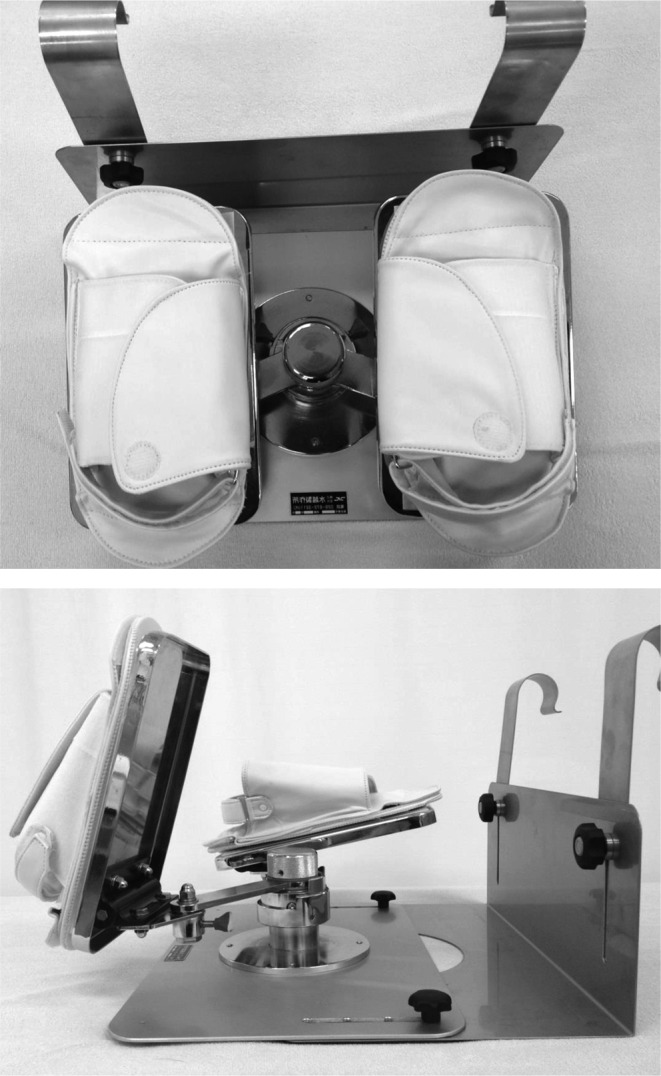
The Leg Exercise Apparatus (LEX).
Figure 2.
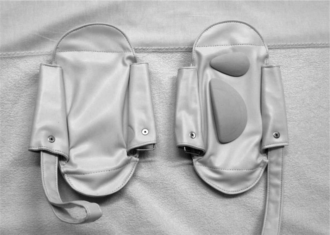
Sole of the Leg Exercise Apparatus with the arch support pad.
Figure 3.

Lateral views of lower limb movement using the Leg Exercise Apparatus. Maximum dorsiflexion (left), maximum plantar flexion (right).
The subjects were eight healthy adult volunteers (five men and three women) aged 20–34 years (mean 27.0 years) (Table 1). All subjects provided written informed consent, and the study protocol was approved by the ethics committee of Tsukuba University Hospital.
Table 1. Characteristics of subjects.
| Subject | Sex | Age | Height | Weight |
|---|---|---|---|---|
| 1 | Male | 20 | 167 | 60 |
| 2 | Male | 24 | 174 | 57 |
| 3 | Male | 27 | 168 | 60 |
| 4 | Male | 28 | 177 | 110 |
| 5 | Male | 32 | 184 | 79 |
| 6 | Female | 22 | 160 | 54 |
| 7 | Female | 29 | 148 | 50 |
| 8 | Female | 34 | 155 | 48 |
The experiment was conducted in a specified room kept at a constant temperature of 25°C with a humidity level of 50–57%. Blood flow was measured using the ultrasonic Doppler method (EUB 7500®, Hitachi, Tokyo, Japan) using a 5 to 13-MHz linear probe, as used in previous studies9, 11). Measurements included blood flow, mean blood flow velocity, maximum blood flow velocity, and vessel diameter of the right common femoral vein. The examination was performed by a single, trained cardiovascular surgeon.
There were two different exercise methods. Exercise 1 (Ex-[1]) consisted of leg exercises using the LEX. With the patient in a supine resting position, the examiner supported the legs of each subject and fitted them to the LEX so that the knee joint could extend to 30°. Complex exercises incorporating plantar flexion/dorsiflexion, inversion/eversion, and knee and hip flexion/extension were performed for 1 minute at 30 cycles/minute. Exercise-2 (Ex-[2]) consisted of in-bed active plantar flexion/dorsiflexion exercises without the device. In supine, subjects positioned the hip in slight flexion with the knee in 30° of flexion and performed maximum-effort plantar flexion/dorsiflexion exercises at 30 cycles/minute (Figure 4). A cycle for each ankle was defined as going from maximum dorsiflexion to maximum plantar flexion and back to the starting position, and the pace was controlled using a metronome. Subjects performed the experiment while wearing compression stockings and were trained in both Ex-[1] and [2] in advance until they were able to acclimate to the exercise methods. Subjects were randomly assigned to perform either Ex-[1] or Ex-[2] first.
Figure 4.

Exercises using the novel active Leg Exercise Apparatus (upper) and traditional simple active ankle movements (lower).
Subjects rested in a supine position for at least 30 minutes, and resting blood flow measurements were obtained after blood flow parameters of the common femoral vein had stabilized. Ex-[1] or [2] was performed immediately after resting blood flow measurements. After completing the 1-minute exercise, measurements were obtained 1, 5, 10, 20, and 30 minutes after exercise. Subjects then took a 90-minute break, and again rested in the supine position for 30 minutes. And resting blood flow measurements were obtained after blood flow parameters of the common femoral vein had stabilized, after which they performed the remaining exercise, and blood flow measurements were repeated in the same manner.
For Ex-[1] and [2], post-exercise values for blood flow, peak blood flow velocity, mean blood flow velocity, and blood vessel diameter were divided by the pre-exercise values to calculate the respective ratios of each. Differences between exercises were then tested using these ratios.
Statistical analysis included timed measurement data analysis using a linear mixed model, and the Bonferroni correction was used for multiple comparisons (p < 0.05).
Results
Blood flow reached a maximum value 1 minute after exercise for both Ex-[1] and [2], at which time blood flow measured 0.65 ± 0.3 L/min and 0.54 ± 0.2 L/min, respectively (Table 2). These values were 1.76 and 1.44 times greater than resting blood flow for Ex-[1] and [2] respectively (F(1, 7) = 16.498, p = 0.005). Moreover, a significant difference was observed between the two exercises for all values 1 minute to 30 minutes after exercise in a two-way linear mixed model (F(1, 77) = 9.138, p = 0.03). No interaction between exercise method and time was observed (Figure 5).
Table 2. Mean (± SD) hemodynamic response to Ex [1] and Ex [2].
| time | Blood flow volume (L/min) | Vessel diameter (mm) | Peak velocity (cm/sec) | Mean velocity (cm/sec) | |||||||
|---|---|---|---|---|---|---|---|---|---|---|---|
| Ex [1] | Ex [2] | Ex [1] | Ex [2] | Ex [1] | Ex [2] | Ex [1] | Ex [2] | ||||
| 0 | 0.39 ± 0.2 | 0.40 ± 0.2 | 7.2 ± 2.4 | 7.8 ± 3.1 | 20.6 ± 7.0 | 20.8 ± 7.4 | 9.7 ± 5.2 | 9.0 ± 4.5 | |||
| 1 | 0.65 ± 0.3 | 0.54 ± 0.2 | 9.5 ± 2.6 | 9.3 ± 3.9 | 21.9 ± 5.9 | 20.7 ± 5.9 | 11.4 ± 3.3 | 9.9 ± 2.1 | |||
| 5 | 0.61 ± 0.2 | 0.48 ± 0.1 | 10.2 ± 2.7 | 9.1 ± 3.8 | 20.0 ± 5.2 | 18.6 ± 5.5 | 10.2 ± 2.9 | 9.1 ± 1.5 | |||
| 10 | 0.61 ± 0.3 | 0.48 ± 0.2 | 9.3 ± 2.2 | 9.0 ± 4.1 | 21.3 ± 5.7 | 21.0 ± 7.3 | 10.8 ± 3.2 | 9.5 ± 3.0 | |||
| 20 | 0.53 ± 0.3 | 0.53 ± 0.2 | 9.1 ± 2.8 | 8.9 ± 3.4 | 19.5 ± 4.4 | 22.7 ± 7.7 | 8.9 ± 3.3 | 10.2 ± 3.2 | |||
| 30 | 0.51 ± 0.2 | 0.48 ± 0.2 | 8.7 ± 2.9 | 8.3 ± 3.6 | 20.7 ± 8.3 | 23.0 ± 10.0 | 10.0 ± 4.4 | 11.1 ± 6.5 | |||
Figure 5.

Blood flow relative to resting levels. Ex-[1]: Exercises using the Leg Exercise Apparatus. Ex-[2]: In-bed active plantar flexion/dorsiflexion exercises without the device. A significant difference was observed between the two exercises for all values 1 minute to 30 minutes after exercise in a two-way linear mixed model (α = 0.05). A significant difference using post-hoc Bonferroni correction was observed at 1 min. ** p < 0.05; GLM, general linear model.
Maximum blood vessel diameter after Ex-[1] reached 10.2 ± 2.7 mm, 1.47-fold greater than resting values at 5 minutes after exercise, while maximum vessel diameter following Ex-[2] reached 9.3 ± 3.9 mm, 1.21-fold greater than resting values at 1 minute after exercise. Blood vessel diameter for Ex-[1] exceeded that of Ex-[2] at all measured post-exercise time points; the difference between exercise methods was significant from 1 minute to 30 minutes after exercise (F(1, 77) = 17.006, p < 0.001). Again, there was no interaction between exercise method and time (Figure 6).
Figure 6.

Blood vessel diameter relative to resting measurements. Ex-[1]: Exercises using the Leg Exercise Apparatus. Ex-[2]: In-bed active plantar flexion/dorsiflexion exercises without the device. A significant difference was observed between the two exercise methods for all values 1 minute to 30 minutes after exercise in a two-way linear mixed model. * (α = 0.05).
For Ex-[1], peak blood flow velocity reached a maximum value 1 minute after exercise, at 1.10 times the resting peak blood flow velocity. For Ex-[2], peak blood flow velocity reached a maximum value 20 minutes after exercise, at 1.10 times the resting peak blood flow velocity (Figure 7). Mean blood flow velocity reached a maximum value 1 minute after exercise for both Ex-[1] and [2], at 1.36 times and 1.22 times the resting mean blood flow velocity, respectively (Figure 8). There were no significant differences between exercises for peak blood flow velocity or mean blood flow velocity.
Figure 7.

Rate of peak blood flow velocity increase relative to resting measurements. Ex-[1]: Exercises using the Leg Exercise Apparatus. Ex-[2]: In-bed active plantar flexion/dorsiflexion exercises without the device. Peak blood flow velocity reached a maximum value 1 minute after exercise for Ex-[1] and 20 minutes after exercise for Ex-[2]. There was no significant difference between exercises.
Figure 8.

Time course of changes in mean blood flow velocity relative to resting measurements. Ex-[1]: Exercises using the Leg Exercise Apparatus. Ex-[2]: In-bed active plantar flexion/dorsiflexion exercises without the device. Mean blood flow reached a maximum value 1 minute after exercise for both Ex-[1] and [2]. There was no significant difference between exercises.
Discussion
Various DVT prevention guidelines recommend active in-bed plantar flexion/dorsiflexion exercises to prevent DVT. The causes of DVT are not clearly understood. Generally, Virchow’s triad (abnormalities in blood flow, blood constituents, and the vessel wall) is thought to be relevant13). To better understand blood flow during active ankle exercises, Sochart et al. assessed blood flow during in-bed active ankle exercise in healthy individuals and found that the mean blood flow velocity of the common femoral vein was 1.38 times greater during exercise than at rest and that the maximum velocity was 1.58 times greater during exercise than at rest9). McNally et al. had patients perform one minute of active plantar flexion/dorsiflexion exercises on day 4 following hip replacement surgery and used strain gauge plethysmography to measure the maximum blood flow volume over time. The maximum blood flow was reached 12 minutes after exercise and was 1.22 times greater than at rest10). Values from these previous studies are similar to those in the present study for Ex-[2], in which blood flow volume and mean blood flow velocity one minute after exercise were 1.44 times and 1.22 times greater than resting levels, respectively. Thus, the active plantar flexion/dorsiflexion exercises increased blood flow similarly to previous studies. By comparison, blood flow following LEX was 1.76 times greater than at rest, which is significantly different from the blood flow increase that occurred following conventional exercises. Blood flow volume is thought to be related to blood viscosity, which is a risk factor for blood coagulation14). Therefore, increased blood flow following LEX could be effective for DVT prevention.
In contrast, peak blood flow velocity in both Ex-[1] and [2] had low rates of elevation, peaking at 1.10 times greater than at rest. In the study by Sochart et al.9), blood flow velocity was measured during active plantar flexion/dorsiflexion exercises same as Ex-[2], resulting in 1.58 times higher compared to the rest at peak blood flow velocity. Alternatively, in the study by McNally et al.10) which measurements were taken after active plantar flexion/dorsiflexion exercises, peak blood flow occurred later rather than immediately after exercise. Peak blood flow velocity was expected to be highest during exercise, but it was not possible to measure blood flow during exercise because of properties of LEX. It’s a limitation of this study.
Of note, quantitative differences in the increase in blood flow volume was significant but maximum mean blood flow velocity was not significant; therefore we could not determine the relationship between these two parameters and no research has been performed to assess blood vessel diameter. The current study demonstrates greater increases in blood vessel diameter following exercises using LEX than with conventional exercises. Since mean and peak velocity of blood flow did not change following exercise, we concluded that the increase in blood flow was related to blood vessel diameter.
Possible mechanisms for the increased blood flow include muscle pumping action15) or autonomic nervous system impact on hemodynamics. However, during exercise using the LEX, cardiac pumping ability may also contribute. One limitation of this experiment is that it was conducted in healthy individuals, and it is possible that these exercises might be difficult for bedridden individuals to perform. The feasibility and outcomes of the exercises should also be verified in older individuals.
Conclusions
Exercise using the LEX increased lower leg venous blood flow and blood vessel diameter. These findings support LEX use as a DVT prevention tool.
Acknowledgement
This study was supported by a “Japan Science and Technology Agency” grant provided through “A-STEP high risk challenging type (revitalization promotion type)”.
We thank Mr. Toshiji Mizukoshi, executive advisor and executive director at Nemoto Co., Ltd., and Tsutomu Mizukoshi, co-producer of LEX, for their advice on production of the LEX. We would also like to express our gratitude to the late Kiyoshi Eguchi, former professor of the University of Tsukuba Hospital, for his general support and warm encouragement.
References
- 1.Kobayashi T. Thromboprophylaxis of venous thromboembolism. Nihon Rinsho 2014; 72: 1303–1308(in Japanese). [PubMed] [Google Scholar]
- 2.JCS Joint Working GroupGuidelines for the diagnosis, treatment and prevention of pulmonary thromboembolism and deep vein thrombosis (JCS 2009). Circ J 2011; 75: 1258–1281. doi: 10.1253/circj.CJ-88-0010 [DOI] [PubMed] [Google Scholar]
- 3.Mont M, Jacobs J, Lieberman J. Members of 2007 and 2011 AAOS Guideline Development Work Groups on PE/VTED ProphylaxisPreventing venous thromboembolic disease in patients undergoing elective total hip and knee arthroplasty. J Bone Joint Surg Am 2012; 94: 673–674. [DOI] [PMC free article] [PubMed] [Google Scholar]
- 4.Falck-Ytter Y, Francis CW, Johansson NA. Prevention of VTE in orthopedic surgery patients. Antithrombotic Therapy and Prevention of Thrombosis, 9th ed. American College of Chest Physicians Evidence-Based Clinical Practice Guidelines. Chest 2012; 141(2 Suppl): e278S-e325S. [DOI] [PMC free article] [PubMed] [Google Scholar]
- 5.Kakkos SK, Szendro G, Griffin M. The efficacy of the new SCD response compression system in the prevention of venous stasis. J Vasc Surg 2000; 32: 932–940. doi: 10.1067/mva.2000.110358 [DOI] [PubMed] [Google Scholar]
- 6.Kakkos SK, Griffin M, Geroulakos G. The efficacy of a new portable sequential compression device (SCD Express) in preventing venous stasis. J Vasc Surg 2005; 42: 296–303. doi: 10.1016/j.jvs.2005.03.031 [DOI] [PubMed] [Google Scholar]
- 7.Griffin M, Nicolaides AN, Bond D. The efficacy of a new stimulation technology to increase venous flow and prevent venous stasis. Eur J Vasc Endovasc Surg 2010; 40: 766–771. doi: 10.1016/j.ejvs.2010.06.019 [DOI] [PubMed] [Google Scholar]
- 8.Charles T, Mackintosh D, Healy B. Merino wool graduated compression stocking increases lower limb venous blood flow: a randomized controlled trial. Adv Ther 2011; 28: 227–237. doi: 10.1007/s12325-010-0107-5 [DOI] [PubMed] [Google Scholar]
- 9.Sochart DH, Hardinge K. The relationship of foot and ankle movements to venous return in the lower limb. J Bone Joint Surg Br 1999; 81: 700–704. doi: 10.1302/0301-620X.81B4.8909 [DOI] [PubMed] [Google Scholar]
- 10.McNally MA, Cooke EA, Mollan RA. The effect of active movement of the foot on venous blood flow after total hip replacement. J Bone Joint Surg Am 1997; 79: 1198–1201. [DOI] [PubMed] [Google Scholar]
- 11.Shimizu Y, Kamada H, Tanaka K. The effect of the ankle active exercise apparatus for improving the venous blood flow in the lower extremities. J Jpn Orthop Assoc 2014; 88: S628.(Abstract). [Google Scholar]
- 12.Shimizu Y, Kamada H, Sakane M. A novel apparatus for active leg exercise improves venous flow in the lower extremity. J Sports Med Phys Fitness 2015; (in press). [PubMed] [Google Scholar]
- 13.Summers JA, Clinch J, Radhakrishnan M. The geko™ electro-stimulation device for venous thromboembolism prophylaxis: a NICE medical technology guidance. Appl Health Econ Health Policy 2015; 13: 135–147. doi: 10.1007/s40258-014-0139-0 [DOI] [PMC free article] [PubMed] [Google Scholar]
- 14.Makin A, Silverman SH, Lip GY. Peripheral vascular disease and Virchow’s triad for thrombogenesis. QJM 2002; 95: 199–210. doi: 10.1093/qjmed/95.4.199 [DOI] [PubMed] [Google Scholar]
- 15.Miller BF, Gruben KG, Morgan BJ. Circulatory responses to voluntary and electrically induced muscle contractions in humans. Phys Ther 2000; 80: 53–60. [PubMed] [Google Scholar]