Abstract
Peroxisome proliferator-activated receptor gamma (PPARγ) is a critical regulator of carbohydrate and lipid metabolism, adipocyte differentiation and inflammatory response. Post-translational modification of PPARγ and its degradation involve several pathways, including the ubiquitin–proteasome system. Here, we identified F-box only protein 9 (FBXO9) as an E3 ubiquitin ligase of PPARγ. We screened interacting partners of PPARγ using immunoprecipitation and mass spectrometric analysis and identified FBXO9 as an E3 ubiquitin ligase of PPARγ. FBXO9 directly interacted with PPARγ through the activation function-1 domain and ligand-binding domain. FBXO9 decreased the protein stability of PPARγ through induction of ubiquitination. We found that the F-box motif of FBXO9 was required for its ubiquitination function. The activity of PPARγ was significantly decreased by FBXO9 overexpression. Furthermore, FBXO9 overexpression in 3T3-L1 adipocytes resulted in decreased levels of endogenous PPARγ and suppression of adipogenesis. These results suggest that FBXO9 is an important enzyme that regulates the stability and activity of PPARγ through ubiquitination.
Introduction
Peroxisome proliferator-activated receptor gamma (PPARγ) is a member of the nuclear hormone receptor superfamily and is a master regulator of adipocyte differentiation.1 There are two isoforms of PPARγ: PPARγ1 and PPARγ2. Thirty extra amino acids are added to the N-terminal of PPARγ2, but the other sequences are identical in both isoforms. While PPARγ1 is expressed in a number of tissues, PPARγ2 is only observed in adipocytes.2 PPARγ activity is upregulated by binding of its ligands, such as thiazolidinediones (TZDs).2 TZDs increase fat accumulation and improve insulin sensitivity via activation of PPARγ. The activity of PPARγ is also regulated by modulating the protein level of PPARγ. PPARγ (+/−) heterozygous mice, which have lower levels of PPARγ than wild-type mice, are protected from high-fat diet-induced weight gain and insulin resistance.3, 4, 5 These findings suggest that obesity and insulin resistance can be ameliorated by controlling the abundance of PPARγ proteins in adipose tissue, independent of TZD activation.
Several reports have demonstrated that PPARγ is degraded by the ubiquitin–proteasome system.6, 7 The ligand-binding domain (LBD) of PPARγ contains major ubiquitination sites, and both the N-terminal activation function-1 (AF-1) domain and the LBD are important for recognition by the proteasome.8 Interestingly, PPARγ ubiquitination and degradation are increased by its specific ligands, TZDs, in adipocytes.9, 10 The reduction in PPARγ protein levels by TZDs is not observed in other cell types, such as myotubes or liver cells. A report has demonstrated that Drosophila seven-in-absentia homolog 2 (Siah2) is an E3 ubiquitin ligase that regulates PPARγ ubiquitination in adipocytes, and its expression is increased by TZDs.11 Recently, other E3 ligases, such as makorin ring finger protein 1 (MKRN1) and neural precursor cell-expressed developmentally downregulated 4 (NEDD4-1)12, were reported to be involved in PPARγ ubiquitination and proteasome-dependent degradation.13
F-box proteins are substrate-recruiting subunits of the Skp1-Cullin-F-box (SCF) ubiquitin ligase complex.14 Little is known about the regulation and functional role of F-box only protein 9 (FBXO9), a member of the F-box proteins. Expression of FBXO9 was increased in vascular smooth muscle cells under high-glucose culture conditions and in streptozotocin (STZ)-induced diabetic rat vessels.15 FBXO9 mediated ubiquitination of telomere maintenance 2 (Tel2) and Tel2 interacting protein 1 (Tti1), integral components of mTORC1, in response to growth factor withdrawal, which inactivates mTORC1 and leads to cell survival in myeloma.16
In this study, we analyzed PPARγ-interacting proteins using co-immunoprecipitation in the presence of MG132, a specific proteasome inhibitor, and mass spectrometric analysis to find a novel E3 ubiquitin ligase specific to PPARγ. FBXO9 was shown to interact with PPARγ and to facilitate ubiquitination and subsequent degradation of PPARγ, suggesting that FBXO9 regulates PPARγ at the protein level as an E3 ubiquitin ligase.
Materials and methods
Antibodies and chemicals
Antibodies against FBXO9, PPARγ, ubiquitin and HA were purchased from Santa Cruz Biotechnology (Santa Cruz, CA, USA). Antibodies against γ-tubulin and FLAG were purchased from Sigma-Aldrich (St Louis, MO, USA). Cycloheximide, 3-isobutyl-1-methylxanthine (IBMX), dexamethasone, insulin and dimethyl sulfoxide (DMSO) were also purchased from Sigma-Aldrich. MG132 was purchased from Calbiochem (Boston, MA, USA).
Cell culture
COS7 cells were cultured in Dulbecco's modified Eagle's medium (DMEM) supplemented with 10% fetal bovine serum (FBS) in a 5% CO2 incubator. 3T3-L1 preadipocytes were maintained in DMEM containing 10% calf serum in a 5% CO2 incubator. Adipocyte differentiation was induced by incubation in DMEM containing 10% FBS, 0.5 μM 3-isobutyl-1-methylxanthine (IBMX), 0.25 μM dexamethasone and 5 μg ml−l insulin for 2 days. Cells were incubated in DMEM with 10% FBS and 1 μg ml−1 insulin for the following 2 days and further cultured in DMEM with 10% FBS for four additional days.
Construction of plasmids and adenoviruses
The mouse FBXO9 (FLAG-FBXO9) expression vector was constructed by subcloning the corresponding cDNAs into the pFLAG-CMV vector using the HindIII and NotI sites. The F-box deletion mutant of mouse FBXO9 (FLAG-FBXO9 ΔF) was generated by subcloning as follows. The region encoding amino acids 1–178 of mouse FBXO9 was ligated into pFLAG-CMV using the HindIII and NotI sites, and the region encoding amino acids 230–437 of mouse FBXO9 was ligated using the NotI and BglII sites. The preparation of the HA-tagged mouse PPARγ2 expression vector was previously described.17 The His-ubiquitin expression vector was kindly provided by CH Chung (Seoul National University, Seoul, South Korea). Adenoviruses encoding FLAG-tagged mouse FBXO9 (Ad-FLAG-FBXO9) were prepared following a previously reported method using the GFP co-expression adenovirus system. Briefly, FLAG-tagged mouse FBXO9 was inserted into pAdTrack-CMV. Ad-FLAG-FBXO9 was generated by homologous recombination between pAdTrack-CMV-Flag-FBXO9 and pAdEasy and by adenovirus packaging in HEK-293. The deletion mutants of mouse PPARγ2 were cloned into HA-tagged pcDNA3.1 as follows. The cDNAs encoding the AF1 domain from amino acids 1 to 138, the DNA-binding domain and the hinge domain from amino acids 139 to 279, and ligand-binding domain (LBD/AF2) from amino acids 279 to 505 of mouse PPARγ2 were generated using PCR. The cDNAs were then ligated into HA-tagged pcDNA3.1 using the KpnI and XbaI sites.
Glutathione-S-transferase (GST) pull-down assay
Glutathione Sepharose 4B (20 μl) (GE Healthcare, Pittsburgh, PA, USA), GST-FBXO9 (1 μg) (Antibodies, Atlanta, GA, USA) and PPARγ LBD (2 μg) (Cayman Chemical, Ann Arbor, MI, USA) were mixed and incubated in 20 mM Tris-HCl, pH 7.4, 10 mM Na4P2O7, 100 mM NaF, 2 mM Na3VO4, 1% NP-40 buffer supplemented with protease inhibitors (10 μg μl−1 aprotinin, 10 μg μl−1 leupeptin and 1 mM phenylmethylsulfonyl fluoride) overnight at 4 °C. The precipitates were washed four times and eluted using Laemmli sample buffer (62.5 mM Tris-HCl, pH 6.8, 2% sodium dodecyl sulfate, 25% glycerol, 5% 2-mercaptoethanol, 0.01% bromophenol blue) (Sigma, St Louis, MI, USA). The precipitates were blotted with either anti-PPARγ antibody or anti-FBXO9 antibody.
Immunoprecipitation and western blot analysis
To evaluate the interaction between PPARγ and FBXO9, cell lysates (500 μg) were prepared with lysis buffer containing 20 mM Tris-HCl (pH 7.4), 1% NP-40, 5 mM EDTA, 2 mM Na3VO4, 100 mM NaF, 10 mM Na4P2O7, 1 mM phenylmethylsulfonyl fluoride, 7 μg ml−1 aprotinin and 7 μg ml−1 leupeptin. To assess the FBXO9-dependent ubiquitination of PPARγ, COS7 cells were transfected with pHA-PPARγ, pHis-Ub and pFLAG-FBXO9 for 12 h. MG132 (10 μM) was added for 4 h before harvesting. Cell lysates (500 μg) were prepared in denaturing condition using lysis buffer containing 20 mM Tris-HCl (pH 7.4), 150 mM NaCl, 1 mM EDTA, 1% Triton X-100, 1% Na-deoxycholate, 0.1% sodium dodecyl sulfate, 10 mM Na4P2O7, 2 mM Na3VO4, 100 mM NaF, 7 μg ml−1 aprotinin, 7 μg ml−1 leupeptin and 1 mM phenylmethylsulfonyl fluoride. Cell lysates were used for immunoprecipitation with anti-HA antibody-coupled agarose beads (Roche, Basel, Switzerland) for 4 h at 4 °C. The precipitates were washed five times and subjected to sodium dodecyl sulfate–polyacrylamide gel electrophoresis and then blotted with specific antibodies. All blots were developed using an enhanced chemiluminescence kit (Thermo Fisher Scientific, Boston, MA, USA).
Transient transfection and reporter assay
COS7 cells were seeded 1 day before transfection and transfected with expression vectors using LipofectAMINE Plus reagent (Invitrogen, Carlsbad, CA, USA). Reporter activity was detected using a luciferase assay system kit (Promega, Fitchburg, WI, USA) and Lumat LB9507 (Berthold Technologies, Bad Wildbad, Germany). Luciferase activity was normalized to β-galactosidase activity.
Real-time PCR
Total RNA was isolated using TRIzol (Invitrogen). cDNAs were synthesized by reverse transcription with 1 μg of total RNA. Real-time PCR was performed using the SYBR master mix (Takara, Otsu, Shiga, Japan) with specific primers for each gene and an ABI 7500 Real-time PCR system (Applied Biosystems, Carlsbad, CA, USA). Primer sets are as follows: mouse PPARγ forward primer, 5′-TCAGGGCTGCCACTTTCG-3′ reverse primer, 5′-GTAATCAGCAACCATTGGGTCA-3′ mouse 18 S rRNA forward primer, 5′-CGCGGTTCTATTTTGTTGGT-3′ reverse primer, 5′-AGTCGGCATCGTTTATGGTC-3′. 18S rRNA was used as an endogenous control for normalization.
Staining of lipid droplets
Cells were fixed with 3.5% paraformaldehyde for 10 min and washed with phosphate-buffered saline three times. Cells were stained using oil red O solution (1.5% oil red O in 60% isopropanol) for 1 h and washed with phosphate-buffered saline three times. Cells were mounted with 50% glycerol and observed.
Mass spectrometry analysis
To isolate interacting proteins of PPARγ, Plat-E cells were overexpressed with FLAG-PPARγ2 or pcDNA, as a negative control, for 20 h. Cells were treated with MG132 (10 μM) for 6 h prior to harvesting. Cell lysates were prepared with lysis buffer containing 50 mM Tris-HCl (pH 7.4), 150 mM NaCl, 1 mM EDTA, 1% Triton X-100 and protease inhibitors. A total of 8 mg of cell lysates was subjected to immunoprecipitation with an anti-FLAG M2 affinity gel (Sigma-Aldrich, 30 μl) for 1.5 h at 4 °C. Beads were washed three times using wash buffer containing 50 mM Tris-HCl (pH 7.4), 150 mM NaCl, 0.2% TritonX-100 followed by two washes using wash buffer without TritonX-100. The eluate for interacting proteins of PPARγ was prepared by a competitive elution using 3X FLAG elution buffer containing 50 mM Tris-HCl (pH 7.4), 150 mM NaCl and 3X FLAG peptides (225 ng μl−1 final concentration; Sigma-Aldrich), and mass spectrometry analysis was then performed.
Statistics
Statistical analyses were performed using SPSS Statistics 20 (SPSS Inc., Chicago, IL, USA). Data are expressed as the mean±s.e., and the difference between the means was analyzed using the Mann–Whitney U-test. A P-value less than 0.05 indicated a statistically significant difference.
Results
FBXO9 interacts with PPARγ and regulates PPARγ protein levels
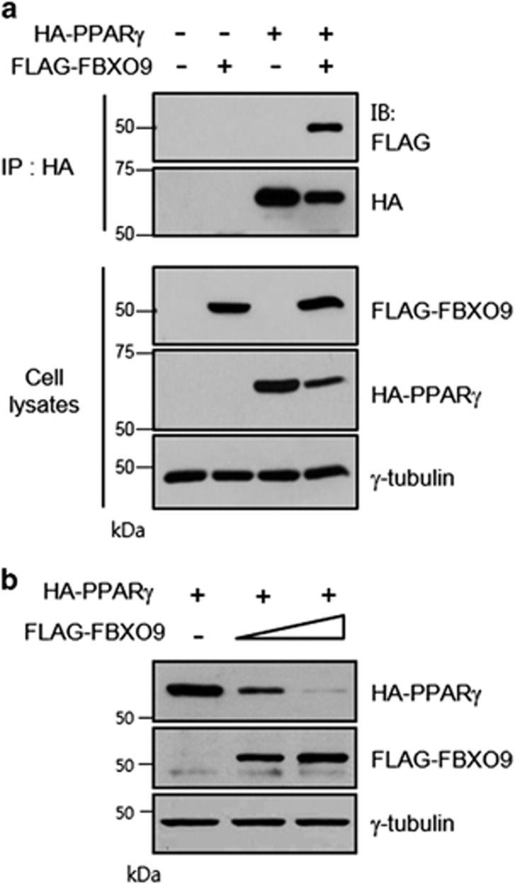
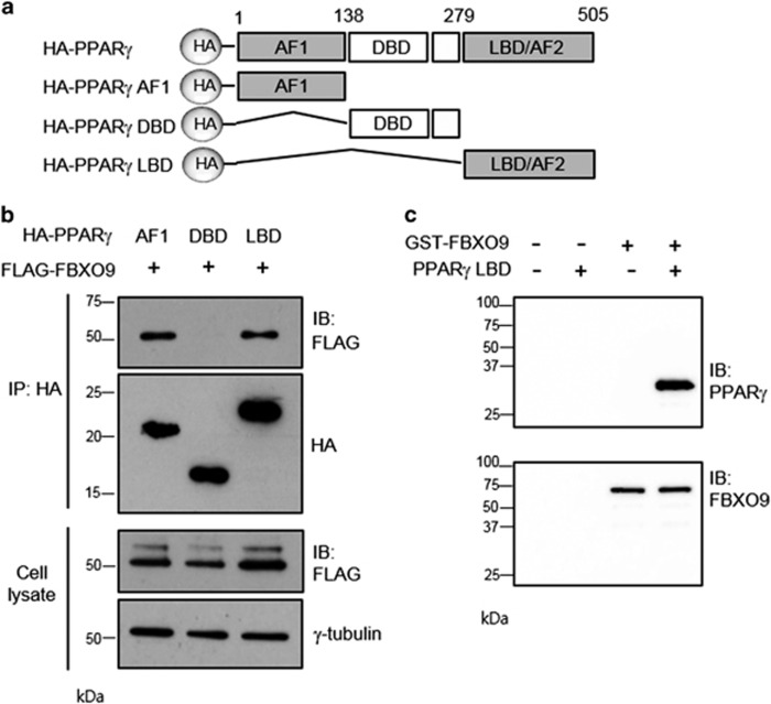
Plat-E cells were transfected with FLAG-PPARγ, and MG132 was added 6 h before harvesting. Cell lysates were immunoprecipitated with FLAG antibody and then the precipitated proteins were subjected to mass spectrometry analysis to identify PPARγ-interacting proteins. Forty proteins were identified (Supplementary Table 1), and we chose an E3 ubiquitin ligase, FBXO9, to further determine whether it is specific to PPARγ. Interaction between PPARγ and FBXO9 was confirmed by transfection of expression vectors for HA-PPARγ and FLAG-FBXO9 into COS7 cells and immunoprecipitation with an anti-HA antibody. FLAG-FBXO9 was co-immunoprecipitated with HA-PPARγ, demonstrating a physical interaction between FBXO9 and PPARγ (Figure 1a). When FBXO9 was co-expressed, the protein level of PPARγ was decreased (Figure 1a). This finding was more apparent with increasing amounts of FBXO9: higher expression levels of FBXO9 led to lower levels of PPARγ (Figure 1b). Next, we identified the PPARγ domain that interacts with FBXO9 using deletion mutants of PPARγ: HA-PPARγ AF1, HA-PPARγ DNA-binding domain and HA-PPARγ LBD (Figures 2a and b). HA-PPARγ AF1 and HA-PPARγ LBD interacted with FBXO9, but HA-PPARγ DNA-binding domain did not. In addition, we performed a GST pull-down assay using the recombinant proteins GST-FBXO9 and PPARγ LBD. PPARγ LBD was co-precipitated with GST-FBXO9, confirming the direct interaction between these proteins (Figure 2c). To summarize, FBXO9 interacts with PPARγ and may regulate PPARγ protein levels.
Figure 1.
FBXO9 interacts with PPARγ. (a) COS7 cells were seeded in a 60-mm dish. The next day, HA-PPARγ2 (1 μg) and FLAG-FBXO9 (1 μg) were co-transfected for 20 h. Whole-cell lysates were subjected to immunoprecipitation with anti-HA antibody-conjugated beads and blotted with anti-FLAG or anti-PPARγ antibody. As a loading control, γ-tubulin was detected using anti-γ-tubulin antibodies. (b) COS7 cells were co-transfected with HA-PPARγ (100 ng) and FLAG-FBXO9 (100 ng, 300 ng) or an empty vector (CMV) as a negative control. The protein levels of PPARγ, FBXO9 or γ-tubulin were detected using anti-HA, anti-FLAG or anti-γ-tubulin antibodies, respectively.
Figure 2.
The domain of PPARγ involved in the interaction with FBXO9. (a) Structure of the deletion mutants of PPARγ. (b) pHA-PPARγ AF1 (3 μg), pHA-PPARγ DBD (4 μg) or pHA-PPARγ LBD (3 μg) was overexpressed with pFLAG-FBXO9 (1 μg) for 24 h and treated with MG132 (10 μM) for 5 h before harvesting. The deletion mutants of PPARγ were immunoprecipitated using anti-HA antibody-conjugated beads and immunoblotted using anti-FLAG and anti-HA antibodies. (c) GST-FBXO9 (1 μg) and PPARγ LBD (2 μg) were mixed and incubated. Co-precipitates were immunoblotted using anti-PPARγ or anti-FBXO9 antibodies.
FBXO9 regulates PPARγ stability by facilitating PPARγ ubiquitination
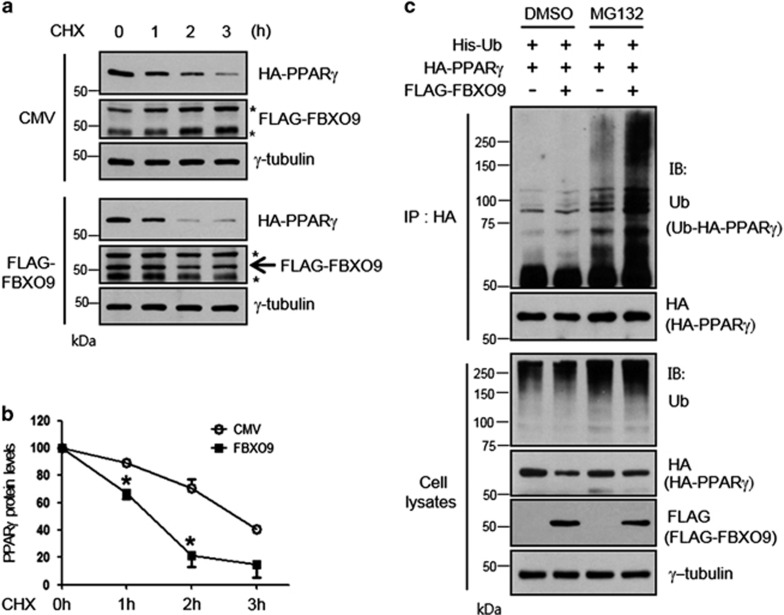
To determine whether FBXO9 overexpression affects the stability of PPARγ protein, COS7 cells were transfected with HA-PPARγ and FLAG-FBXO9 expression vectors and then treated with cycloheximide, a protein synthesis inhibitor. When FBXO9 was overexpressed, PPARγ protein levels decreased more rapidly (Figures 3a and b), suggesting that FBXO9 accelerates PPARγ degradation. Because FBXO9 is considered to be a ubiquitin ligase, we tested whether FBXO9 is involved in ubiquitination of PPARγ. In the presence of MG132, a proteasome inhibitor, ubiquitin-conjugated PPARγ was dramatically increased by FBXO9 overexpression (Figure 3c). These results suggest that FBXO9 is an E3 ubiquitin ligase targeting PPARγ.
Figure 3.
FBXO9 decreases the levels of PPARγ protein by induction of ubiquitination. (a) The day before transfection, COS7 cells were plated in 12-well plates. CMV (100 ng) or FLAG-FBXO9 (100 ng) was co-transfected with HA-PPARγ (100 ng) for 24 h, and cycloheximide (5 μM) was added for the indicated times. Whole-cell lysates were then analyzed using western blotting. Stars indicate non-specific bands. (b) The densities of the PPARγ bands at time point 0 were set to 100%, and the remaining densities were expressed as relative values. Data represent the mean±s.e.m. of three independent experiments. *P<0.05 vs CMV. (c) COS7 cells were subcultured in 60 mm dishes one day before transfection. HA-PPARγ (1 μg), His-Ub (0.2 μg) and FLAG-FBXO9 (0.5 μg) or CMV (0.5 μg) were overexpressed for 12 h and treated with DMSO or MG132 (10 μM) 5 h before harvesting. PPARγ was precipitated with anti-HA antibody-conjugated beads and immunoblotted with anti-Ub, anti-PPARγ, anti-FLAG or anti-γ-tubulin antibodies.
The F-box motif of FBXO9 is important for the regulation of PPARγ protein levels
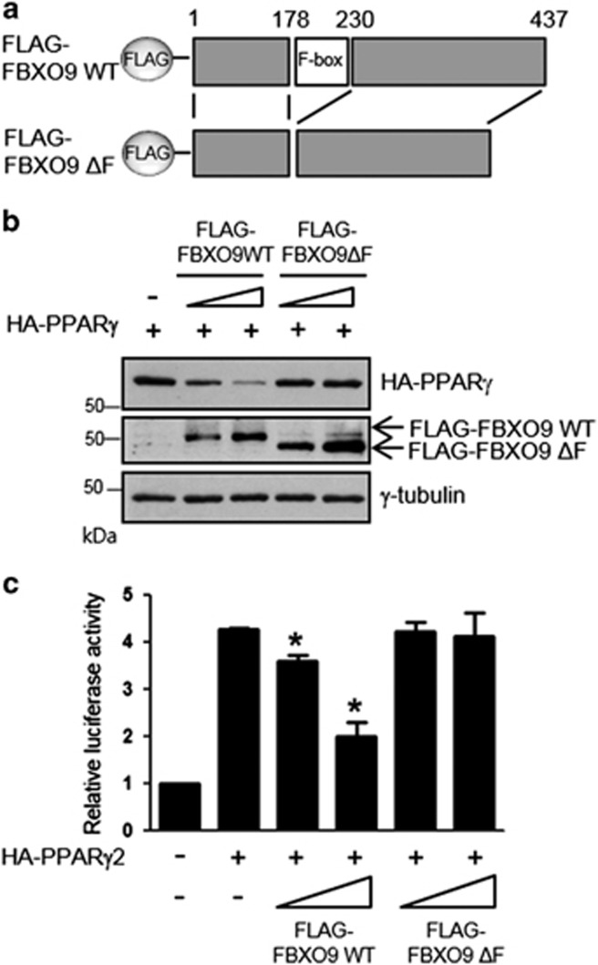
The F-box motif in F-box family proteins is involved in the association with Skp1, which acts as bridge between F-box protein and Cullin. Therefore, F-box proteins lacking the F-box motif can bind to their substrates but are unable to induce ubiquitination of the substrates. We prepared a mutant form of FBXO9 lacking the F-box motif (Figure 4a) and tested whether the motif is important for the regulation of PPARγ protein levels. As expected, the FBXO9 mutant form (FBXO9 ΔF) did not affect PPARγ protein levels when it was overexpressed, while the FBXO9 wild type (FBXO9 WT) effectively decreased PPARγ levels (Figure 4b). The activity of PPARγ was also measured using transfection of pPPRE-TK-luc and expression vectors for the FBXO9 WT or FBXO9 ΔF. Luciferase activity was dramatically reduced when FBXO9 WT was co-transfected but was not affected by FBXO9 ΔF (Figure 4c). These results confirm that FBXO9 reduces PPARγ protein level and activity through PPARγ ubiquitination and that the F-box motif is required for its action.
Figure 4.
The requirement for the F-box motif on the effect of FBXO9 on PPARγ stability. (a) Structure of FBXO9. (b) COS7 cells were seeded in 12-well plates. The next day, cells were transfected with pHA-PPARγ2 (100 ng) and pFLAG-FBXO9 WT (100 ng, 300 ng) or pFLAG-FBXO9 ΔF (25 ng, 50 ng) for 24 h. The amount of transfected DNA of FBXO9 WT and ΔF was determined by similarly expressed protein levels. Whole-cell lysates were then subjected to western blot analysis. (c) COS7 cells were transfected with pFLAG-FBXO9 WT or pFLAG-FBXO9 ΔF in the presence of pPPRE-Tk-Luc (1 μg), pHA-PPARγ (0.3 μg) and pCMV-βgal (0.5 ug) for 18 h. Luciferase activities were normalized to β-galactosidase activity. The relative values for the luciferase activity were calculated from three independent experiments (mean±s.e.m.). *P<0.05 vs the transfection of HA-PPARγ alone.
Overexpression of FBXO9 regulates endogenous PPARγ levels in adipocytes
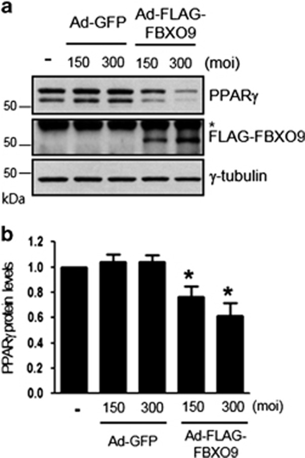
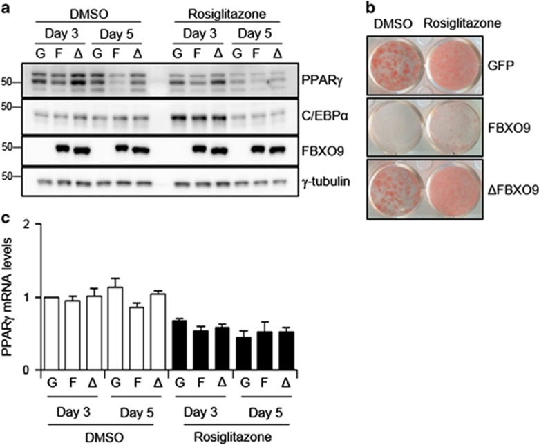
To clarify whether FBXO9 overexpression affects endogenous PPARγ protein levels, FBXO9 was overexpressed using an adenoviral system in 3T3-L1 adipocytes, in which PPARγ is abundantly expressed. Two days after the viral infection, PPARγ protein levels were determined using immunoblotting with an anti-PPARγ antibody. PPARγ protein levels were reduced after overexpression of FBXO9 in a dose-dependent manner (Figures 5a and b). In contrast, PPARγ protein levels were not affected by the control virus (Ad-GFP). These results suggest that endogenous PPARγ levels were regulated by FBXO9 in the adipocytes. Furthermore, we investigated the effect of FBXO9 overexpression on adipogenesis. FBXO9 was overexpressed 2 days after initiation of differentiation, when endogenous PPARγ expression starts to rise. Adipocyte differentiation was inhibited by overexpression of FBXO9 (Figures 6a and b). However, FBXO9 ΔF did not affect the levels of PPARγ and adipogenesis. The effect of FBXO9 on adipogenesis was not influenced by the treatment of TZD. The PPARγ mRNA level was not influenced by FBXO9 overexpression (Figure 6c). These findings indicate that overexpression of FBXO9 inhibits adipogenesis by reducing PPARγ protein stability.
Figure 5.
The effect of FBXO9 overexpression on endogenous PPARγ protein levels in adipocytes. (a) Control adenovirus (Ad-GFP) or adenovirus-expressing FLAG-FBXO9 (Ad-Flag-FBXO9) was infected into 3T3-L1 adipocytes using the indicated multiplicity of infection (MOI). Whole-cell lysates were prepared 48 h after infection. Endogenous PPARγ, FLAG-FBXO9, GFP or γ-tubulin was detected by specific antibodies. Stars indicate non-specific bands. (b) The intensity of PPARγ bands in (a) was normalized to γ-tubulin. The mean value was calculated from six independent experiments (mean±s.e.m.). *P<0.05 vs control.
Figure 6.
Adipogenesis is inhibited by overexpression of FBXO9. Adipogenesis was induced using differentiation medium containing IBMX, dexamethasone and insulin either with or without rosiglitazone. Cells were transfected with Ad-GFP (100 MOI), Ad-FBXO9 (100 MOI) or Ad-FBXO9 ΔF (100 MOI) 2 days after induction of differentiation. (a) Cell lysates were harvested at the indicated day of adipogenesis and subjected to western blot analysis. (b) Cells were stained with oil red O at day 8 of adipogenesis. (c) Total RNA was harvested at the indicated day of adipogenesis, and PPARγ mRNA levels were determined by real-time PCR. The PPARγ mRNA level of the Ad-GFP-infected cells harvested on day 3 without rosiglitazone treatment was set as a reference value of 1, and others are presented as relative values (mean±s.e.m., n=4). G, Ad-GFP; F, Ad-FBXO9; Δ, Ad-FBXO9ΔF.
Discussion
In this study, using co-immunoprecipitation in the presence of MG132 and mass spectrometry analysis, we identified FBXO9 as a novel interacting protein of PPARγ with E3 ligase activity. We showed that FBXO9 decreased the protein stability of PPARγ by inducing ubiquitination and proteasomal degradation. In addition, the PPARγ activity was also decreased by overexpression of FBXO9. The F-box motif of FBXO9 was crucial for modulating PPARγ protein levels, and FBXO9 interacted with the AF1 and LBD of PPARγ. Finally, we demonstrated that FBXO9 was important in regulating endogenous PPARγ levels in 3T3-L1 adipocytes and overexpression of FBXO9 impaired adipogenesis.
Independent of activation by TZD, the level of PPARγ protein per se is important in exerting its effect.5 Because the half-life of PPARγ is relatively short (t1/2=2 h), the process of degradation is important in controlling the metabolic effects of PPARγ.9 It is well known that PPARγ is ubiquitinated and degraded by the proteasomal pathway.9 However, the precise mechanism and molecules involved in this process are not fully understood. We have identified a novel E3 ligase, FBXO9, which is involved in the process of PPARγ degradation. Overexpression of FBXO9 not only decreased the protein stability of PPARγ but also decreased its transcriptional activity. One of the limitations of our study is that FBXO9 knockdown in 3T3-L1 adipocytes did not result in meaningful changes in PPARγ expression (data not shown). This might be because endogenous FBXO9 expression is relatively low in the basal state of adipocytes. It is speculated that FBXO9 might play an important role when FBXO9 expression is induced by certain conditions. It would be of interest to find treatments that modulate the FBXO9 level and regulate the activity and protein level of PPARγ, which might have clinical implications.
In our previous report, we showed that FBXO9 expression is increased at the very early stage of adipogenesis of 3T3-L1 cells, and knockdown of FBXO9 at the early stage (day 0 to day 1) significantly inhibited adipogenesis.18 Because PPARγ is not expressed at this early stage of adipogenesis, FBXO9 might target other proteins, which could be inhibitory factors of adipogenesis. In this study, we further investigated the effect of FBXO9 overexpression on adipogenesis. We observed that overexpression of FBXO9 in 3T3-L1 cells also resulted in decreased levels of PPARγ and impaired adipogenesis. Therefore, it is suggested that normal levels of FBXO9 are required for normal adipocyte differentiation. As we only investigated the role of FBXO9 on adipogenesis, it would be of interest to investigate the functional role of endogenous FBXO9 in differentiated adipocytes.
Several different E3 ligases, Siah2, NEDD4-1 and MKRN1, have been reported to be involved in PPARγ ubiquitination. In the report by Kilroy et al.,11 TZDs increased expression of Siah2, and it was involved in TZD-dependent PPARγ ubiquitination and degradation in adipocytes. However, it was suggested that the nuclear hormone receptor corepressor NCoR might be the direct target of Siah2. Han et al.12 identified NEDD4-1 as an E3 ligase that leads to PPARγ ubiquitination and degradation. NEDD4-1 was shown to delay cellular senescence by decreasing PPARγ expression. A study by Kim et al.13 showed that adipogenesis was suppressed in stable cells overexpressing MKRN1, and conversely, adipogenesis was increased when MKRN1 was knocked down. These results suggest that there are several ubiquitin ligases, including FBXO9, that are involved in the ubiquitination of PPARγ.
There are several distinct features of FBXO9 compared with the other suggested E3 ligases of PPARγ. It has been reported that Siah2 gene expression is increased by TZDs, and Siah2 is involved in TZD-dependent PPARγ ubiquitination and degradation. In contrast, FBXO9 expression was not regulated by TZDs, and FBXO9 was not involved in TZD-dependent PPARγ degradation. It has been reported that MKRN1 overexpression suppressed adipogenesis. Regarding FBXO9, we showed that both inhibition and overexpression resulted in impaired adipogenesis, and normal levels of FBXO9 are required for normal adipocyte differentiation. These results suggest that each E3 ligase regulates PPARγ stability in response to different cellular signaling or conditions in adipocytes. Further studies are required to elucidate the relative contribution of different E3 ligases to PPARγ ubiquitination and their distinct roles in different settings.
F-box proteins usually recognize specific phosphorylation sites of the substrate proteins and facilitate ubiquitination of the target proteins.14 PPARγ has several sites that are phosphorylated by various kinases.19, 20, 21 We tested whether two well-known phosphorylation sites of PPARγ, S112 and S273, are required for FBXO9-mediated ubiquitination. FBXO9 was able to efficiently reduce the stability of two mutant forms of PPARγ, PPARγ S112A and PPARγ S273A (data not shown), suggesting that these two phosphorylation sites are not important for the recognition by FBXO9. Regarding MKRN1, it has been shown that two lysine sites at 184 and 185 of PPARγ were critical for ubiquitination. Unfortunately, these two sites were not tested for their role in PPARγ ubiquitination in our study. Further investigation is required to understand the specific target sites of PPARγ by FBXO9.
In conclusion, we have identified FBXO9, an E3 ligase, as a novel PPARγ ubiquitination enzyme that regulates protein stability and transcriptional activity of PPARγ. We hope this information on post-translational modification of PPARγ will extend our understanding of the regulation of PPARγ and possibly lead to the development of novel therapeutic targets to ameliorate obesity and diabetes.
Acknowledgments
This research was supported by the Basic Science Research Program (2012R1A1A2005546) of the National Research Foundation of Korea (NRF) funded by the Ministry of Education, Science and Technology (MEST) and the BK21Plus Program (10Z20130000017) of the NRF funded by the MEST.
The authors declare no conflict of interest.
Footnotes
Supplementary Information accompanies the paper on Experimental & Molecular Medicine website (http://www.nature.com/emm)
Supplementary Material
References
- Corzo C, Griffin PR. Targeting the peroxisome proliferator-activated receptor-gamma to counter the inflammatory milieu in obesity. Diabetes Metab J 2013; 37: 395–403. [DOI] [PMC free article] [PubMed] [Google Scholar]
- Semple RK, Chatterjee VK, O'Rahilly S. PPAR gamma and human metabolic disease. J Clin Invest 2006; 116: 581–589. [DOI] [PMC free article] [PubMed] [Google Scholar]
- Kubota N, Terauchi Y, Miki H, Tamemoto H, Yamauchi T, Komeda K et al. PPAR gamma mediates high-fat diet-induced adipocyte hypertrophy and insulin resistance. Mol Cell 1999; 4: 597–609. [DOI] [PubMed] [Google Scholar]
- Yamauchi T, Kamon J, Waki H, Murakami K, Motojima K, Komeda K et al. The mechanisms by which both heterozygous peroxisome proliferator-activated receptor gamma (PPARgamma) deficiency and PPARgamma agonist improve insulin resistance. J Biol Chem 2001; 276: 41245–41254. [DOI] [PubMed] [Google Scholar]
- Miles PD, Barak Y, He W, Evans RM, Olefsky JM. Improved insulin-sensitivity in mice heterozygous for PPAR-gamma deficiency. J Clin Invest 2000; 105: 287–292. [DOI] [PMC free article] [PubMed] [Google Scholar]
- Waite KJ, Floyd ZE, Arbour-Reily P, Stephens JM. Interferon-gamma-induced regulation of peroxisome proliferator-activated receptor gamma and STATs in adipocytes. J Biol Chem 2001; 276: 7062–7068. [DOI] [PubMed] [Google Scholar]
- Floyd ZE, Stephens JM. Interferon-gamma-mediated activation and ubiquitin-proteasome-dependent degradation of PPARgamma in adipocytes. J Biol Chem 2002; 277: 4062–4068. [DOI] [PubMed] [Google Scholar]
- Kilroy GE, Zhang X, Floyd ZE. PPAR-gamma AF-2 domain functions as a component of a ubiquitin-dependent degradation signal. Obesity 2009; 17: 665–673. [DOI] [PMC free article] [PubMed] [Google Scholar]
- Hauser S, Adelmant G, Sarraf P, Wright HM, Mueller E, Spiegelman BM. Degradation of the peroxisome proliferator-activated receptor gamma is linked to ligand-dependent activation. J Biol Chem 2000; 275: 18527–18533. [DOI] [PubMed] [Google Scholar]
- Floyd ZE, Wang ZQ, Kilroy G, Cefalu WT. Modulation of peroxisome proliferator-activated receptor gamma stability and transcriptional activity in adipocytes by resveratrol. Metabolism 2008; 57(Suppl 1): S32–S38. [DOI] [PMC free article] [PubMed] [Google Scholar]
- Kilroy G, Kirk-Ballard H, Carter LE, Floyd ZE. The ubiquitin ligase Siah2 regulates PPARgamma activity in adipocytes. Endocrinology 2012; 153: 1206–1218. [DOI] [PMC free article] [PubMed] [Google Scholar]
- Han L, Wang P, Zhao G, Wang H, Wang M, Chen J et al. Upregulation of SIRT1 by 17beta-estradiol depends on ubiquitin-proteasome degradation of PPAR-gamma mediated by NEDD4-1. Protein Cell 2013; 4: 310–321. [DOI] [PMC free article] [PubMed] [Google Scholar]
- Kim JH, Park KW, Lee EW, Jang WS, Seo J, Shin S et al. Suppression of PPARgamma through MKRN1-mediated ubiquitination and degradation prevents adipocyte differentiation. Cell Death Diff 2014; 21: 594–603. [DOI] [PMC free article] [PubMed] [Google Scholar]
- Ho MS, Ou C, Chan YR, Chien CT, Pi H. The utility F-box for protein destruction. Cell Mol Life Sci 2008; 65: 1977–2000. [DOI] [PMC free article] [PubMed] [Google Scholar]
- Zhang DM, He T, Katusic ZS, Lee HC, Lu T. Muscle-specific f-box only proteins facilitate bk channel beta(1) subunit downregulation in vascular smooth muscle cells of diabetes mellitus. Circ Res 2010; 107(12): 1454–1459. [DOI] [PMC free article] [PubMed] [Google Scholar]
- Fernandez-Saiz V, Targosz BS, Lemeer S, Eichner R, Langer C, Bullinger L et al. SCFFbxo9 and CK2 direct the cellular response to growth factor withdrawal via Tel2/Tti1 degradation and promote survival in multiple myeloma. Nat Cell Biol 2013; 15: 72–81. [DOI] [PubMed] [Google Scholar]
- Chung SS, Ahn BY, Kim M, Kho JH, Jung HS, Park KS. SUMO modification selectively regulates transcriptional activity of peroxisome-proliferator-activated receptor gamma in C2C12 myotubes. Biochem J 2010; 433: 155–161. [DOI] [PubMed] [Google Scholar]
- Lee KW, Kwak SH, Ahn BY, Lee HM, Jung HS, Cho YM et al. F-box only protein 9 is required for adipocyte differentiation. Biochem Biophys Res Commun 2013; 435: 239–243. [DOI] [PubMed] [Google Scholar]
- Zhang B, Berger J, Zhou G, Elbrecht A, Biswas S, White-Carrington S et al. Insulin- and mitogen-activated protein kinase-mediated phosphorylation and activation of peroxisome proliferator-activated receptor gamma. J Biol Chem 1996; 271: 31771–31774. [DOI] [PubMed] [Google Scholar]
- Camp HS, Tafuri SR, Leff T. c-Jun N-terminal kinase phosphorylates peroxisome proliferator-activated receptor-gamma1 and negatively regulates its transcriptional activity. Endocrinology 1999; 140: 392–397. [DOI] [PubMed] [Google Scholar]
- Choi JH, Banks AS, Kamenecka TM, Busby SA, Chalmers MJ, Kumar N et al. Antidiabetic actions of a non-agonist PPARgamma ligand blocking Cdk5-mediated phosphorylation. Nature 2011; 477: 477–481. [DOI] [PMC free article] [PubMed] [Google Scholar]
Associated Data
This section collects any data citations, data availability statements, or supplementary materials included in this article.


