Abstract
Background
Ankle fragility fractures are difficult to treat due to poor bone quality and soft tissues as well as the near ubiquitous presence of comorbidities including diabetes mellitus and peripheral neuropathy. Conventional open reduction and internal fixation in this population has been shown to lead to a significant rate of complications. Given the high rate of complications with contemporary fixation methods, the present study aims to critically evaluate the use of acute hindfoot nailing as a percutaneous fixation technique for high-risk ankle fragility fractures.
Methods
In this study, we retrospectively evaluated 31 patients treated with primary retrograde tibiotalocalcaneal nail without joint preparation for a mean of 13.6 months postoperatively from an urban Level I trauma center during the years 2006-2012.
Results
Overall, there were two superficial infections (6.5%) and three deep infections (9.7%) in the series. There were 28 (90.3%) patients that went on to radiographic union at a mean of 22.2 weeks with maintenance of foot and ankle alignment. There were three cases of asymptomatic screw breakage observed at a mean of 18.3 months postoperatively, which were all treated conservatively..
Conclusions
This study shows that retrograde hindfoot nailing is an acceptable treatment option for treatment of ankle fragility fractures. Hindfoot nailing allows early weightbearing, limited soft tissue injury, and a relatively low rate of complications, all of which are advantages to conventional open reduction internal fixation techniques. Given these findings, larger prospective randomized trials comparing this treatment with conventional open reduction internal fixation techniques are warranted.
Introduction
The incidence of low-energy ankle fragility fractures has been increasing rapidly due to the increasing age and activity levels of the elderly population1. However, patient-related factors and comorbidities pose several management challenges while increasing complication rates. By definition, fragility fractures occur in patients with osteoporotic bone, making traditional open reduction internal fixation techniques difficult in this population2. Along with difficult fixation, the soft-tissue envelope in these patients is frequently compromised at the time of injury and poor host factors limit healing potential.. The treatment of fragility fractures includes non-operative management as well as conventional and variations of conventional open reduction internal fixation (ORIF) techniques with varying results reported1,3-5.
Use of a transarticular intramedullary Steinmann pin has been previously documented as a treatment option for unstable ankle fractures in the elderly3. We have used a modification of this technique, with the use of a retrograde intramedullary tibiotalocalcaneal (TTC) nail in this series of patients with fragility fractures3. Despite iatrogenically limiting motion of the tibiotalar and subtalar joints, we hypothesize that treatment with this biomechanically sound device allows early mobilization and return to function with adequate union and minimal wound complications
The goal of the present study is to retrospectively evaluate the use of the retrograde TTC nail in the setting of ankle fragility fractures both clinically and radiographically. We hypothesize that primary fixation with a TTC nail is a safe surgical option that not only leads to satisfactory fracture alignment and union, but also decreases the overall perioperative complication rate in this high-risk cohort.
Materials and Methods
Data Collection
We retrospectively reviewed the database of a single urban, Level I trauma center for all ankle and pilon fractures treated from January 2006-December 2012. We included all patients over 18 years of age treated with retrograde TTC nail for primary treatment of any hindfoot or ankle injury, without formal arthrodesis of the tibiotalar or subtalar joints. After initial review, we identified 38 patients that met inclusion criteria. Four patients were excluded for follow up less than one year, while another patient passed away 10 days following surgery due to complications related to a polytrauma. Two patients were also excluded as they were treated for high-energy, non-reconstructible hindfoot fractures. This left 31 patients available for analysis.
Patient Demographics
Through retrospective chart and radiograph review, we evaluated this cohort for demographic data, type and severity of injury, comorbidities, employment status, ambulatory status, and operative details. We included both rotational ankle fractures and pilon fractures, but all were the result of low-energy mechanisms.
Operative Technique
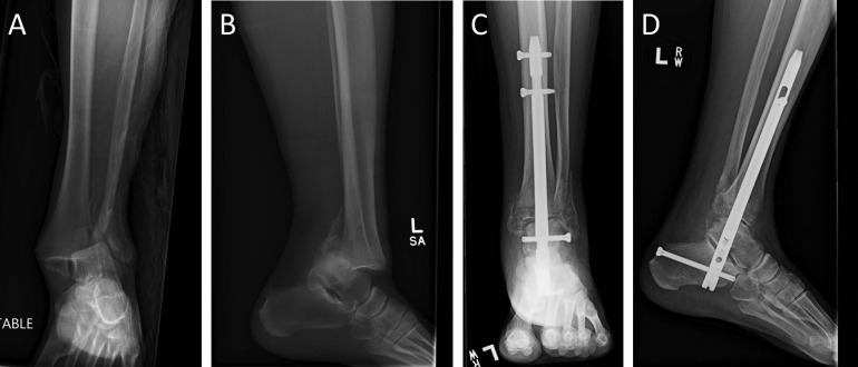
All patients were primarily treated with retrograde TTC nail without joint preparation as primary and definitive fixation for their injuries. The ankle fractures were all reduced in a closed fashion and provisional retrograde pinning from the calcaneus to the tibia was performed in all cases. Pin placement was either anterior or posterior to the tract of the definitive nail, to ensure no difficulties with tract preparation or nail insertion occurred. A starting guidewire was advanced in a retrograde fashion from the calcaneus into the talus and subsequently the tibia. The opening reamer was then advanced over this wire to the distal tibial physeal scar. The wire and opening reamer were then removed and a ball-tipped guide wire was placed into the tract; this was advanced to the middiaphyseal level of the tibia. Minimal reaming was then performed, as most of the patient’s canal diameters were large enough for easy passage of nails of 9 millimeters or more. However, reaming to 1 millimeter greater than the eventual nail size was executed in efforts to minimize risk of nail incarceration. All but two patients were treated with the Phoenix ankle arthrodesis nail (Biomet, Warsaw, IN), while the remaining two were treated with a short Synthes retrograde supracondylar femoral nail (Synthes, West Chester, PA); both of these nails are straight nails without any valgus bend. In all cases, two interlocking screws were placed proximally in the tibia, and at least one interlocking screw was placed through both the talus and calcaneus. No tourniquets were utilized during these procedures (Figure 1). Postoperatively, patients were allowed partial or full weightbearing according to surgeon preference, with all patients progressing to unrestricted ambulation as tolerated by six weeks after surgery.
Figure 1.
A trimalleolar ankle fracture-dislocation in an osteoporotic, poorly controlled diabetic patient is shown in A and B. Successful union is noted in Images C and D at three-month follow up after immediate postoperative mobilization and weightbearing.
Outcomes
The primary outcome of our study was union rate, with infection and implant-related complications being two other primary variables of interest. Superficial infection was defined as anything requiring local wound care or antibiotics, while deep infection was defined as the need to return to the operating room for formal debridement.
Statistics
Mean, range and confidence intervals were calculated for continuous variables and compared using Student’s t-tests. Frequencies were calculated for continuous variables and compared using Fisher’s exact test for increased accuracy in small proportion analysis. A significance level of P < 0.05 was set as significant, with a trend being defined as a P value being between 0.05 and 0.10.
Results
Table I shows demographic data on the patients included in our review. Only approximately 2/3 of the series were unassisted community ambulators (67.6%) prior to their injury, with over half of the series carrying a diagnosis of diabetes or peripheral neuropathy (54.8% and 51.6, respectively). Eight patients (25.8%) sustained open injuries. No patient underwent any concurrent procedures, and there were no intensive care unit admissions postoperatively.
Table I.
Patient Characteristics
| Variable | Results |
| Age (years) | 63.0 ± 16.7 (37-90) |
| Sex (female) | 17 (54.8%) |
| Body Mass Index (kg/m2) | 35.6 ± 10.8 (21.3-65.5) |
| Mechanism | Fall 77.4%, Motor Vehicle Collision 18.8%, Gunshot 3.2% |
| Fracture Pattern | |
| OTA 43A1 | 1 |
| OTA 43B3 | 2 |
| OTA 43C1 | 1 |
| OTA 43C2 | 3 |
| OTA 43C3 | 3 |
| OTA 44A2 | 1 |
| OTA 44B1 | 1 |
| OTA 44B2 | 6 |
| OTA 44B3 | 12 |
| OTA 44C2 | 1 |
| Open Injury | 8 (25.8%) |
| Diabetes Mellitus | 54.8% |
| Hgb A1C (%) | 7.6 ± 1.9 (5.5-13.0) |
| Peripheral Neuropathy | 16 (51.6%) |
| Paraplegia | 1 (3.2%) |
| Tobacco Use | 7 (22.6%) |
| Employment | 5 (16.1%) |
| Ambulatory Status | |
| Unassisted | 21 (67.7%) |
| Cane | 1 (3.2%) |
| Walker | 8 (25.8%) |
| Wheelchair | 1 (3.2%) |
| Operative Variables | |
| Time to fixation (days) | 2.4 ± 2.7 (0.1 - 10) |
| Operative Room Time (minutes) | 76.7 ± 24.3 (43 - 140) |
| Estimated Blood Loss (mL) | 65.6 ± 67.9 (5-250) |
| Hospital Length of Stay (days) | 7.6 ± 4.4 (1-16) |
Average length of follow-up was 407.9 days in our series (Table II). There were two superficial and three deep infections in this cohort. All of the patients developing deep infections sustained open ankle fractures. These three patients subsequently underwent operative debridement, nail removal and antibiotic-impregnated cement rod placement. At most recent follow-up, there had been no recurrence of infection using this treatment algorithm. For the entire cohort, union was observed in 90.3% of patients at an average of 22.2 weeks postoperatively. There were three instances of broken proximal interlocking screws at average follow-up of 18.3 months postoperatively; however, all hardware failures remained asymptomatic at final follow-up.
Table II.
Patient Outcomes
| Variable | Outcome |
|---|---|
| Follow-up Length (days) | 407.89 ± 219.03 |
| Bony Union | 90.3% |
| Time to Union (weeks) | 22.2 ± 6.2 |
| Implant Failure | 3 (9.7%) |
| Infection | |
| Superficial | 2 (6.5%) |
| Deep | 3 (9.7%) |
| Wound Dehiscence | 0 (0%) |
| Amputation | 1 (3.2%) |
Discussion
Treatment of ankle and hindfoot fragility fractures by conventional means poses several challenges. There are numerous studies showing significantly worse outcomes in elderly patients treated with ORIF of ankle fractures when compared to younger cohorts6-8. Studies have shown several predictive risk factors for poor outcome with conventional ORIF of ankle fractures, including open injuries, diabetes mellitus, peripheral neuropathy, and peripheral vascular disease4,5,9-14. Wukich and Kline found that diabetic patients with concomitant peripheral vascular disease and neuropathy are at an even greater risk15. Additionally, elderly patients who are forced to remain immobile after these injuries are at a much higher risk of developing perioperative complications, including pressure ulcers, pneumonia and deep vein thrombosis1. In the present study, we describe the use of a TTC nail for treatment of ankle fragility fractures and found a low rate of overall complications.
Union rate of fragility ankle fractures is an increasing concern, and we were able to obtain an ankle fracture union rate of 90.3% with the current series. We feel that this is important, as a decreased union rate and increased complication rate in this population has been associated with decreased quality of life and self-reported functional outcomes at one year post-injury1. Infection rates in this patient population are also increased, with patients greater than 80 years of age undergoing operative fixation of an unstable ankle fracture sustaining a 7% superficial infection rate and 4.6% deep infection rate, while only 86% of patients returned to pre-injury mobility5,6.
Our study shows retrograde TTC nail can be a very useful treatment option in this difficult population. Some advantages of this procedure, as compared to traditional open reduction and fixation are: operative time and blood loss are decreased, soft-tissue dissection is kept to a minimum, and patients are allowed to mobilize and bear full weight earlier. We were able to show that primary hindfoot nailing is successful in treating fragility ankle fractures, especially in the setting of diabetes and peripheral neuropathy. There was a risk of deep infection as a complication in this group, but these were all in the setting of an open fracture, and we hypothesize that these infections were the result of the open injury as well as patient comorbidities. However, the majority of patients went on to radiographic union with minimal complications.
There are several limitations with this study. It is a non-randomized retrospective cohort study from a single institution. As such, we provide a description of our experience with the use of TTC in ankle fragility fractures. As there was no control group, comparisons are limited to those values that have been previously reported in the literature. The number of patients included in the study is small. However, as this is a relatively novel technique description, we feel that reporting our early outcomes is important. Further inclusion of functional outcome scores and prospective comparisons to a matched cohort with traditional fixation constructs would also be helpful to provide data to the practicing surgeon.
In conclusion, we found that retrograde TTC nail is a safe and effective treatment option for ankle fragility fractures. The use of retrograde TTC in this population provides the advantages of early mobilization, limited soft tissue injury, and relatively few complications as compared to previous studies evaluating the use of conventional ORIF in similar cohorts. Given these promising findings, future research comparing retrograde TTC and conventional ORIF using prospective methods and randomization should be considered.
References
- 1.Nilsson G, Jonsson K, Ekdahl C, Eneroth M. Outcome and quality of life after surgically treated ankle fractures in patients 65 years or older. BMC Musculoskelet Disord. 2007 Dec;20(8):127. doi: 10.1186/1471-2474-8-127. [DOI] [PMC free article] [PubMed] [Google Scholar]
- 2.Amirfeyz R, Bacon A, Ling J, Blom A, Hepple S, Winson I, Harries W. Fixation of ankle fragility fractures by tibiotalocalcaneal nail. Arch Orthop Trauma Surg. 2008 Apr;128(4):423–8. doi: 10.1007/s00402-008-0584-z. [DOI] [PubMed] [Google Scholar]
- 3.Lemon M, Somayaji HS, Khaleel A, Elliott DS. Fragility fractures of the ankle: stabilisation with an expandable calcaneotalotibial nail. J Bone Joint Surg Br. 2005 Jun;87(6):809–13. doi: 10.1302/0301-620X.87B6.16146. [DOI] [PubMed] [Google Scholar]
- 4.SooHoo NF, Krenek L, Eagan MJ, Gurbani B, Ko CY, Zingmond DS. Complication rates following open reduction and internal fixation of ankle fractures. J Bone Joint Surg Am. 2009 May;91(5):1042–9. doi: 10.2106/JBJS.H.00653. [DOI] [PubMed] [Google Scholar]
- 5.Shivarathre DG, Chandran P, Platt SR. Operative fixation of unstable ankle fractures in patients aged over 80 years. Foot Ankle Int. 2011 Jun;32(6):599–602. doi: 10.3113/FAI.2011.0599. [DOI] [PubMed] [Google Scholar]
- 6.Anderson SA, Li X, Franklin P, Wixted JJ. Ankle fractures in the elderly: initial and long-term outcomes. Foot Ankle Int. 2008 Dec;29(12):1184–8. doi: 10.3113/FAI.2008.1184. [DOI] [PubMed] [Google Scholar]
- 7.Beauchamp CG, Clay NR, Thexton PW. Displaced ankle fractures in patients over 50 years of age. J Bone Joint Surg Br. 1983 May;65(3):329–32. doi: 10.1302/0301-620X.65B3.6404905. [DOI] [PubMed] [Google Scholar]
- 8.Litchfield JC. The treatment of unstable fractures of the ankle in the elderly. Injury. 1987 Mar;18(2):128–32. doi: 10.1016/0020-1383(87)90189-6. [DOI] [PubMed] [Google Scholar]
- 9.Chaudhary SB, Liporace FA, Gandhi A, Donley BG, Pinzur MS, Lin SS. Complications of ankle fracture in patients with diabetes. J Am Acad Orthop Surg. 2008 Mar;16(3):159–70. doi: 10.5435/00124635-200803000-00007. [DOI] [PubMed] [Google Scholar]
- 10.Dellenbaugh SG, Dipreta JA, Uhl RL. Treatment of ankle fractures in patients with diabetes. Orthopedics. 2011 May;34(5):385. doi: 10.3928/01477447-20110317-19. [DOI] [PubMed] [Google Scholar]
- 11.Ganesh SP, Pietrobon R, Cecílio WA, Pan D, Lightdale N, Nunley JA. The impact of diabetes on patient outcomes after ankle fracture. J Bone Joint Surg Am. 2005 Aug;87(8):1712–8. doi: 10.2106/JBJS.D.02625. [DOI] [PubMed] [Google Scholar]
- 12.Jones KB, Maiers-Yelden KA, Marsh JL, Zimmerman MB, Estin M, Saltzman CL. Ankle fractures in patients with diabetes mellitus. J Bone Joint Surg Br. 2005 Apr;87(4):489–95. doi: 10.1302/0301-620X.87B4.15724. [DOI] [PubMed] [Google Scholar]
- 13.McCormack RG, Leith JM. Ankle fractures in diabetics Complications of surgical management. J Bone Joint Surg Br. 1998 Jul;80(4):689–92. doi: 10.1302/0301-620x.80b4.8648. [DOI] [PubMed] [Google Scholar]
- 14.Wukich DK, Joseph A, Ryan M, Ramirez C, Irrgang JJ. Outcomes of ankle fractures in patients with uncomplicated versus complicated diabetes. Foot Ankle Int. 2011 Feb;32(2):120–30. doi: 10.3113/FAI.2011.0120. [DOI] [PubMed] [Google Scholar]
- 15.Wukich DK, Kline AJ. The management of ankle fractures in patients with diabetes. J Bone Joint Surg Am. 2008 Jul;90(7):1570–8. doi: 10.2106/JBJS.G.01673. [DOI] [PubMed] [Google Scholar]


