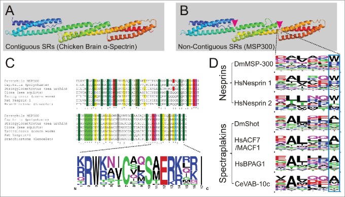
Figure 3.

Highly conserved non-canonical motifs in spectrin-repeat-containing proteins. (A) Ribbon diagram of the chicken brain α-spectrin fragment (1u4q) exemplifies 3 contiguous SRs. (B) Homology modeling of MSP300 isoform D (5140-5460) based on 1u4q exemplifies 3 non-contiguous SRs. The magenta arrowheads indicate the position of the linkers. (C) A highly conserved but non-canonical SR is found in nesprins of different animal groups. (D) The hexapeptide linker regions between 2 consecutive SRs are conserved in nesprins and spectraplakins. The blue frame demonstrates the typical tryptophan (W) / histidine (H) residue variants at the -1 of helix C.
