Summary
Background
Eagle syndrome is a condition caused by an elongated styloid process. Unilateral face, neck and ear pain, stinging pain, foreign body sensation and dysphagia can be observed with this syndrome. Rarely, the elongated styloid process may cause pain by compressing the cervical segment of the internal carotid and the surrounding sympathetic plexus, and that pain spreading along the artery can cause neurological symptoms such as vertigo and syncope.
Case Report
In this case report we presented a very rare eagle syndrome with neurological symptoms that occurred suddenly with cervical rotation. The symptoms disappeared as suddenly as they occurred, with the release of pressure in neutral position. We also discussed CT angiographic findings of this case.
Conclusions
Radiological diagnosis of the Eagle syndrome that is manifested with a wide variety of symptoms and causes diagnostic difficulties when it is not considered in the differential diagnosis is easy in patients with specific findings. CT angiography is a fast and effective examination in terms of showing compression in patients with the Eagle syndrome that is considered to be atypical and causes vascular compression.
MeSH Keywords: Angiography, Eagles, Multidetector Computed Tomography, Vertigo
Background
Eagle syndrome is a pathology caused by an elongated styloid process or a calcified stylohyoid and stylomandibular ligament [1,2]. Styloid process is 20 to 25 mm in length and has a cylindrical form with an incidence of 4%, and about 4 to 10.3% of them are symptomatic [3–5]. Clinical and radiological findings, as well as treatment of the elongated styloid process was first mentioned by W. Eagle in 1937 [6]. Eagle mentions two clinical entities, i.e. classic stylocarotid syndrome and stylocarotid syndrome [7,8]. In the Classic Stylohyoid Syndrome, the following clinical findings are distinctive: post-tonsillectomy pain in the operation site, stinging pain, dysphagia, painful swallowing, and foreign body sensation. In the Stylocarotid Syndrome, the elongated styloid process deviated laterally or medially, regardless of tonsillectomy, causes compression on the internal and external carotid arteries and perivascular sympathetic fibres. There are also similar symptoms triggered or exacerbated by neck rotation and compression. However, cases with neurological symptoms secondary to vascular compression of the styloid processes have been reported subsequently [9–11]. In this study, we aimed to present a rare case with a complaint of vertigo that was secondary to internal carotid artery (ICA) compression by an elongated styloid process which completely disappeared in the neutral position, and to review the literature.
Case Report
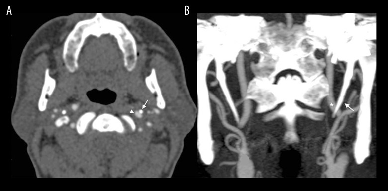
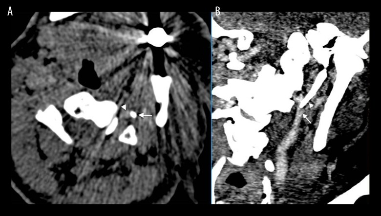
No distinctive feature was found during physical examination of the patient. It was found in history-taking that the patient experienced vertigo when he turned his head to the left, which indicated that it was caused by cervical rotation, and the patient’s complaints disappeared completely in the neutral position. The patient stated that he had been suffering from this complaint for about two (2) years. The patient had no complaints on his neck region, such as pain, foreign body sensation, dysphagia or visual impairment. No finding was observed on the patient’s two-way cervical x-ray. A CT angiography scan was performed in order to assess the neck and cerebral vascular structures. The CT angiography scan was performed by obtaining continuous sections on the axial plane of 3 mm to include the region starting from the aortic arch and reaching the supraventricular level. The obtained images were reconstructed on coronal and sagittal planes with thin sections, of 1 mm. No intraluminal-extraluminal pathologies were detected on the common carotid artery, ICA, external carotid artery or intra-cerebral vascular structures during the CT angiography examination. No pathologies were found on soft tissues in the extra-vascular region. However, the styloid processes were found to be longer than normal when bone structures were examined (3.2 cm on the right, 3.4 cm on the left). Also, it was discovered that the left process was adjacent to the ICA and caused a slight compression on the ICA (Figure 1). The CT angiography scan was re-performed with a lower dose and contrast agent in the left cervical rotation position considering that the elongated styloid processes might be the cause of vertigo secondary to vascular compression when taking patient’s complaints and CT angiography findings together. As a result, it was observed that the compression on the ICA became evident (Figure 2). Based on the existing finding, the patient was diagnosed with the Eagle syndrome aggravating with cervical rotation and was referred for surgery.
Figure 1.
(A) The contrast-enhanced MDCT angiography shows the styloid process causing a light pressure on the left ICA (arrowhead: ICA, arrow: styloid process). (B) Bilateral elongated styloid processes are seen on the coronal section MIP (Maximum Intensity Projection) images. (star: ICA, arrow: styloid process).
Figure 2.
(A) The MDBT angiography shows the evident compression of the styloid processes on the left cervical rotational position (arrowhead: ICA, arrow: styloid process). (B) Compression finding on the coronal section and MIP images (arrowhead: styloid process, arrow: ICA). Imaging in the cervical rotation was performed with lower kV and lower contrast agent volume in order to reduce the amount of radiation and contrast agent-related burden, which is why the image quality is lower.
Discussion
Although the classification of the styloid process was first defined by Watt W. Eagle in 1937, Weinlecher mentioned symptoms caused by elongation of the styloid process and performed styloid resection in 1872 [6,12,13]. Different values were reported as concerns the length of the styloid process in the literature. Moffat et al. in their cadaver studies found the mean values of styloid processes to be 1.52 cm and 4.77 cm [12]. The upper value of the styloid process was reported to be 30 mm by Kaufman et al. and 40 mm by Monsour et al. Reviews on the literature and radiological studies suggest that the length of the styloid process should not exceed 25 mm and its upper limit should be accepted as 3 cm in general [4,14,15]. In our case, the length of the right styloid was 32 mm, while the length of the left styloid was 34 mm.
Although the incidence of elongated styloid processes varies between 1% and 30% (mean 24%), most elongated styloid process cases are asymptomatic and only about 4% are symptomatic [4]. Although it has been reported that it is generally more common in women than men and occurs at the age of 30 [16], in our case it was rather late in the age. Elongation of the styloid process is bilateral in most cases. However, even though the majority of symptomatic cases have a bilaterally elongated process, symptoms appear to be unilateral [2,5]. Our case also had a bilaterally elongated styloid process, as stated in the literature. However, the symptoms and findings were observed to be associated with the left side.
The occurrence of symptoms in the Classic Stylohyoid Syndrome is considered to result from a traumatic fracture of the elongated styloid process and direct compression on the pharyngeal mucosa and cranial nerves [1,5]. However, it is considered to result from the congenital length of styloid processes or the embryonic ossification of the stylohyoid ligament in those with no trauma or surgical history [5].
Findings in the stylocarotid syndrome occur as a result of the medial or the lateral deviation of the elongated styloid process. In this case, head and eye pain could be observed as a result of vascular compression and mechanical irritation of the plexus adjacent to the vascular structures [1,2,5]. In addition, pain can be observed along the course of ICA. Patients can be mistakenly diagnosed with a cluster-type headache or migraine [5]. The external carotid artery involvement causes under-eye pain. In addition, and more rarely, symptoms may appear as a result of direct compression on the carotid artery. Cases in which vascular compression is more evident and with clinical findings, such as aphasia secondary to interruption of the affected artery blood flow, visual symptoms, transient ischaemic attack, vertigo and syncope, have been reported [9,10]. Such patients with this condition state that their symptoms start and disappear suddenly as their occurrence is a synchronized finding due to changes in the flow of arteries. We believe that the complaint of vascular compression and vertigo of our case had the same course as described above. Irritation of the sympathetic plexus can lead to dizziness and tinnitus [10]. This irritation is usually accompanied by pain and symptoms can aggravate with turning of the head [1,2,9]. Our case did not have tinnitus or pain. We considered vascular compression due to sudden occurrence and disappearance of vertigo.
Palpating the styloid process in the tonsillar region can facilitate Eagle syndrome diagnosis. However, objective findings are revealed by radiographic imaging [4,17]. Lateral head-neck radiography, anteroposterior radiography, towne radiography, panoramic mandible radiography and computed tomography are the imaging methods used for diagnosing the elongated styloid process [17,18]. Although plain X-rays provide us with valuable anatomical information, there may be diagnostic difficulties due to super-positions. For example, styloid process and calcification of the stylohyoid ligament can be overlooked due to the mandible and shading caused by teeth. A CT scan would exclude these negative situations [17,18]. In particular, multi-detector CT shows the length, the direction of the styloid process and its relationship with anatomical structures very well. Three-dimensional imaging, coronal and sagittal reformatted images allow for assessment of the process as a whole, measurement of its length and offer preoperative guidance to the surgeon [17,18]. We considered that the best approach would be CT angiography in our case because its scanning duration is shorter and it shows vascular structures and bone calcifications very well. Measuring artery flow in neutral position and rotation using Doppler ultrasonography and comparing the results can be considered theoretically in cases with suspected vascular compression, as in our case. However, it is not easy to implement this in practice because it is difficult to scan the distal part of the artery during rotation. Moreover, scanning would become more difficult to tolerate by the patients when the vertigo or other complaints start.
Glossopharyngeal neuralgia, trigeminal neuralgia, temporomandibular joint arthritis, chronic tonsillar and pharyngeal infections, migraine, cluster-type headache, temporal arthritis, cervical vertebral pathologies, cervical oesophageal pathologies should be considered in the differential diagnosis of the Eagle syndrome [2,17]. Surgical shortening of the elongated styloid process is the only effective treatment. However, there is a large number of treatment options as an alternative to surgical treatment such as the use of non-steroidal anti-inflammatory drugs, injection of transpharyngeal steroids or long-acting local anaesthetics, use of oral carbamazepine [1,5]. We offered the surgical treatment option to our patient.
Conclusions
In conclusion, radiological diagnosis of the Eagle syndrome that is manifested with a wide variety of symptoms and causes diagnostic difficulties when it is not considered in the differential diagnosis, is easy in patients with specific findings. CT angiography is a fast and effective examination in terms of showing compression in patients with the Eagle syndrome that is considered to be atypical and causes vascular compression.
References
- 1.Gervickas A, Kubilius R, Sabalys G. Clinic, diagnostics and treatment pecularities of Eagle’s syndrome. Stomatologija, Baltic Dental and Maxillofacial Journal. 2004;6:11–13. [Google Scholar]
- 2.Yavuz H, Çakmak Ö, Akkuzu B, Özlüoğlu L. Eagles syndrome: A case report. K.B.B. and BBC Journal. 2002;(10):97–101. [Google Scholar]
- 3.Gök U, Yıldız M. Eagle Sendromu. Fırat Medical Journal. 2004;9(3):79–81. [Google Scholar]
- 4.Murtagh RD, Caracciolo JT, Fernandez G. CT findings associated with Eagle syndrome. Am J Neuroradiol. 2001;22(7):1401–2. [PMC free article] [PubMed] [Google Scholar]
- 5.Hekimoğlu C. Eagle’s Syndrome. Clinical Dentistry and Research. 2005;(3):27–32. [Google Scholar]
- 6.Eagle WW. Elongated styloid process: Report of two cases. Arch Otolaryngol. 1937;25:584–86. doi: 10.1001/archotol.1949.03760110046003. [DOI] [PubMed] [Google Scholar]
- 7.Fini G, Gasparini G, Filippini F, et al. The long styloid process syndrome or Eagle syndrome. J Craniomaxillofac Surg. 2000;28:123–27. doi: 10.1054/jcms.2000.0128. [DOI] [PubMed] [Google Scholar]
- 8.Eagle WW. Symptomatic elongated styloid process – carotid artery syndrome with operation. Arch Otolaryngol. 1949;49:490–503. doi: 10.1001/archotol.1949.03760110046003. [DOI] [PubMed] [Google Scholar]
- 9.Farhat HI, Elhammady MS, Ziayee H, et al. Eagle syndrome as a cause of transient ischemic attacks: Case report. J Neurosurg. 2009;110(1):90–93. doi: 10.3171/2008.3.17435. [DOI] [PubMed] [Google Scholar]
- 10.Chuang WC, Short JH, McKinney AM, et al. Reversible left hemispheric ischemia secondary to carotid compression in Eagle syndrome: Surgical and CT angiographic correlation. Am J Neuroradiol. 2007;28:143–45. [PMC free article] [PubMed] [Google Scholar]
- 11.Infante-Cossío P, García-Perla A, González-García A, et al. Compression of the internal carotid artery due to elongated styloid process. Rev Neurol. 2004;39(4):339–43. [PubMed] [Google Scholar]
- 12.Moffat DA, Ramsden RT, Shaw HJ. The styloid process syndrome: aetiological factors and surgical management. J Laryngol Otol. 1977;91:279–94. doi: 10.1017/s0022215100083699. [DOI] [PubMed] [Google Scholar]
- 13.Fini G, Gasparini G, Filippini F, et al. The long styloid process syndrome or Eagle’s syndrome. J Craniomaxillofac Surg. 2000;28:123–27. doi: 10.1054/jcms.2000.0128. [DOI] [PubMed] [Google Scholar]
- 14.Akpinar YZ, Yilmaz B, Tatar N, Demirtag Z. Eagle’s Syndrome: A case report. Acta Medica Anatolia. 2014;2(4):145–46. [Google Scholar]
- 15.Montalbetti L, Ferrandi D, Pergami P, Savaldi F. Elongated styloid process and Eagle’s syndrome. Cephalgia. 1995;15:80–93. doi: 10.1046/j.1468-2982.1995.015002080.x. [DOI] [PubMed] [Google Scholar]
- 16.Gossman JR, Tarsitano JJ. The styloid-stylohyoid syndrome. J Oral Surg. 1977;35:555–60. [PubMed] [Google Scholar]
- 17.Savranlar A, Uzun L, Uğur MB, Özer T. Three-dimensional CT of Eagle’s syndrome. Diagn Interv Radiol. 2005;11:206–9. [PubMed] [Google Scholar]
- 18.Nakamaru Y, Fukuda S, Miyashita S, Ohashi M. Diagnosis of the elongated styloid process by three-dimensional computed tomography. Auris Nasus Larynx. 2002;29:55–57. doi: 10.1016/s0385-8146(01)00102-x. [DOI] [PubMed] [Google Scholar]


