Abstract
Butterfly eyespot colour patterns are a key example of how a novel trait can appear in association with the co-option of developmental patterning genes. Little is known, however, about how, or even whether, co-opted genes function in eyespot development. Here we use CRISPR/Cas9 genome editing to determine the roles of two co-opted transcription factors that are expressed during early eyespot determination. We found that deletions in a single gene, spalt, are sufficient to reduce or completely delete eyespot colour patterns, thus demonstrating a positive regulatory role for this gene in eyespot determination. Conversely, and contrary to previous predictions, deletions in Distal-less (Dll) result in an increase in the size and number of eyespots, illustrating a repressive role for this gene in eyespot development. Altogether our results show that the presence, absence and shape of butterfly eyespots can be controlled by the activity of two co-opted transcription factors.
 Transcription factors Distal-less(Dll) and spalt were co-opted during the evolution of butterfly eyespots. Here, Zhang and Reed use CRISPR/Cas9 genome editing to show that while spalt is a positive regulator of eyespots as predicted, Dll knockouts have positive effects on both eyespot size and number, thus suggesting that Dll is an eyespot repressor, not an activator as previously thought.
The eyespot patterns found on butterfly wings are among the most striking colour patterns in the natural world, and there is a large literature concerning their ecology, evolution and development1,2. Across different species eyespots play a number of ecological roles, including in such disparate phenomena as mate choice and predation avoidance, and thus provide a powerful example of how a single trait can adapt to serve many different functions under different types of selective pressures. In 1994, Carroll et al. found that the transcription factor gene Dll, which plays an ancestral role in animal appendage formation3, is expressed in early eyespot determination4. This discovery provided one of the most surprising and marked examples of evolutionary gene co-option, that is, the redeployment of an ancestral gene for a novel function5, and remains among the most widely cited case studies in evolutionary developmental biology. Subsequent work has identified a number of other apparently co-opted genes expressed at different times during eyespot development, some of which show compelling phylogenetic patterns of expression gain-and-loss that suggest evolutionary dynamicity in the eyespot regulatory network1,6,7. A major challenge in eyespot development work, however, has been a lack of functional data. Although candidate gene expression patterns are clearly associated with eyespots both developmentally and evolutionarily, we still do not know how, or even whether, these genes function in colour pattern formation. Because of this it has not been possible to assess the morphological or adaptive significance of eyespot-associated gene co-option.
Here we address to what extent co-opted genes expressed during early butterfly eyespot determination function in eyespot development. Comparative expression studies show that the transcription factor spalt is one of the earliest factors expressed in eyespot foci, and phylogenetic work suggests that this early spalt expression is a conserved ancestral trait across nymphalid butterflies6,8. Because of these observations it has been proposed that spalt may be a key upstream regulator of eyespot determination1. Another transcription factor expressed in association with eyespots across many species is Dll6,9. Various lines of evidence, primarily comparative expression studies, but also including some overexpression and sequence association work10,11,12, have led to the hypothesis that Dll is also a positive regulator of eyespots1. Here we use CRISPR/Cas9 genome editing to assess the developmental functions of spalt and Dll in eyespot colour pattern development, and, more broadly, to show that the co-option of these transcription factors has played a role in the evolution of butterfly wing patterns.
Results
Pigmentation gene deletion demonstrates wing mosaics
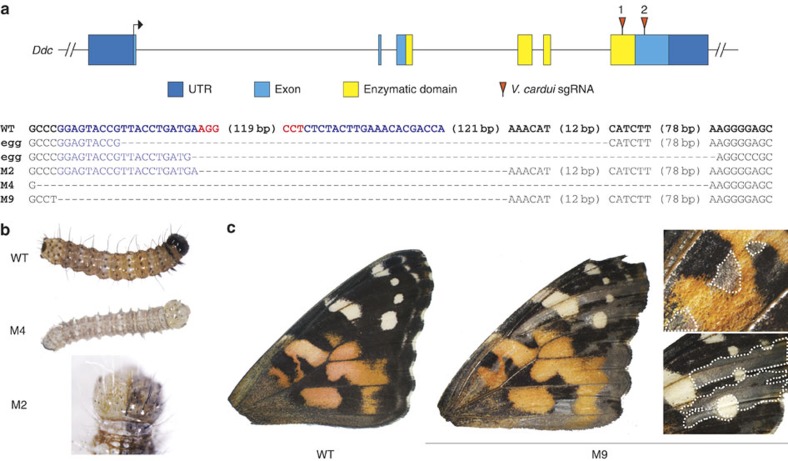
To test the function of candidate eyespot determination genes we opted for a loss-of-function approach using CRISPR/Cas9 targeted deletions13,14. To test the utility of CRISPR/Cas9 for making G0 deletion mosaic butterflies, we optimized a protocol in the painted lady butterfly Vanessa cardui by targeting the melanin pigmentation gene Dopa decarboxylase (Ddc), reasoning that pigmentation defects should allow for easy visualization of potential mosaicism in wings (Fig. 1). On the basis of genotyping a panel of 81 embryos we found that our optimized double-guide RNA method resulted in a 69% rate of long deletions at the target locus. Furthermore, across all deletion clones, including for spalt and Dll as mentioned below, we observed a 66% frameshift rate downstream of deletions. Consistent with Ddc phenotypes observed in Drosophila melanogaster mutants, the majority of injected animals that survived to hatching age were unable to emerge from their eggs without assistance, likely due to deficiencies in mouthpart sclerotization15. Larvae showed mosaic pigmentation defects, including bilateral mosaics, which ranged in severity (Fig. 1b), and adult wings were mosaic for melanin-deficient clones (Fig. 1c). In sum, by using a pigment marker gene we demonstrate that CRISPR/Cas9 can be used to produce butterflies with adult wings that are mosaic for targeted deletions. This ability to produce somatic deletion mosaics is an important new tool in the butterfly experimental system, as it permits the analysis of tissue-specific deletion effects that would otherwise be embryonic lethal in pure mutant lines.
Figure 1. CRISPR/Cas9 deletions in the melanin pigmentation gene Ddc result in mosaic depigmentation phenotypes in V. cardui.
(a) Location of sgRNAs relative to the V. cardui Ddc locus. Sequences of selected deletion alleles from eggs and the butterflies shown (M2, M4 and M9) confirm disruption of Ddc. Blue: sgRNA targets. Red: PAM sequences. (b) Ddc deletions result in depigmentation of larvae, including bilateral mosaics. (c) An example of mosaic melanin-specific depigmentation in adult wings.
Spalt is a positive regulator of eyespot colour patterns
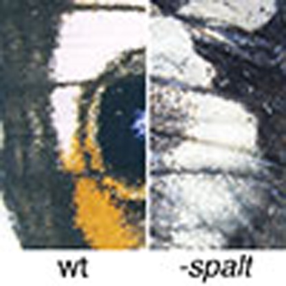
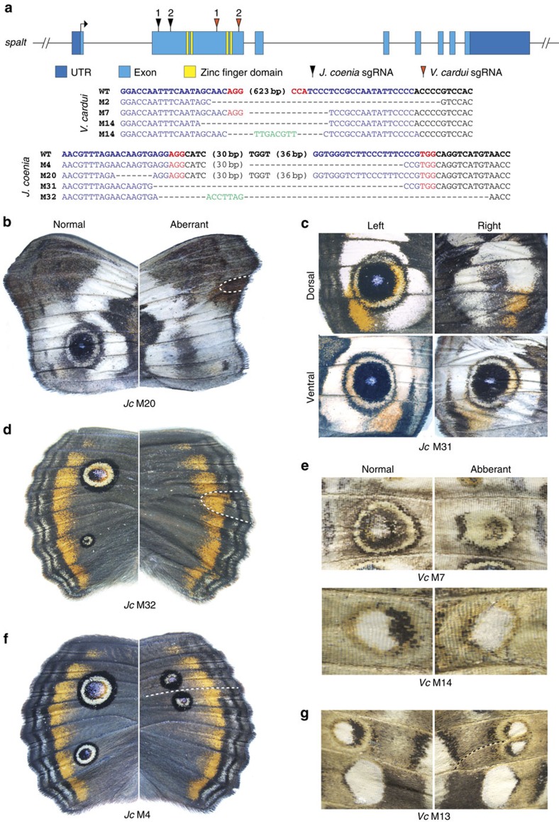
To test the hypothesis that spalt is a regulator of eyespot colour patterns, we used CRISPR/Cas9 to produce deletions, and related frameshifts, in the spalt coding region of two nymphalid species that display eyespots (Fig. 2a): Junonia coenia and V. cardui. In both the species inducing deletions in spalt was sufficient to produce aberrant mosaic-wing phenotypes not seen in natural populations (Table 1). Of greatest interest was the frequent reduction and/or complete loss of eyespot colour patterns in wings of injected animals of both species (Fig. 2, Supplementary Fig. 1). This effect was particularly marked in J. coenia where spalt deletions produced many individuals completely missing eyespots from the forewing and/or hindwing (Fig. 2b–d,f). We also noted that the loss of an eyespot on one wing surface (that is, dorsal or ventral) could occur without affecting an eyespot on the opposing surface (Fig. 2c), demonstrating that even though eyespots occur in the same position on opposing wing surfaces, their determination is dorsoventrally decoupled to a large degree.
Figure 2. CRISPR/Cas9 spalt deletions result in reduction and loss of eyespot colour patterns.
(a) Location of sgRNAs relative to the V. cardui spalt locus. Sequences of spalt alleles from the animals shown confirm lesions in target regions. Blue: sgRNA targets. Red: PAM sequences. Green: novel sequences not observed in wild-type alleles. Somatic deletions in spalt cause complete loss of forewing eyespots in J. coenia: (b) ventral forewing; (c) dorsal forewing; (d) dorsal hindwing. (e) spalt deletions reduce eyespots in V. cardui ventral hindwings (top) and forewings (bottom). Ectopic wing veins (dashed lines) resulting from spalt deletion subdivide eyespot patterns in (f) J. coenia (also showing a missing posterior eyespot), and (g) V. cardui. All comparisons shown are left–right asymmetrical phenotypes from individual injected butterflies. ‘Normal' phenotypes are as wild-type, ‘aberrant' phenotypes are non-wild type and vary asymmetrically from the normal patterns on the same animal.
Table 1. Summary of CRISPR/Cas9 injection results.
|
spalt | ||||||||||
|---|---|---|---|---|---|---|---|---|---|---|
| Species | Injected eggs | Hatched eggs | Hatch ratio | Total adults | Mutant phenotypes | Eyespot reduction | Eyespot loss | Abnormal veins | Vein-related discal spot distortion | |
| V. cardui | 421 | 281 | 66.27% | 25 | 14 (56%) | 11 | 1 | 3 | 0 | |
| J. coenia | 508 | 90 | 17.71% | 63 | 21 (33.3%) | 16 | 20 | 8 | 8 | |
| Distal-less | ||||||||||
| Distal-less | |||||||||||
|---|---|---|---|---|---|---|---|---|---|---|---|
| Species | Injected eggs | Hatched eggs | Hatch ratio | Total adults | Mutant phenotypes | Antenna, leg, or palp reduction | Eyespot expansion | Additional eyespots | White HW eyespot foci | Parafocal and margin distortion | Dark patches |
| V. cardui | 772 | 609 | 78.80% | 85 | 44 (51.7%) | 44 | 16 | 16 | 5 | 16 | 0 |
| J. coenia | 323 | 129 | 38.15% | 95 | 39 (41.0%) | 39 | 1 | 0 | 0 | 4 | 3 |
We did not observe the same type of complete eyespot loss in V. cardui as we saw in J. coenia, possibly due to different deletion positions (V. cardui deletion alleles retained two unaffected 5′ zinc finger domains relative to J. coenia). Nonetheless, we still observed many clear cases of mosaic eyespot reduction in V. cardui (Fig. 2e, Supplementary Fig. 1). In all cases in both species, eyespot diminution due to spalt deletion had a highly specific effect only on eyespot patterns, leaving neighbouring colour patterns unaffected. In addition to eyespot reduction phenotypes, spalt deletions in both J. coenia and V. cardui frequently resulted ectopic wing veins (Fig. 2b,d,f,g), consistent with spalt mutant phenotypes in D. melanogaster16. Interestingly, these ectopic veins often traced through the middle of eyespots, dividing eyespots into two (Fig. 2f,g). In J. coenia, we also observed several cases where vein abnormalities around the discal cell were associated with reduction of discal spot patterns (Supplementary Fig. 1); however, we tentatively speculate that these colour pattern defects are due primarily to vein-related pattern disruption since spalt expression has not been observed in discal spot patterns. Altogether our results show that spalt is a positive regulator of eyespots, and that it is required for eyespot determination in J. coenia.
Dll is an eyespot repressor and organizes distal colour patterns
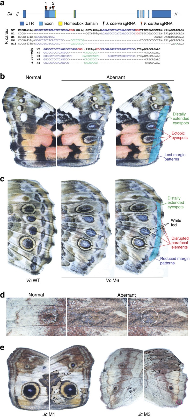
To test the hypothesis that Dll is an activator of eyespot determination we used CRISPR/Cas9 to induce deletions, and related frameshifts, in the Dll coding region in J. coenia and V. cardui (Fig. 3a). Consistent with the known function of Dll in D. melanogaster appendage development17, we observed a high frequency of missing or reduced legs, antennae, and labial palps in injected animals of both species (Supplementary Fig. 2, Table 1). The resulting eyespot phenotypes were surprising, however, because Dll deletions had marked positive effects on both eyespot size and number. In both J. coenia and V. cardui, Dll deletions were sufficient to promote spatial expansion of eyespots towards the wing margin, causing the eyespots to be distally elongated, and, consequently, larger (Fig. 3b–d). These eyespot elongation phenotypes were reminiscent of Bicyclus anynana comet mutants18, as well as natural eyespot phenotypes seen in some other butterflies such Chloreuptychia spp., suggesting that Dll downregulation may be implicated in these other phenotypes as well. Overall, we saw the most extreme Dll phenotypes in V. cardui, in which we recovered individuals with rows of multiple novel, ectopic eyespots on the forewing (Fig. 3b). These ectopic spots were distally expanded, as was the case for all aberrant eyespots resulting from Dll deletion.
Figure 3. CRISPR/Cas9 Dll deletions result in expansion and gain of eyespot colour patterns.
(a) Location of sgRNAs relative to the V. cardui Dll locus. Sequences of Dll alleles from the animals shown confirm lesions in target regions. Blue: sgRNA targets. Red: PAM sequences. Green: novel sequences not observed in wild-type alleles. (b) Dll deletion causes multiple effects on distal colour patterns of the V. cardui forewing, including an increase in size and number of eyespots. Both wings shown are from the same individual (Vc M15), where the ‘normal' wing shows a wild-type phenotype, while the ‘aberrant' wing shows several abnormalities as annotated to the right. (c) Effects of Dll mutation on the V. cardui hindwing. (d) A J. coenia Dll mutant (Jc M3) shows distal elongation of a hindwing eyespot. The white dotted circle shows the size of the wild-type eyespot on the opposing wing of the same individual. (e) J. coenia Dll deletion result in patches of dark pigmentation on both the forewing (left) and hindwing (right). The individual on the left also shows disruption of margin patterns and anterior eyespot size asymmetry.
In addition to changes in shape, size and number of eyespots, Dll deletions also resulted in several other colour pattern defects (Table 1), including the disruption and loss of marginal bands and parafocal elements in both species (Fig. 3b–e, Supplementary Fig. 2), loss of pigmentation in V. cardui eyespot foci (Fig. 3c), and the appearance of hyperpigmented clones across the J. coenia wing surface (Fig. 3d,e). It was notable that Dll deletions had a different range of wing pattern effects in V. cardui versus J. coenia despite the fact that the efficiency of the Dll mutagenesis appeared to be similar, as evidenced by similar rates of appendage mutations (52% in V. cardui and 41% in J. coenia; Table 1). For instance in J. coenia we did not observe the marked gain-of-eyespot phenotypes observed in V. cardui, but conversely, V. cardui mutants did not show hyperpigmented clones as seen in J. coenia. This leads us to speculate that Dll may play some divergent colour patterning functions in the two genera we examined, in addition to its shared functions in eyespot and margin pattern determination. This idea, however, is presented with the caveat that we cannot rigorously confirm lack-of-function (or cell autonomy) without a clone boundary marker, as is standard in D. melanogaster work19. Nonetheless, our strong Dll deletion phenotypes in both species clearly show that Dll plays a variety of roles in colour pattern development, including shaping and repressing eyespots, organizing parafocal and margin colour patterns, and regulating pigmentation.
Discussion
In this study we used CRISPR/Cas9 somatic mosaics to show that the transcription factor genes spalt and Dll play key roles in butterfly wing pattern development, and by extension, that the co-option of these genes has played a role in colour pattern evolution. The effect of spalt deletion—that is, the loss and/or reduction of eyespots—clearly shows that this gene plays a role in promoting eyespots. spalt is one of the earliest known factors to presage eyespots in imaginal disc development; therefore, it is reasonable to hypothesize that this gene is a positive upstream regulator of eyespot determination, as is consistent with earlier models based on expression associations.
Dll deletion phenotypes, on the other hand, were both varied and surprising. Perhaps the greatest surprise was finding that Dll acts as a repressor of eyespot patterns, contrary to expectations arising from decades of comparative expression work. Despite observing many strong Dll deletion phenotypes that showed clear eyespot effects, we saw no evidence in our experiments that Dll deletion had any diminishing effects on eyespots. All aberrant eyespot phenotypes we observed involved an increase in eyespot size and/or number. We are now challenged to reconcile this repressive function with previous expression studies that show a positive association between Dll expression and the presence and size of eyespots. In this regard it is likely important that in V. cardui and J. coenia expression of early eyespot-associated factors, including spalt, occurs in eyespot foci before Dll, at a time when only fine, transient lines of Dll expression extend from the eyespot foci to the wing margin6,9 (for example, Fig. 4). If these lines of Dll expression play an early role to inhibit distal expression of eyespot determination genes, then loss of these Dll lines could result in extension of eyespot-activating gene expression towards the margin, as is consistent with our observed phenotypes. We therefore speculate that a primary colour patterning role of Dll is to inhibit eyespot determination genes in the distal wing margin during an early stage of pattern formation, before Dll activation in eyespot foci (for example, Fig. 4c). It is still unclear, however, why some spots are completely repressed by Dll (for example, V. cardui posterior forewing spots), while others are only partially repressed (for example, V. cardui anterior forewing spots)—further work will be required to address this. It is also unclear why Dll is expressed in imaginal disc eyespot foci in the species we examined, since our results do not support a role for Dll in focal determination. We speculate that Dll focal expression may instead play a later role in pigmentation, as implied by the loss of pigmentation in V. cardui hindwing eyespot foci in response to Dll deletion (Fig. 3c).
Figure 4. Eyespot determination precedes Dll focal expression.
(a) Dll expression in line patterns in a mid-last instar V. cardui forewing imaginal disc. The red box highlights the area of the wing shown in subsequent panels. (b) Eyespot markers Notch and spalt are expressed in foci at a time point when Dll is only expressed in a line extending from the eyespot focus to the wing margin. (c) A time series of Dll expression shows the progression of line, to line-plus-focus (corresponding to b), to focus only.
The effects of Dll deletion on non-eyespot traits including parafocal and margin colour pattern elements, and pigmentation, were surprising and of significant interest. Although previous studies have not focused much attention on the possible role of Dll in the development of these other colour pattern traits, these effects are in line with earlier observations. Importantly, strong wing margin expression of Dll (Fig. 4a) is consistent with an organizing role for marginal and parafocal colour patterns9. Indeed, a developmental link between eyespots, marginal bands and parafocal elements is observed in coldshock experiments in V. cardui that cause eyespots and parafocal elements to merge and marginal bands to expand20. We further speculate that the pigment-altering effects of Dll mutagenesis are likely related to pupal-stage Dll expression in eyespot foci and more widely across the wing epithelium21. Pigment regulation by Dll is also supported by work in B. anyana and Junonia orithya showing that ectopic expression of Dll causes localized changes in pigmentation, but is insufficient to induce distinct patterns elements10,12. Future models of wing pattern development must consider the complex role of Dll as both a promoter and repressor of multiple colour pattern traits, especially along the distal wing margin.
Altogether, our results show that the early stages of butterfly eyespot determination involve the activity of both promoting and repressing regulators, and that the presence, absence, shape and colour of eyespots can be modulated by the activity of two co-opted genes. Our surprising finding that Dll is a repressor of eyespot determination requires a significant revision of current models of eyespot development and provides a cautionary tale about interpreting correlations between traits and candidate gene expression levels. In addition to providing the first unambiguous functional validation of the role of specific genes in butterfly wing pattern determination, our study also demonstrates the power and potential of CRISPR/Cas9 somatic mosaics for work in new model systems.
Methods
Animals
V. cardui caterpillars were obtained from Carolina Biological Supply (Burlington, NC, USA) and J. coenia eggs were obtained from Fred Nijhout (Duke University, NC, USA). All butterflies were reared on artificial diet at 16/8-h light/darkness at 28 °C and 70% humidity.
Preparation of guide-RNAs
Our strategy for producing long deletions in target loci was to generate two Cas9 cut sites flanking the desired lesion interval using two single guide-RNAs (sgRNAs), then to rely on nonhomologous end joining to close the resulting gap22. sgRNAs were designed by manually searching genomic regions for GGN18NGG or N20NGG protospacer adjacent motif (PAM) sequences on the sense or antisense strands (Supplementary Table 1). sgRNA template was generated by PCR using Phusion polymerase (New England Biolabs, Ipswich, MA, USA). In vitro transcription was conducted using Megascript T7 Kit (Ambion, Waltham, MA, USA) and purified by phenol–chloroform extraction and isopropanol precipitation.
Microinjection of butterfly embryos
Butterfly eggs were collected from host plant leaves within an hour of being laid and arranged on double-sided adhesive tape on a microscope slide. To soften the egg chorion J. coenia eggs were dipped in 5% benzalkonium chloride (Sigma-Aldrich, St Louis, MO, USA) for 90 s, and then washed in water for 2 min before mounting on microscope slide. Eggs were then dried for 15 min in a desiccant chamber. 0.5 μg of Cas9 protein (PNA Bio Inc., Thousand Oaks, CA, USA) and 250 ng of each sgRNA were mixed in a 2.5 μl volume and injected into eggs using a 0.5-mm borosilicate needle (Sutter Instruments, Novato, CA, USA).
Mutation genotyping and trait scoring
For Ddc larval genotyping genomic DNA was extracted from caterpillars using proteinase K in digestion buffer13. For all other genotyping DNA was extracted from wing muscle tissue using a QIAamp DNA mini kit (Qiagen, Hilden, Germany). Fragments flanking the CRISPR target regions were amplified by PCR, gel-purified, subcloned into a TOPO TA vector (Invitrogen, Carlsbad, CA, USA), and sequenced on an ABI 3730 sequencer. Genotyping primers are provided in Supplementary Table 1. To rule out non-CRISPR variation when scoring phenotypes, we only called an individual as having an aberrant deletion phenotype if a trait showed a strong left–right asymmetry. Although there is a small degree of individual wing pattern variation in species we surveyed, this variation is typically symmetrical. We observed asymmetrical, mosaic variation only in individuals injected with sgRNAs and Cas9.
Immunohistochemistry
Larval wing discs were dissected, staged, fixed, stained and imaged as previously described8,21 using a rabbit anti-spalt polyclonal antibody23 at a 1:200 dilution, a rabbit anti-Dll polyclonal antibody24 at a 1:100 dilution, and/or a mouse anti-Notch monoclonal antibody (C17.9C6)25 at a 1:200 dilution.
Data availability
Images of all wings from butterflies showing presumptive deletion phenotypes. along with a reference spreadsheet, are available on Dryad: http://dx.doi.org/10.5061/dryad.tj45p.
Additional information
How to cite this article: Zhang, L. & Reed, R. D. Genome editing in butterflies reveals that spalt promotes and Distal-less represses eyespot colour patterns. Nat. Commun. 7:11769 doi: 10.1038/ncomms11769 (2016).
Supplementary Material
Supplementary Figures 1-2 and Supplementary Table 1
Acknowledgments
This work was supported by National Science Foundation grant DEB-1354318 to R.D.R. We thank R. Barrio for spalt antibody, G. Panganiban for Dll antibody, the University of Iowa Developmental Studies Hybridoma Bank for Notch antibody and F. Nijhout for J. coenia eggs. We thank J. Lewis, A. Martin, A. Mazo-Vargas, M. Sheehan, K. van der Burg and M. Wolfner for discussions and comments on the manuscript.
Footnotes
Author contributions R.D.R. and L.Z.: designed the study and wrote the paper. L.Z.: CRISPR/Cas9 genome editing. R.D.R.: immunohistochemistry.
References
- Monteiro A. Origin, development, and evolution of butterfly eyespots. Ann. Rev. Entomol. 60, 253–271 (2015). [DOI] [PubMed] [Google Scholar]
- Nijhout H. F. The Development and Evolution of Butterfly Wing Patterns Smithsonian Institution Press (1991). [Google Scholar]
- Panganiban G. et al. The origin and evolution of animal appendages. Proc. Natl Acad. Sci. USA 94, 5162–5166 (1997). [DOI] [PMC free article] [PubMed] [Google Scholar]
- Carroll S. B. et al. Pattern formation and eyespot determination in butterfly wings. Science 265, 109–114 (1994). [DOI] [PubMed] [Google Scholar]
- True J. R. & Carroll S. B. Gene co-option in physiological and morphological evolution. Ann. Rev. Cell Dev. B 18, 53–80 (2002). [DOI] [PubMed] [Google Scholar]
- Oliver J. C., Tong X. L., Gall L. F., Piel W. H. & Monteiro A. A single origin for nymphalid butterfly eyespots followed by widespread loss of associated gene expression. PLoS Genet. 8, 1002893 (2012). [DOI] [PMC free article] [PubMed] [Google Scholar]
- Shirai L. T. et al. Evolutionary history of the recruitment of conserved developmental genes in association to the formation and diversification of a novel trait. BMC Evol. Biol. 12, 21 (2012). [DOI] [PMC free article] [PubMed] [Google Scholar]
- Reed R. D., Chen P. H. & Nijhout H. F. Cryptic variation in butterfly eyespot development: the importance of sample size in gene expression studies. Evol. Dev. 9, 2–9 (2007). [DOI] [PubMed] [Google Scholar]
- Reed R. D. & Serfas M. S. Butterfly wing pattern evolution is associated with changes in a Notch/Distal-less temporal pattern formation process. Curr. Biol. 14, 1159–1166 (2004). [DOI] [PubMed] [Google Scholar]
- Monteiro A. et al. Distal-less regulates eyespot patterns and melanization in Bicyclus butterflies. J. Exp. Zool. Part B 320B, 321–331 (2013). [DOI] [PubMed] [Google Scholar]
- Beldade P., Brakefield P. M. & Long A. D. Contribution of Distal-less to quantitative variation in butterfly eyespots. Nature 415, 315–318 (2002). [DOI] [PubMed] [Google Scholar]
- Dhungel B. et al. Distal-less induces elemental color patterns in Junonia butterfly wings. Zool. Lett. 2, 4 (2016). [DOI] [PMC free article] [PubMed] [Google Scholar]
- Bassett A. R., Tibbit C., Ponting C. P. & Liu J. L. Highly efficient Targeted mutagenesis of Drosophila with the CRISPR/Cas9 system. Cell Rep. 6, 1178–1179 (2014). [DOI] [PMC free article] [PubMed] [Google Scholar]
- Li X. Y. et al. Outbred genome sequencing and CRISPR/Cas9 gene editing in butterflies. Nat. Commun. 6, 9212 (2015). [DOI] [PMC free article] [PubMed] [Google Scholar]
- Wright T. R. F., Bewley G. C. & Sherald A. F. The genetics of dopa decarboxylase in Drosophila melanogaster. II. Isolation and characterization of dopa-decarboxylase-deficient mutants and their relationship to the a-methyl-dopa-hypersensitive mutants. Genetics 84, 187–310 (1976). [DOI] [PMC free article] [PubMed] [Google Scholar]
- Organista M. F. & De Celis J. F. The Spalt transcription factors regulate cell proliferation, survival and epithelial integrity downstream of the Decapentaplegic signalling pathway. Biol. Open 2, 37–48 (2013). [DOI] [PMC free article] [PubMed] [Google Scholar]
- Gorfinkiel N., Morata G. & Guerrero I. The homeobox gene Distal-less induces ventral appendage development in Drosophila. Genes Dev. 11, 2259–2271 (1997). [DOI] [PMC free article] [PubMed] [Google Scholar]
- Brakefield P. M. The evolution-development interface and advances with the eyespot patterns of Bicyclus butterflies. Heredity 80, 265–272 (1998). [Google Scholar]
- Rodriguez A. D., Didiano D. & Desplan C. Power tools for gene expression and clonal analysis in Drosophila. Nat. Methods 9, 47–55 (2012). [DOI] [PMC free article] [PubMed] [Google Scholar]
- Nijhout H. F. The developmental physiology of color patterns in Lepidoptera. Adv. Insect Physiol. 18, 181–247 (1985). [Google Scholar]
- Brunetti C. R. et al. The generation and diversification of butterfly eyespot color patterns. Curr. Biol. 11, 1578–1585 (2001). [DOI] [PubMed] [Google Scholar]
- Gratz S. J. et al. Genome engineering of Drosophila with the CRISPR RNA-guided Cas9 nuclease. Genetics 194, 1029–1035 (2013). [DOI] [PMC free article] [PubMed] [Google Scholar]
- Barrio R., de Celis T. F., Bolshakov S. & Kafatos F. C. Identification of regulatory regions driving the expression of the Drosophila spalt complex at different developmental stages. Dev. Biol. 215, 33–47 (1999). [DOI] [PubMed] [Google Scholar]
- Panganiban G., Sebring A., Nagy L. & Carroll S. The development of crustacean limbs and the evolution of arthropods. Science 270, 1363–1366 (1995). [DOI] [PubMed] [Google Scholar]
- Fehon R. G. et al. Molecular interactions between the protein products of the neurogenic loci Notch and Delta, 2 EGF-homologous genes in Drosophila. Cell 61, 523–534 (1990). [DOI] [PubMed] [Google Scholar]
Associated Data
This section collects any data citations, data availability statements, or supplementary materials included in this article.
Supplementary Materials
Supplementary Figures 1-2 and Supplementary Table 1
Data Availability Statement
Images of all wings from butterflies showing presumptive deletion phenotypes. along with a reference spreadsheet, are available on Dryad: http://dx.doi.org/10.5061/dryad.tj45p.


