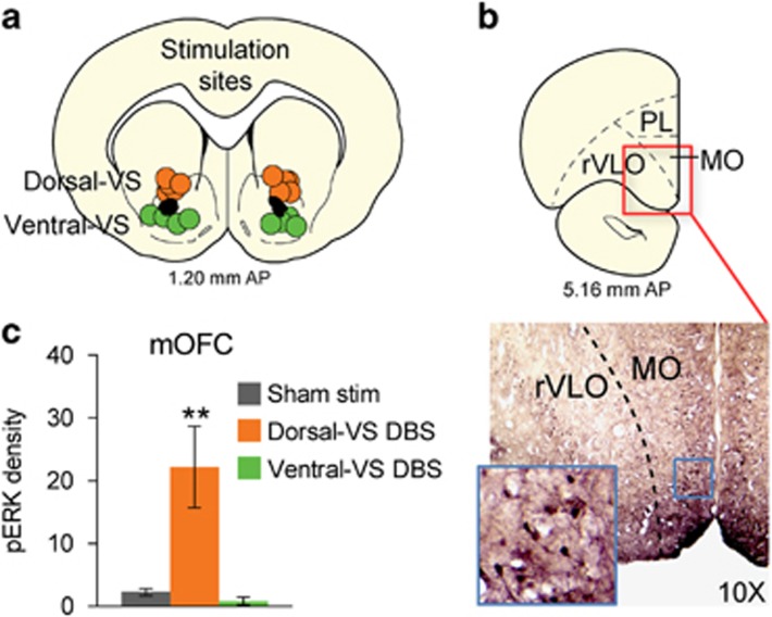
Figure 4.
Deep brain stimulation (DBS) of dorsal–ventral striatum (VS) increases pERK expression in medial OFC (mOFC) neurons. (a) DBS electrode placements in ventral striatum. Circle diameter indicates the estimated spread of current from electrode tip. DBS of dorsal–VS was applied dorsal to the anterior commissure (orange circles) and DBS of ventral–VS was applied ventral to the anterior commissure (green circles). (b) Representative micrograph showing pERK labeling in mOFC after DBS of dorsal-VS. (c) Comparison of pERK density (counts/0.1 mm2) after rats underwent 3 h of Sham DBS (n=9), dorsal–VS DBS (n=4) or ventral–VS DBS (n=4). Data presented as mean±SEM. **p<0.01.

