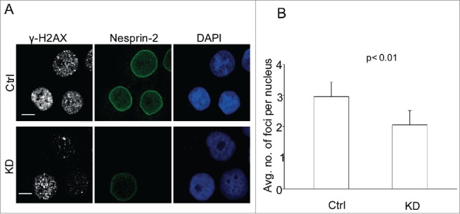
Figure 2.

Nesprin-2 KD cells have a hypoactive DDR. (A) Immunofluorescence analysis after UV treatment of control and Nesprin-2 KD HaCaT cells with antibodies against Nesprin-2 (pAbK1), γ-H2AX and DAPI. Scale bar, 10 µm. (B) Graphical representation of the average number of γ-H2AX foci per nucleus. Foci ≥ 0.7 µm were counted, analysis was with cellprofiler image analysis software (mean ± s.d, p value, 0.0084). 100 cells were analyzed.
