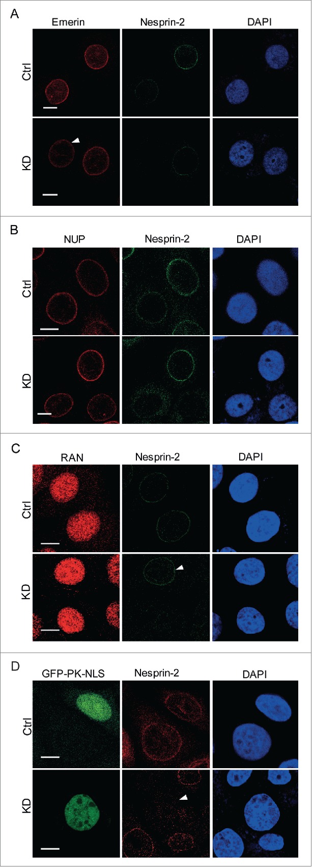
Figure 3.

Nesprin-2 knockdown does not affect the localization of Emerin, the nuclear pore complex, RAN, and the trafficking of SV40 NLS-GFP. (A) Immunofluorescence study of HaCaT control and Nesprin-2 KD cells using anti-Emerin and anti-Nesprin-2 antibodies (pAbK1). Arrow indicates a Nesprin-2 KD cell with emerin nuclear envelope localization. (B) Presence of nuclear pore proteins (NUP) at the NE as detected with mAb414. (C) Distribution of RAN. Immunofluorescence study using RAN (shown in red) and pAbK1 anti-Nesprin-2 antibodies (shown in green). Arrow indicates a control cell with Nesprin-2 nuclear envelope localization for comparison with surrounding KD cells. (D) Immunofluorescence analysis of double transfected cells either with Ctrl shRNA+GFP-PK-SV40 NLS or with Nesprin-2 shRNA+GFP-PK-SV40 NLS to detect localization of GFP-NLS in both control and Nesprin-2 KD cells. Nesprin-2 was detected with pAbK1. Arrow points to a KD cell having nuclear GFP-PK-SV40 NLS. Scale bar, 10 µm.
