Abstract
Fracture risk is significantly increased in both type 1 and type 2 diabetes, and individuals with diabetes experience worse fracture outcomes than normoglycemic individuals. Factors that increase fracture risk include lower bone mass in type 1 diabetes and compromised skeletal quality and strength despite preserved bone density in type 2 diabetes, as well as the effects of comorbidities such as diabetic macro- and microvascular complications. In this Perspective, we assess the developing scientific knowledge regarding the epidemiology and pathophysiology of skeletal fragility in patients with diabetes and the emerging data on the prediction, treatment, and outcomes of fractures in individuals with type 1 and type 2 diabetes.
Introduction
Fractures are a significant health issue for patients with diabetes. In type 1 diabetes (T1D), improvements in life expectancy are increasing the number of patients who are living to older age. In addition, over a quarter of adults aged 65 years and older in the U.S. have type 2 diabetes (T2D). In this older age-group, fractures are a common event; a 60-year-old white woman has a 44% probability of having at least one fracture in her remaining lifetime (1). The cost of treating fractures in the U.S. exceeded $17 billion in 2005 and is predicted to increase by 50% by 2025 (2).
Importantly, hip fractures result in a very high risk of mortality and disability. Mortality rates increase five- to eightfold in the 3 months following a hip fracture and remain elevated even 5 years after fracture (3). Furthermore, approximately 29% of hip fracture patients never return to their prefracture status for activities of daily living (4). Extended recovery time and disability are also common after a vertebral fracture.
Individuals with diabetes are at higher fracture risk and have even worse fracture outcomes than normoglycemic individuals. However, strategies to reduce fracture risk appear underutilized in this population, possibly related to challenges of identifying high-risk patients and concerns regarding effective treatments for prevention. The pathophysiology of increased skeletal fragility is complex, differs between T1D and T2D, and is the subject of intense investigation. In this Perspective, we review the current knowledge regarding the epidemiology and pathophysiology of diabetes-induced bone disease. We also discuss current issues pertaining to the prediction, treatment, and outcomes of fractures in individuals with T1D and T2D.
Epidemiology of Fractures in Individuals With T1D and T2D
Individuals with T1D have double the risk of any fracture and four to five times higher hip fracture risk compared with those without diabetes (5). Higher fracture risk in T1D is evident in childhood and extends throughout the life span, affecting both sexes similarly. T1D is characterized by modest deficits in bone mineral density (BMD) that account for some, but not all, of the increased fracture risk (6). Reduced bone quality also appears to contribute to increased fracture risk in T1D, as discussed later. In contrast, T2D is associated with overweight and higher bone density, factors that are associated with lower fracture risk in normoglycemic individuals. However, among older adults with T2D, the risk of hip fracture is increased 40–70% compared with normoglycemic individuals (6,7). Among individuals over the age of 65 years participating in the National Health and Nutrition Examination Survey (NHANES), the risk of any fracture in non-Hispanic white adults was similar in those with and without diabetes, as assessed by self-report or HbA1c ≥6.5% (hazard ratio [HR] 1.17 [95% CI 0.89–1.52]) (8). Given the age at diabetes diagnosis and use of insulin, it was estimated that 97% of participants had T2D in this analysis. Risks were higher compared with non-Hispanic black (HR 1.86 [95% CI 1.05–3.30]) and Mexican American (HR 2.29 [95% CI 1.41–3.73]) adults without diabetes. Higher fracture risk does not appear to extend to those with prediabetes, defined by fasting glucose or 2-h glucose (8,9).
Individuals with either T1D or T2D, particularly those with diabetes complications, are more likely to experience delayed healing and postsurgical complications, such as wound infection (10–14). Mortality following hip fracture is 1.44 times higher in those with diabetes (15). Several large case-control studies among individuals admitted with hip fractures have shown an increased risk of postoperative cardiac events among those patients with diabetes and an increased length of stay of 1–4 days (10,16,17).
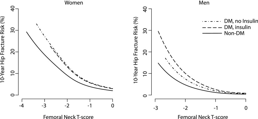
Prediction of fracture risk in patients with T2D is challenging. Older adults with T2D have fractures at a higher bone density than individuals who do not have diabetes. As a result, while lower BMD does predict fracture risk in patients with diabetes, the BMD T-score underestimates fracture risk (18) (Fig. 1). For example, hip fracture risk in a woman with diabetes and a femoral neck BMD T-score of −1.9 is similar to the risk in a normoglycemic woman of the same age with a T-score of −2.5.
Figure 1.
Femoral neck BMD T-score and 10-year fracture risk at age 75 years by diabetes status and insulin use. Estimated 10-year cumulative fracture risk at age 75 years in men and women, calculated using the Cox proportional hazards regression model baseline survival function raised to the power of the relative hazard for each combination of diabetes group and T-score. DM, diabetes. Adapted with permission from Schwartz et al. (18).
The World Health Organization Fracture Risk Assessment Tool (FRAX) takes into account additional risk factors besides BMD in fracture prediction, including age, sex, race, BMI, previous fracture, parental history of hip fracture, smoking, alcohol consumption, rheumatoid arthritis, and use of glucocorticoids. Glucocorticoid use in particular is higher among those with T2D. However, even with these additional risk factors taken into account, FRAX underestimates fracture risk in patients with T2D; it has been calculated that the effect of diabetes on FRAX estimated fracture risk is equivalent to adding 10 years of age (18).
Traditional risk factors for fracture, including lower BMD, older age, female sex, and glucocorticoid use, predict fractures in patients with diabetes (5,19). In addition, in patients with T2D, longer duration of diabetes and poor glycemic control are each associated with higher fracture risk (20–24). Among participants with diabetes in a U.S. cohort, those with baseline HbA1c >8% had a 1.63 (95% CI 1.09–2.44) higher rate of any fracture than those with lower HbA1c (23). Recent evidence suggests that poor glycemic control is a risk factor for fracture in T1D as well (5). There is evidence that microvascular complications (5,25), stroke, and cardiovascular disease (26) are also risk factors for fracture in T1D and T2D, although current data are limited (25).
The majority of fractures in older adults are the result of a fall with relatively modest trauma. Evidence regarding falls in patients with T1D is lacking, but a recent meta-analysis reported a modestly increased rate of falls in patients with T2D (HR 1.19 [95% CI 1.08–1.31]), with an even higher fall rate in insulin-treated patients with T2D (27). Although the increased propensity for falling likely contributes to the increased fracture risk at a given BMD, observational studies have found that falls do not fully account for the increased risk in T2D (9,21,28,29), suggesting that reduced bone quality is an important contributor. This epidemiological evidence is limited by the imprecision in measuring fall frequency by self-report. Rodent models of diabetic bone, discussed below, provide another indication that bone quality is reduced in diabetes.
Pathogenesis of T1D Effects on the Skeleton
Determinants of Reduced Bone Strength in T1D
The pathogenesis of impaired bone strength and increased fragility fractures in T1D is not fully understood. Skeletal health in this condition is highly variable and, as in normoglycemic individuals, depends on physical activity, lifestyle, and genetic factors. The age at diagnosis of T1D, disease duration and control, and the presence of microvascular complications affect bone mass and strength (30). Patients with T1D onset at childhood, i.e., before the peak bone mass is acquired, have a BMD measured by DXA that is, on average, 0.5–1.0 SD lower (30). Moreover, bones in children with T1D tend to be smaller, translating into an unfavorable geometry to resist fractures. These bone remodeling defects have been linked to a relative lack of the anabolic effects of insulin on osteoblastic bone formation (31) and alterations of the growth hormone/IGF-I axis as a result of poor metabolic control (32). However, bone size may only be transiently decreased; among 10-year-old children with T1D with a duration of 4 years, bone size was normal 5 years later (33). The exact biological basis underlying this “vulnerable phase” for bone development in some children with T1D is unclear.
In adults with T1D, most studies indicate a BMD of approximately 0.5–1.0 SD below subjects without diabetes of the same age, i.e., a Z-score of −0.5 to −1.0 when bone density is measured by DXA. Although the duration of the disease or HbA1c level was not commonly associated with low BMD, diabetic polyneuropathy, retinopathy, and nephropathy have been consistently linked to lower BMD in T1D (30). Trabecular bone score, an indirect assessment of bone microarchitecture derived from DXA scans, has also been shown to be lower in patients with T1D with vertebral fractures (34).
Lower BMD is an important contributing factor to fracture risk in T1D; however, the relatively modest reduction in BMD relative to normoglycemic individuals does not fully account for the increased fracture risk in patients with diabetes (6), suggesting that other aspects of bone quality not captured by DXA are compromised in T1D. Hypothesized mechanisms for reduced bone quality, in both T1D and T2D, include direct effects of hyperglycemia on bone cells, accumulation of advanced glycation end products (AGEs) in bone collagen, and damage to bone vasculature.
Two recent studies have linked microvascular disease to altered bone microarchitecture measured with high-resolution peripheral quantitative computed tomography (HR-pQCT) in T1D. Bone volume/total volume of the proximal tibia was lower in subjects with T1D and retinopathy was associated with lower serum IGF-I levels (35). Similarly, patients with T1D and retinopathy displayed lower total and trabecular volumetric BMD and substantial microarchitectural abnormalities, including lower trabecular thickness and estimated bone strength and greater trabecular separation and network inhomogeneity compared with patients without microvascular disease (36). Bone microarchitecture in the T1D patients without evidence of microvascular complications did not differ from those without diabetes (35,36).
Alterations at the Bone Cell and Tissue Level
Rodent models of T1D do not fully recapitulate the bone alterations seen in humans; however, such models are useful to study the interactions between bone and energy metabolism. Commonly used models include the two spontaneous NOD mice (37,38) and BioBreeding diabetes-prone rats (39), as well as streptozotocin-induced diabetes in rats and mice (37,40,41). Studies in these animals consistently demonstrate reduced trabecular and cortical bone mass, reduced bone formation rate, and low bone turnover based on gene expression and histomorphometry analysis, possibly the result of increased oxidative stress. Insulin-treated animals showed no differences compared with control animals. Rodent models of diabetes also show a greater accumulation of AGEs in bone collagen, resulting in alterations in the material properties of the bone (42).
In vitro, high glucose levels and AGEs suppress bone formation by increasing sclerostin expression in osteocytes and AGEs inhibit bone resorption by decreasing RANKL expression; both effects can be prevented by pretreatment with parathyroid hormone (43). Osteoblast function has been shown to depend on glucose uptake via the transporter GLUT1, whose expression precedes that of Runx2, the earliest osteoblast transcription factor (44). In the absence of normal glucose uptake, Runx2 does not induce osteoblast differentiation, whereas increased serum glucose levels rescue osteoblast functions in Runx2 deficiency (44).
In humans, T1D is associated with lower serum levels of bone formation markers and vitamin D and results for bone resorption markers are equivocal (45). In contrast, a histomorphometry study of iliac crest biopsies found no major differences in bone formation rates, comparing 18 otherwise healthy patients with T1D and age- and sex-matched control subjects (46). However, this study only included T1D patients without any evidence of microvascular or macrovascular complications. Patients with T1D and a history of fracture showed subtle abnormalities in bone microarchitecture by micro–computed tomography and dynamic histomorphometry. In these T1D patients with fractures, the presence of pentosidine, an AGE, in the bone matrix, along with a higher degree of mineralization, was associated with a reduced modulus of elasticity, thus rendering the bone less flexible (47). In a separate study, serum levels of pentosidine were associated with prevalent fractures in T1D independently of BMD (48).
Bone Vasculature in Diabetes
Bone vasculature is critical for bone growth, remodeling, and fracture healing as it provides sustained blood supplies of oxygen, nutrients, and regulatory factors and removal of metabolic waste. Bone receives up to 10% of cardiac output, which is distributed within the mineral compartment and the bone marrow by a complex system of sinusoid and classic capillaries. Vasculature also provides a niche for the development of osteoblast progenitors, and capillaries present in Haversian canals deliver osteoclasts and are a source of skeletal stem cells (pericytes) (49). The vascular complications in diabetes include impairment in endothelium-dependent vasodilation, vascular calcification, and defective angiogenesis, and it is conceivable that the same pathological changes develop in bone. Thus, reduction in bone and marrow blood flow and impairment in new vessel formation may have significant consequences for the osteoblast-dependent hematopoietic niche and decrease bone remodeling activity, consequently decreasing bone quality and delaying fracture healing.
Direct studies of diabetes and bone vasculature in humans are not available. However, indirect evidence provides some intriguing clues that vascular damage may be an important component of diabetic bone disease in both T1D and T2D. As discussed earlier, deficits in bone microarchitecture are associated with microvascular complications. The hip is particularly prone to fractures in T1D, which may be related to the peculiar vascular supply of the femoral head by an end artery (A. capitis femoris). In addition, microvascular complications are associated with lower BMD (30) and fracture risk in those with T1D (5).
Pathogenesis of T2D Effects on the Skeleton
BMD in T2D
As reviewed earlier, fracture risk is increased in patients with T2D despite preserved or even increased BMD by DXA. A meta-analysis reported high Z-scores of 0.41 at the spine and 0.27 at the hip in patients with T2D, primarily associated with the higher BMI in these patients (6). Data from a cohort of Chinese postmenopausal women with T2D showed that although obese patients with diabetes and control subjects had similar BMD T- and Z-scores at various skeletal sites, nonobese women with T2D had lower BMD than control subjects matched on BMI (50).
Even though obese patients with T2D have increased BMD by DXA, there is evidence that older white women, but not men or black women, with diabetes have more rapid bone loss at the femoral neck and total hip (51). In part, this was associated with weight loss over time in the white women, which did not occur in men or black women. However, the association between T2D and bone loss persisted at the femoral neck in white women even after adjusting for weight loss. Thus, despite having higher baseline BMD, white women with T2D have increased rates of bone loss, particularly at the femoral neck, which may contribute to their increase in fracture risk. This seemingly contradictory finding of higher cross-sectional BMD with more rapid bone loss may reflect the net result of the positive effects of overweight and hyperinsulinemia on bone combined with the negative effects of longer duration of diabetes, including the development of microvascular complications and accumulation of AGEs. In prediabetes and newly diagnosed diabetes, positive effects predominate, whereas with longer duration of diabetes, the negative effects become increasingly significant.
Bone Turnover in T2D
Similar to T1D, most studies have reported reductions in biochemical markers of bone formation and bone resorption in patients with T2D (45). Whether there is a disproportionate reduction in bone formation relative to bone resorption remains unclear. A potential limitation with the use of serum markers of bone resorption (e.g., serum COOH-terminal telopeptide of type I collagen) in patients with diabetes is that, based on animal data, diabetes may be associated with a reduction in enzymatic cross-links, leading to an underestimation of the bone resorption rate (42). Although bone histomorphometry remains the definitive approach to assess bone remodeling, bone biopsy studies in T2D patients are sparse and have examined relatively small numbers of subjects. Significantly reduced indices of bone formation were found among 8 subjects with diabetes (2 with T1D and 6 with T2D) as compared with 23 control subjects (52). Data on bone resorption were not explicitly provided in this report, though Krakauer et al. (52) commented that “eroded surface was high-normal but osteoclast surface was low-normal (data not shown), probably reflecting prior resorptive activity that was not followed by formation.” In another study, reduced dynamic indices of bone formation (bone formation rate and osteoblast numbers/bone surface) were found in 5 subjects with T2D relative to 4 control subjects (53). Eroded surfaces and osteoclast numbers/bone surface did not differ between the T2D and control subjects. Thus, bone formation appears to be reduced in T2D patients, while data regarding bone resorption are less clear.
Increased serum sclerostin, which inhibits bone formation, has been reported in patients with T2D relative to control subjects (54,55); however, the role of sclerostin in mediating impaired bone formation in T2D remains to be established.
Bone Quality in T2D
Considering that BMD by DXA is preserved, other components of skeletal strength generally categorized as “bone quality” may be abnormal in T2D patients. As high glucose levels lead to the accumulation of AGEs in the organic bone matrix by nonenzymatic glycation, it is possible that the accumulation of AGEs in bone leads to impaired biomechanical properties in T2D patients (56). Support for this hypothesis comes from studies showing that urinary pentosidine is associated with increased fracture incidence in T2D patients (57) and that serum pentosidine is increased in T2D patients with vertebral fractures (58). Thus, the accumulation of AGEs may be a common pathophysiological mechanism of reduced bone quality in T1D and T2D.
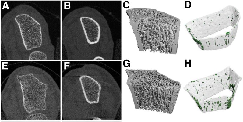
Further supporting the notion of defective bone quality (and strength) in T2D, one study using HR-pQCT (59) demonstrated that cortical porosity was markedly increased (by 124%) in 19 T2D postmenopausal women relative to an equal number of control subjects without diabetes (59) (Fig. 2). None of the trabecular parameters (e.g., trabecular bone volume fraction, trabecular number or thickness) differed between the T2D and control subjects. An increase in cortical porosity, albeit of lesser magnitude (26%), was also reported in 22 African American women with T2D relative to 78 control women (60), again with no significant differences in trabecular parameters. Thus, increased cortical porosity, an element of bone quality not assessed by DXA, may contribute to increased fracture risk in T2D patients.
Figure 2.
Median (by total volumetric BMD) HR-pQCT images of the distal radius from control (top) and T2D (bottom) subjects: distal-most slices (A and E), proximal-most slices (B and F), three-dimensional visualization of the mineralized bone structure (C and G), and three-dimensional visualization of cortical bone (transparent gray) and cortical porosity (dark gray dots) (D and H). Reprinted with permission from Burghardt et al. (59).
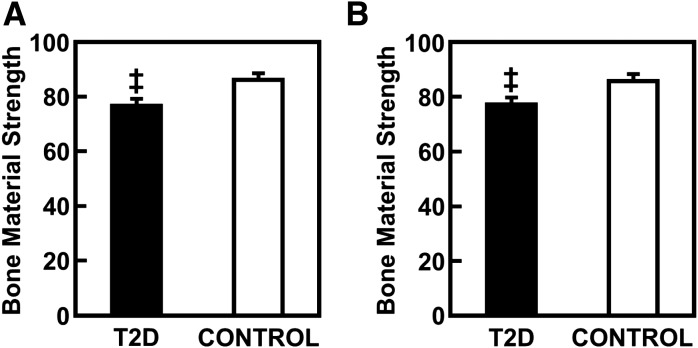
In addition to bone microarchitecture, the material properties of bone also contribute to bone quality. Recently, microindentation of the cortex has gained acceptance as a research tool for estimating bone material strength in humans. Following local anesthesia, this device creates microindents over the shaft of the tibia, which provides a measure of bone material strength (bone material strength index [BMSi]). This technique (61) showed that postmenopausal women with T2D have significant reductions in BMSi as compared with control subjects without diabetes (Fig. 3). This study also found that HbA1c levels were inversely correlated with BMSi in the T2D patients, suggesting that the abnormal bone material properties in these patients may be related to chronic hyperglycemia, perhaps mediated by AGEs.
Figure 3.
Unadjusted (A) and BMI-adjusted (B) comparisons of bone material strength between patients with T2D and age-matched control subjects without diabetes. Values are shown as mean ± SE. ‡P < 0.001. Reprinted with permission from Farr et al. (61).
Bone Turnover and Insulin Signaling
Hyperinsulinemia and hyperglycemia may affect bone remodeling by either directly modulating activities of bone cells or changing the milieu of the bone marrow environment. Osteoblasts, osteoclasts, and osteocytes express insulin receptors, and animal studies imply that increased insulin signaling correlates positively with bone turnover and bone formation, whereas insulin resistance attenuates bone remodeling (62,63). Bone dependence on insulin and glucose metabolism poses the question as to whether bone develops insulin resistance and whether such resistance is manifested with decreased bone turnover. Clarification of this issue may have significant implications for the development of therapies to treat diabetic bone disease associated with low bone turnover.
Bone and Fat Relationships in T2D
Impairment in adipose tissue function is one of the consequences of T2D that directly affects carbohydrate and lipid metabolism and insulin sensitivity. Increase in visceral adiposity, which relates to increased inflammation and metabolic syndrome, is negatively associated with lumbar volumetric BMD (64) and with bone volume and bone formation in iliac crest biopsies from premenopausal women (65). Fat infiltration in muscles is increased in diabetes and is associated with incident fractures, although this association does not account for the higher fracture risk in diabetes (66). Diabetes is associated with higher marrow fat in rodent models, although definitive human studies are lacking (37,67). Increased marrow adiposity and decreased levels of unsaturated fatty acids in the bone marrow correlate positively with fractures (68,69). Marrow adipose tissue accumulates in long bones and vertebrae and constitutes up to 10% of total body fat. Marrow adipose tissue is both unique and similar to extramedullary fat in respect to origin, metabolism, and function and possesses characteristics of both white and beige fat (70). Studies in rodents show that the beige phenotype, which is characterized by the production of bone anabolic factors, is attenuated with diabetes despite an expansion of this fat depot (71).
Bone and Antidiabetes Medications
As bone is involved with energy metabolism, it can be a target for certain antidiabetes therapies (72). Thiazolidinediones (TZDs), high-affinity ligands and activators of peroxisome proliferator–activated receptor γ, target hematopoietic and mesenchymal cells in the bone marrow, resulting in unbalanced bone remodeling with high bone resorption and low bone formation and consequent bone loss and accumulation of large quantities of fat in the bone marrow cavity. Increased bone loss at the lumbar spine, total hip, and femoral neck in women on TZD therapy emerged in the recent meta-analysis of 10 randomized clinical trials (73). Therapy with either rosiglitazone or pioglitazone also increased fracture risk by approximately twofold in women, but not in men. Risk appeared to increase with duration of treatment.
Recently, a novel class of antidiabetes medications, sodium–glucose cotransporter 2 inhibitors, has been scrutinized by the U.S. Food and Drug Administration for a potential harmful effect on bone. It has been reported that patients receiving canagliflozin have an increased fracture rate as early as 12 weeks after initiating therapy (74). However, there was no difference in fracture rates for empagliflozin (75). The bone risk associated with sodium–glucose cotransporter 2 inhibitor therapy may include alterations in calcium and phosphate homeostasis or more direct effects on cells involved in bone remodeling owing to the glucose dependence of their metabolism. There is no evidence for negative effects on bone of other antidiabetes therapies, including biguanides, glucagon-like peptide 1 analogs, and dipeptidyl peptidase 4 inhibitors; in fact, some of these therapies may even be protective against fractures (72).
Management of Low Bone Mass and Fracture Prevention
Effects of Diabetes Complications and Improving Glycemic Control as a Means to Reduce Fracture Risk
Diabetic microvascular complications, such as neuropathy, nephropathy, and retinopathy, have been associated with an increased risk of falls and fractures. Additionally, poor glycemic control, generally defined as an HbA1c value >8%, has been shown to increase fracture risk; however, the fracture benefits of reducing HbA1c levels to lower levels have not been established. In randomized trials among individuals with T2D, neither intensive glycemic control (median HbA1c 6.4% in the intensive group vs. 7.5% in the standard glycemic control group) nor intensive blood pressure control affected the risk of falls or fractures, either positively or negatively (76,77). Although there is limited evidence that improved glycemic control may prevent bone loss in T1D (78), significant hypoglycemia has been associated with increased fracture risk in T1D and T2D, possibly related to falls in the older population (79,80). Glycemic control following current guidelines may be helpful to prevent complications, removing the contribution of hyperglycemia and diabetes complications to increased fracture risk; however, hypoglycemia should be avoided, particularly in older individuals. The evidence that TZDs increase fracture risk in postmenopausal women is robust, and these agents should be avoided in postmenopausal women with T2D.
Prevention of Postfracture Complications in Patients With Diabetes
Animal studies demonstrate reduced rates of cellular differentiation, delayed callus formation, and slowed mineralization after fracture in diabetic animals (81–83). Tight glycemic control and local insulin infusion improve these bone properties in animals (84,85). In humans, higher HbA1c levels at the time of surgery for ankle fracture and within 3 months postoperatively have been associated with an increased risk of infection, delayed union, malunion, and nonunion among patients with T1D or T2D (13,86). Although a baseline and postoperative HbA1c level <7% appears to be beneficial in several reports, data are not available on the optimal level of glycemic control in patients with diabetes with fractures. Until such data are available, current guidelines for inpatient and outpatient glycemic control should be followed.
Nutrition and Lifestyle Interventions to Reduce Fracture Risk
Age-appropriate intakes of calcium and vitamin D should be ensured in all individuals with diabetes. Calcium intake and calcium supplements are not associated with the amount of calcified plaque in the carotid, coronary, or aortic arteries in individuals with T2D (87). Weight-bearing physical activity increases bone density in children with T1D to a degree similar to that in normoglycemic children (88). Assessment of fall risk and appropriate fall prevention measures should be included in the care of older patients with diabetes. Among overweight adults with T2D, significant weight loss may result in bone loss, although the magnitude of bone loss appears to be small (less than 1% at 4 years), was seen only in men, and fracture rates were not increased (89,90). The optimal exercise regimen for weight reduction in individuals with diabetes while minimizing bone loss has not been determined.
Effect of Osteoporosis Therapies in Patients With Diabetes
As discussed earlier, serum markers of bone turnover are generally lower in patients with T1D and T2D than in normoglycemic individuals, raising the concern that antiresorptive agents used as osteoporosis therapy may further exacerbate an already decreased remodeling state rather than provide a protective skeletal effect. However, registry data and data from clinical trials of osteoporosis medications support the effectiveness of these agents in individuals with diabetes. Alendronate increased bone density in 297 women with diabetes enrolled in the Fracture Intervention Trial (FIT) equivalently to normoglycemic individuals (91). Raloxifene reduced the risk of vertebral fracture risk by 35% in the Raloxifene Use for The Heart (RUTH) trial, with consistent effects among subgroups including the approximately 4,500 women with diabetes (92). Examination of osteoporosis medication use and fractures in the Danish Registry demonstrated no difference in fracture rates during treatment with bisphosphonates or raloxifene between individuals with T1D or T2D and normoglycemic control subjects (93). No definitive data are available for strontium or teriparatide, for which only case reports exist.
Analysis of bisphosphonate and denosumab randomized fracture trials shows no significant effect of osteoporosis medications on glucose levels or diabetes incidence (94). Observational data among a small number of individuals treated with teriparatide show similar findings (95).
Summary
Fracture risk is increased in individuals with T1D or T2D, and consequences of fracture are more severe. Reduced bone density contributes to fracture risk in T1D. In T2D, BMD is increased, but in both T1D and T2D, bone quality is negatively affected. Abnormalities of bone cells, bone tissue, and microstructure may all contribute, but the precise mechanisms leading to such abnormalities remain unclear. The roles of hyperglycemia, AGEs, and damage to bone vasculature are current areas of research. Increased falls also contribute to the higher fracture risk. Improved glycemic control may reduce fracture risk and is important to fracture healing but must be balanced against negative effects of hypoglycemia. Nutrition and lifestyle measures to improve bone health are appropriate for individuals with diabetes, and osteoporosis medications have generally proven to be equally effective in patients with diabetes compared with euglycemic individuals. Initiation of osteoporosis medications in patients with diabetes with low bone density or low-trauma fracture is appropriate. More data are needed on identifying patients with diabetes with normal bone density and no history of fracture who will benefit from treatment with osteoporosis medications to prevent fractures. If patients with diabetes develop renal insufficiency, particularly when estimated glomerular filtration rate is below 30 mL/min or the patient undergoes transplantation, additional considerations with respect to renal-related metabolic bone disease or glucocorticoid treatment also need to be addressed. Although skeletal health in T1D and T2D is an area of very active investigation, much remains to be learned regarding the pathophysiology of increased fracture risk, how to estimate fracture risk, and effective strategies to reduce fracture risk in patients with diabetes.
Article Information
Funding. L.C.H. received grant support from Deutsche Forschungsgemeinschaft Sonderforschungsbereiche/Transregio 67 (Project B2). S.K. received grant support from the National Institutes of Health (AG004875 and AR027065). B.L.-C. received grant support from the American Diabetes Association (7-13-BS-089).
Duality of Interest. D.E.S. is a member of Data Safety Monitoring Board for Amgen. R.C. received research support from Amgen and has stock ownership in Amgen, Eli Lilly, and Merck & Co. A.V.S. received a speaker honorarium from Chugai Pharmaceutical Co. and served on an advisory board for Janssen Pharmaceuticals. No other potential conflicts of interest relevant to this article were reported.
References
- 1.Nguyen ND, Ahlborg HG, Center JR, Eisman JA, Nguyen TV. Residual lifetime risk of fractures in women and men. J Bone Miner Res 2007;22:781–788 [DOI] [PubMed] [Google Scholar]
- 2.Burge R, Dawson-Hughes B, Solomon DH, Wong JB, King A, Tosteson A. Incidence and economic burden of osteoporosis-related fractures in the United States, 2005-2025. J Bone Miner Res 2007;22:465–475 [DOI] [PubMed] [Google Scholar]
- 3.Haentjens P, Magaziner J, Colón-Emeric CS, et al. . Meta-analysis: excess mortality after hip fracture among older women and men. Ann Intern Med 2010;152:380–390 [DOI] [PMC free article] [PubMed] [Google Scholar]
- 4.Bertram M, Norman R, Kemp L, Vos T. Review of the long-term disability associated with hip fractures. Inj Prev 2011;17:365–370 [DOI] [PubMed] [Google Scholar]
- 5.Weber DR, Haynes K, Leonard MB, Willi SM, Denburg MR. Type 1 diabetes is associated with an increased risk of fracture across the life span: a population-based cohort study using The Health Improvement Network (THIN). Diabetes Care 2015;38:1913–1920 [DOI] [PMC free article] [PubMed] [Google Scholar]
- 6.Vestergaard P. Discrepancies in bone mineral density and fracture risk in patients with type 1 and type 2 diabetes--a meta-analysis. Osteoporos Int 2007;18:427–444 [DOI] [PubMed] [Google Scholar]
- 7.Janghorbani M, Van Dam RM, Willett WC, Hu FB. Systematic review of type 1 and type 2 diabetes mellitus and risk of fracture. Am J Epidemiol 2007;166:495–505 [DOI] [PubMed] [Google Scholar]
- 8.Looker AC, Eberhardt MS, Saydah SH. Diabetes and fracture risk in older U.S. adults. Bone 2016;82:9–15 [DOI] [PMC free article] [PubMed] [Google Scholar]
- 9.de Liefde II, van der Klift M, de Laet CE, van Daele PL, Hofman A, Pols HA. Bone mineral density and fracture risk in type-2 diabetes mellitus: the Rotterdam Study. Osteoporos Int 2005;16:1713–1720 [DOI] [PubMed] [Google Scholar]
- 10.Norris R, Parker M. Diabetes mellitus and hip fracture: a study of 5966 cases. Injury 2011;42:1313–1316 [DOI] [PubMed] [Google Scholar]
- 11.Hernandez RK, Do TP, Critchlow CW, Dent RE, Jick SS. Patient-related risk factors for fracture-healing complications in the United Kingdom General Practice Research Database. Acta Orthop 2012;83:653–660 [DOI] [PMC free article] [PubMed] [Google Scholar]
- 12.Wukich DK, Joseph A, Ryan M, Ramirez C, Irrgang JJ. Outcomes of ankle fractures in patients with uncomplicated versus complicated diabetes. Foot Ankle Int 2011;32:120–130 [DOI] [PubMed] [Google Scholar]
- 13.Humphers JM, Shibuya N, Fluhman BL, Jupiter D. The impact of glycosylated hemoglobin and diabetes mellitus on wound-healing complications and infection after foot and ankle surgery. J Am Podiatr Med Assoc 2014;104:320–329 [DOI] [PubMed] [Google Scholar]
- 14.Shibuya N, Humphers JM, Fluhman BL, Jupiter DC. Factors associated with nonunion, delayed union, and malunion in foot and ankle surgery in diabetic patients. J Foot Ankle Surg 2013;52:207–211 [DOI] [PubMed] [Google Scholar]
- 15.Hu F, Jiang C, Shen J, Tang P, Wang Y. Preoperative predictors for mortality following hip fracture surgery: a systematic review and meta-analysis. Injury 2012;43:676–685 [DOI] [PubMed] [Google Scholar]
- 16.Golinvaux NS, Bohl DD, Basques BA, Baumgaertner MR, Grauer JN. Diabetes confers little to no increased risk of postoperative complications after hip fracture surgery in geriatric patients. Clin Orthop Relat Res 2015;473:1043–1051 [DOI] [PMC free article] [PubMed] [Google Scholar]
- 17.Nirantharakumar K, Toulis KA, Wijesinghe H, et al. . Impact of diabetes on inpatient mortality and length of stay for elderly patients presenting with fracture of the proximal femur. J Diabetes Complications 2013;27:208–210 [DOI] [PubMed] [Google Scholar]
- 18.Schwartz AV, Vittinghoff E, Bauer DC, et al.; Study of Osteoporotic Fractures (SOF) Research Group; Osteoporotic Fractures in Men (MrOS) Research Group; Health, Aging, and Body Composition (Health ABC) Research Group . Association of BMD and FRAX score with risk of fracture in older adults with type 2 diabetes. JAMA 2011;305:2184–2192 [DOI] [PMC free article] [PubMed] [Google Scholar]
- 19.Leslie WD, Morin SN, Lix LM, Majumdar SR. Does diabetes modify the effect of FRAX risk factors for predicting major osteoporotic and hip fracture? Osteoporos Int 2014;25:2817–2824 [DOI] [PubMed] [Google Scholar]
- 20.Hothersall EJ, Livingstone SJ, Looker HC, et al. . Contemporary risk of hip fracture in type 1 and type 2 diabetes: a national registry study from Scotland. J Bone Miner Res 2014;29:1054–1060 [DOI] [PMC free article] [PubMed] [Google Scholar]
- 21.Bonds DE, Larson JC, Schwartz AV, et al. . Risk of fracture in women with type 2 diabetes: the Women’s Health Initiative Observational Study. J Clin Endocrinol Metab 2006;91:3404–3410 [DOI] [PubMed] [Google Scholar]
- 22.Li CI, Liu CS, Lin WY, et al. . Glycated hemoglobin level and risk of hip fracture in older people with type 2 diabetes: a competing risk analysis of Taiwan Diabetes Cohort Study. J Bone Miner Res 2015;30:1338–1346 [DOI] [PubMed] [Google Scholar]
- 23.Schneider AL, Williams EK, Brancati FL, Blecker S, Coresh J, Selvin E. Diabetes and risk of fracture-related hospitalization: the Atherosclerosis Risk in Communities Study. Diabetes Care 2013;36:1153–1158 [DOI] [PMC free article] [PubMed] [Google Scholar]
- 24.Oei L, Zillikens MC, Dehghan A, et al. . High bone mineral density and fracture risk in type 2 diabetes as skeletal complications of inadequate glucose control: the Rotterdam Study. Diabetes Care 2013;36:1619–1628 [DOI] [PMC free article] [PubMed] [Google Scholar]
- 25.Vestergaard P, Rejnmark L, Mosekilde L. Diabetes and its complications and their relationship with risk of fractures in type 1 and 2 diabetes. Calcif Tissue Int 2009;84:45–55 [DOI] [PubMed] [Google Scholar]
- 26.Sennerby U, Melhus H, Gedeborg R, et al. . Cardiovascular diseases and risk of hip fracture. JAMA 2009;302:1666–1673 [DOI] [PubMed] [Google Scholar]
- 27.Schwartz AV, Hillier TA, Sellmeyer DE, et al. . Older women with diabetes have a higher risk of falls: a prospective study. Diabetes Care 2002;25:1749–1754 [DOI] [PubMed] [Google Scholar]
- 28.Strotmeyer ES, Cauley JA, Schwartz AV, et al. . Nontraumatic fracture risk with diabetes mellitus and impaired fasting glucose in older white and black adults: the Health, Aging, and Body Composition study. Arch Intern Med 2005;165:1612–1617 [DOI] [PubMed] [Google Scholar]
- 29.Schwartz AV, Sellmeyer DE, Ensrud KE, et al.; Study of Osteoporotic Features Research Group . Older women with diabetes have an increased risk of fracture: a prospective study. J Clin Endocrinol Metab 2001;86:32–38 [DOI] [PubMed] [Google Scholar]
- 30.Hofbauer LC, Brueck CC, Singh SK, Dobnig H. Osteoporosis in patients with diabetes mellitus. J Bone Miner Res 2007;22:1317–1328 [DOI] [PubMed] [Google Scholar]
- 31.Thrailkill KM, Lumpkin CK Jr, Bunn RC, Kemp SF, Fowlkes JL. Is insulin an anabolic agent in bone? Dissecting the diabetic bone for clues. Am J Physiol Endocrinol Metab 2005;289:E735–E745 [DOI] [PMC free article] [PubMed] [Google Scholar]
- 32.Moyer-Mileur LJ, Slater H, Jordan KC, Murray MA. IGF-1 and IGF-binding proteins and bone mass, geometry, and strength: relation to metabolic control in adolescent girls with type 1 diabetes. J Bone Miner Res 2008;23:1884–1891 [DOI] [PMC free article] [PubMed] [Google Scholar]
- 33.Bechtold S, Putzker S, Bonfig W, Fuchs O, Dirlenbach I, Schwarz HP. Bone size normalizes with age in children and adolescents with type 1 diabetes. Diabetes Care 2007;30:2046–2050 [DOI] [PubMed] [Google Scholar]
- 34.Neumann T, Lodes S, Kästner B, et al. . Trabecular bone score in type 1 diabetes-a cross-sectional study. Osteoporos Int 2016;27:127–133 [DOI] [PubMed] [Google Scholar]
- 35.Abdalrahaman N, McComb C, Foster JE, et al. . Deficits in trabecular bone microarchitecture in young women with type 1 diabetes mellitus. J Bone Miner Res 2015;30:1386–1393 [DOI] [PubMed] [Google Scholar]
- 36.Shanbhogue VV, Hansen S, Frost M, et al. . Bone geometry, volumetric density, microarchitecture, and estimated bone strength assessed by HR-pQCT in adult patients with type 1 diabetes mellitus. J Bone Miner Res 2015;30:2188–2199 [DOI] [PubMed] [Google Scholar]
- 37.Botolin S, McCabe LR. Bone loss and increased bone adiposity in spontaneous and pharmacologically induced diabetic mice. Endocrinology 2007;148:198–205 [DOI] [PubMed] [Google Scholar]
- 38.Thrailkill KM, Liu L, Wahl EC, et al. . Bone formation is impaired in a model of type 1 diabetes. Diabetes 2005;54:2875–2881 [DOI] [PubMed] [Google Scholar]
- 39.Verhaeghe J, Visser WJ, Einhorn TA, Bouillon R. Osteoporosis and diabetes: lessons from the diabetic BB rat. Horm Res 1990;34:245–248 [DOI] [PubMed] [Google Scholar]
- 40.Nyman JS, Even JL, Jo CH, et al. . Increasing duration of type 1 diabetes perturbs the strength-structure relationship and increases brittleness of bone. Bone 2011;48:733–740 [DOI] [PMC free article] [PubMed] [Google Scholar]
- 41.Hamada Y, Kitazawa S, Kitazawa R, Fujii H, Kasuga M, Fukagawa M. Histomorphometric analysis of diabetic osteopenia in streptozotocin-induced diabetic mice: a possible role of oxidative stress. Bone 2007;40:1408–1414 [DOI] [PubMed] [Google Scholar]
- 42.Saito M, Fujii K, Mori Y, Marumo K. Role of collagen enzymatic and glycation induced cross-links as a determinant of bone quality in spontaneously diabetic WBN/Kob rats. Osteoporos Int 2006;17:1514–1523 [DOI] [PubMed] [Google Scholar]
- 43.Tanaka K, Yamaguchi T, Kanazawa I, Sugimoto T. Effects of high glucose and advanced glycation end products on the expressions of sclerostin and RANKL as well as apoptosis in osteocyte-like MLO-Y4-A2 cells. Biochem Biophys Res Commun 2015;461:193–199 [DOI] [PubMed] [Google Scholar]
- 44.Wei J, Shimazu J, Makinistoglu MP, et al. . Glucose uptake and Runx2 synergize to orchestrate osteoblast differentiation and bone formation. Cell 2015;161:1576–1591 [DOI] [PMC free article] [PubMed] [Google Scholar]
- 45.Starup-Linde J, Vestergaard P. Biochemical bone turnover markers in diabetes mellitus - a systematic review. Bone 2016;82:69–78 [DOI] [PubMed] [Google Scholar]
- 46.Armas LA, Akhter MP, Drincic A, Recker RR. Trabecular bone histomorphometry in humans with type 1 diabetes mellitus. Bone 2012;50:91–96 [DOI] [PMC free article] [PubMed] [Google Scholar]
- 47.Farlay D, Armas LA, Gineyts E, et al. . Nonenzymatic glycation and degree of mineralization are higher in bone from fractured patients with type 1 diabetes mellitus. J Bone Miner Res 2016;31:190–195 [DOI] [PMC free article] [PubMed] [Google Scholar]
- 48.Neumann T, Lodes S, Kästner B, et al. . High serum pentosidine but not esRAGE is associated with prevalent fractures in type 1 diabetes independent of bone mineral density and glycaemic control. Osteoporos Int 2014;25:1527–1533 [DOI] [PubMed] [Google Scholar]
- 49.Lafage-Proust MH, Roche B, Langer M, et al. Assessment of bone vascularization and its role in bone remodeling. Bonekey Rep 2015;4:662 [DOI] [PMC free article] [PubMed]
- 50.Zhou Y, Li Y, Zhang D, Wang J, Yang H. Prevalence and predictors of osteopenia and osteoporosis in postmenopausal Chinese women with type 2 diabetes. Diabetes Res Clin Pract 2010;90:261–269 [DOI] [PubMed] [Google Scholar]
- 51.Schwartz AV, Sellmeyer DE, Strotmeyer ES, et al.; Health ABC Study . Diabetes and bone loss at the hip in older black and white adults. J Bone Miner Res 2005;20:596–603 [DOI] [PubMed] [Google Scholar]
- 52.Krakauer JC, McKenna MJ, Buderer NF, Rao DS, Whitehouse FW, Parfitt AM. Bone loss and bone turnover in diabetes. Diabetes 1995;44:775–782 [DOI] [PubMed] [Google Scholar]
- 53.Manavalan JS, Cremers S, Dempster DW, et al. . Circulating osteogenic precursor cells in type 2 diabetes mellitus. J Clin Endocrinol Metab 2012;97:3240–3250 [DOI] [PMC free article] [PubMed] [Google Scholar]
- 54.García-Martín A, Rozas-Moreno P, Reyes-García R, et al. . Circulating levels of sclerostin are increased in patients with type 2 diabetes mellitus. J Clin Endocrinol Metab 2012;97:234–241 [DOI] [PubMed] [Google Scholar]
- 55.Gennari L, Merlotti D, Valenti R, et al. . Circulating sclerostin levels and bone turnover in type 1 and type 2 diabetes. J Clin Endocrinol Metab 2012;97:1737–1744 [DOI] [PubMed] [Google Scholar]
- 56.Vashishth D. The role of the collagen matrix in skeletal fragility. Curr Osteoporos Rep 2007;5:62–66 [DOI] [PubMed] [Google Scholar]
- 57.Schwartz AV, Garnero P, Hillier TA, et al.; Health, Aging, and Body Composition Study . Pentosidine and increased fracture risk in older adults with type 2 diabetes. J Clin Endocrinol Metab 2009;94:2380–2386 [DOI] [PMC free article] [PubMed] [Google Scholar]
- 58.Yamamoto M, Yamaguchi T, Yamauchi M, Yano S, Sugimoto T. Serum pentosidine levels are positively associated with the presence of vertebral fractures in postmenopausal women with type 2 diabetes. J Clin Endocrinol Metab 2008;93:1013–1019 [DOI] [PubMed] [Google Scholar]
- 59.Burghardt AJ, Issever AS, Schwartz AV, et al. . High-resolution peripheral quantitative computed tomographic imaging of cortical and trabecular bone microarchitecture in patients with type 2 diabetes mellitus. J Clin Endocrinol Metab 2010;95:5045–5055 [DOI] [PMC free article] [PubMed] [Google Scholar]
- 60.Yu EW, Putman MS, Derrico N, Abrishamanian-Garcia G, Finkelstein JS, Bouxsein ML. Defects in cortical microarchitecture among African-American women with type 2 diabetes. Osteoporos Int 2015;26:673–679 [DOI] [PMC free article] [PubMed] [Google Scholar]
- 61.Farr JN, Drake MT, Amin S, Melton LJ 3rd, McCready LK, Khosla S. In vivo assessment of bone quality in postmenopausal women with type 2 diabetes. J Bone Miner Res 2014;29:787–795 [DOI] [PMC free article] [PubMed] [Google Scholar]
- 62.Fulzele K, Riddle RC, DiGirolamo DJ, et al. . Insulin receptor signaling in osteoblasts regulates postnatal bone acquisition and body composition. Cell 2010;142:309–319 [DOI] [PMC free article] [PubMed] [Google Scholar]
- 63.Lecka-Czernik B, Stechschulte LA, Czernik PJ, Dowling AR. High bone mass in adult mice with diet-induced obesity results from a combination of initial increase in bone mass followed by attenuation in bone formation; implications for high bone mass and decreased bone quality in obesity. Mol Cell Endocrinol 2015;410:35–41 [DOI] [PubMed]
- 64.Ng AC, Melton LJ 3rd, Atkinson EJ, et al. . Relationship of adiposity to bone volumetric density and microstructure in men and women across the adult lifespan. Bone 2013;55:119–125 [DOI] [PMC free article] [PubMed] [Google Scholar]
- 65.Cohen A, Dempster DW, Recker RR, et al. . Abdominal fat is associated with lower bone formation and inferior bone quality in healthy premenopausal women: a transiliac bone biopsy study. J Clin Endocrinol Metab 2013;98:2562–2572 [DOI] [PMC free article] [PubMed] [Google Scholar]
- 66.Schafer AL, Vittinghoff E, Lang TF, et al.; Health, Aging, and Body Composition (Health ABC) Study . Fat infiltration of muscle, diabetes, and clinical fracture risk in older adults. J Clin Endocrinol Metab 2010;95:E368–E372 [DOI] [PMC free article] [PubMed] [Google Scholar]
- 67.Devlin MJ, Van Vliet M, Motyl K, et al. . Early-onset type 2 diabetes impairs skeletal acquisition in the male TALLYHO/JngJ mouse. Endocrinology 2014;155:3806–3816 [DOI] [PMC free article] [PubMed] [Google Scholar]
- 68.Schwartz AV, Sigurdsson S, Hue TF, et al. . Vertebral bone marrow fat associated with lower trabecular BMD and prevalent vertebral fracture in older adults. J Clin Endocrinol Metab 2013;98:2294–2300 [DOI] [PMC free article] [PubMed] [Google Scholar]
- 69.Patsch JM, Li X, Baum T, et al. . Bone marrow fat composition as a novel imaging biomarker in postmenopausal women with prevalent fragility fractures. J Bone Miner Res 2013;28:1721–1728 [DOI] [PMC free article] [PubMed] [Google Scholar]
- 70.Lecka-Czernik B, Stechschulte LA: Bone and fat: a relationship of different shades. Arch Biochem Biophys 2014;561:124–129 [DOI] [PubMed]
- 71.Krings A, Rahman S, Huang S, Lu Y, Czernik PJ, Lecka-Czernik B. Bone marrow fat has brown adipose tissue characteristics, which are attenuated with aging and diabetes. Bone 2012;50:546–552 [DOI] [PMC free article] [PubMed] [Google Scholar]
- 72.Meier C, Schwartz AV, Egger A, Lecka-Czernik B. Effects of diabetes drugs on the skeleton. Bone 2016;82:93–100 [DOI] [PubMed] [Google Scholar]
- 73.Zhu ZN, Jiang YF, Ding T. Risk of fracture with thiazolidinediones: an updated meta-analysis of randomized clinical trials. Bone 2014;68:115–123 [DOI] [PubMed]
- 74.U.S. Food and Drug Administration. Invokana and Invokamet (Canagliflozin): Drug Safety Communication - New Information on Bone Fracture Risk and Decreased Bone Mineral Density [Internet], 2015. Available from http://www.fda.gov/Safety/MedWatch/SafetyInformation/SafetyAlertsforHumanMedicalProducts/ucm461876.htm. Accessed 2 March 2016
- 75.Zinman B, Wanner C, Lachin JM, et al.; EMPA-REG OUTCOME Investigators . Empagliflozin, cardiovascular outcomes, and mortality in type 2 diabetes. N Engl J Med 2015;373:2117–2128 [DOI] [PubMed] [Google Scholar]
- 76.Schwartz AV, Margolis KL, Sellmeyer DE, et al. . Intensive glycemic control is not associated with fractures or falls in the ACCORD randomized trial. Diabetes Care 2012;35:1525–1531 [DOI] [PMC free article] [PubMed] [Google Scholar]
- 77.Margolis KL, Palermo L, Vittinghoff E, et al. . Intensive blood pressure control, falls, and fractures in patients with type 2 diabetes: the ACCORD trial. J Gen Intern Med 2014;29:1599–1606 [DOI] [PMC free article] [PubMed] [Google Scholar]
- 78.Campos Pastor MM, López-Ibarra PJ, Escobar-Jiménez F, Serrano Pardo MD, García-Cervigón AG. Intensive insulin therapy and bone mineral density in type 1 diabetes mellitus: a prospective study. Osteoporos Int 2000;11:455–459 [DOI] [PubMed] [Google Scholar]
- 79.Johnston SS, Conner C, Aagren M, Ruiz K, Bouchard J. Association between hypoglycaemic events and fall-related fractures in Medicare-covered patients with type 2 diabetes. Diabetes Obes Metab 2012;14:634–643 [DOI] [PubMed] [Google Scholar]
- 80.Majkowska L, Waliłko E, Molęda P, Bohatyrewicz A. Thoracic spine fracture in the course of severe nocturnal hypoglycemia in young patients with type 1 diabetes mellitus--the role of low bone mineral density. Am J Emerg Med 2014;32:816.e5–816.e7 [DOI] [PubMed] [Google Scholar]
- 81.Retzepi M, Donos N. The effect of diabetes mellitus on osseous healing. Clin Oral Implants Res 2010;21:673–681 [DOI] [PubMed] [Google Scholar]
- 82.Kayal RA, Tsatsas D, Bauer MA, et al. . Diminished bone formation during diabetic fracture healing is related to the premature resorption of cartilage associated with increased osteoclast activity. J Bone Miner Res 2007;22:560–568 [DOI] [PMC free article] [PubMed] [Google Scholar]
- 83.Lu H, Kraut D, Gerstenfeld LC, Graves DT. Diabetes interferes with the bone formation by affecting the expression of transcription factors that regulate osteoblast differentiation. Endocrinology 2003;144:346–352 [DOI] [PubMed] [Google Scholar]
- 84.Gandhi A, Beam HA, O’Connor JP, Parsons JR, Lin SS. The effects of local insulin delivery on diabetic fracture healing. Bone 2005;37:482–490 [DOI] [PubMed] [Google Scholar]
- 85.Beam HA, Parsons JR, Lin SS. The effects of blood glucose control upon fracture healing in the BB Wistar rat with diabetes mellitus. J Orthop Res 2002;20:1210–1216 [DOI] [PubMed] [Google Scholar]
- 86.Liu J, Ludwig T, Ebraheim NA. Effect of the blood HbA1c level on surgical treatment outcomes of diabetics with ankle fractures. Orthop Surg 2013;5:203–208 [DOI] [PMC free article] [PubMed] [Google Scholar]
- 87.Raffield LM, Agarwal S, Cox AJ, et al. . Cross-sectional analysis of calcium intake for associations with vascular calcification and mortality in individuals with type 2 diabetes from the Diabetes Heart Study. Am J Clin Nutr 2014;100:1029–1035 [DOI] [PMC free article] [PubMed] [Google Scholar]
- 88.Maggio AB, Rizzoli RR, Marchand LM, Ferrari S, Beghetti M, Farpour-Lambert NJ. Physical activity increases bone mineral density in children with type 1 diabetes. Med Sci Sports Exerc 2012;44:1206–1211 [DOI] [PubMed] [Google Scholar]
- 89.Lipkin EW, Schwartz AV, Anderson AM, et al.; Look AHEAD Research Group . The Look AHEAD Trial: bone loss at 4-year follow-up in type 2 diabetes. Diabetes Care 2014;37:2822–2829 [DOI] [PMC free article] [PubMed] [Google Scholar]
- 90.Wing RR, Bolin P, Brancati FL, et al.; Look AHEAD Research Group . Cardiovascular effects of intensive lifestyle intervention in type 2 diabetes. N Engl J Med 2013;369:145–154 [DOI] [PMC free article] [PubMed] [Google Scholar]
- 91.Keegan TH, Schwartz AV, Bauer DC, Sellmeyer DE, Kelsey JL; Fracture Intervention Trial . Effect of alendronate on bone mineral density and biochemical markers of bone turnover in type 2 diabetic women: the Fracture Intervention Trial. Diabetes Care 2004;27:1547–1553 [DOI] [PubMed] [Google Scholar]
- 92.Ensrud KE, Stock JL, Barrett-Connor E, et al. . Effects of raloxifene on fracture risk in postmenopausal women: the Raloxifene Use for the Heart Trial. J Bone Miner Res 2008;23:112–120 [DOI] [PubMed] [Google Scholar]
- 93.Vestergaard P, Rejnmark L, Mosekilde L. Are antiresorptive drugs effective against fractures in patients with diabetes? Calcif Tissue Int 2011;88:209–214 [DOI] [PubMed] [Google Scholar]
- 94.Schwartz AV, Schafer AL, Grey A, et al. . Effects of antiresorptive therapies on glucose metabolism: results from the FIT, HORIZON-PFT, and FREEDOM trials. J Bone Miner Res 2013;28:1348–1354 [DOI] [PubMed] [Google Scholar]
- 95.Mazziotti G, Maffezzoni F, Doga M, Hofbauer LC, Adler RA, Giustina A. Outcome of glucose homeostasis in patients with glucocorticoid-induced osteoporosis undergoing treatment with bone active-drugs. Bone 2014;67:175–180 [DOI] [PubMed]


