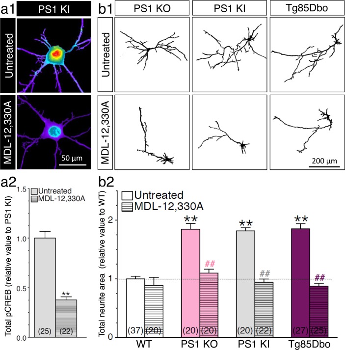
Figure 7. PS1-induced neurite outgrowth and associated CREB signaling require adenylate cyclase activation.
(a1) Representative pseudocolor images of phosphorylated CREB (pCREB) immunofluorescence is shown in 14 DIV neurons generated from PS1 KI mice treated with adenylate cyclase inhibitor MDL-12,330A (100 nM, 30 min) using polyclonal phospho-(Ser133) CREB antibody. (a2) Quantitative analysis of pCREB staining intensity is represented as relative changes as compared to untreated PS1 KI neurons. **p<0.001 compared to PS1 KI. (b1) Representative inverted images of YFP fluorescence in cortical neurons (8 DIV) generated from PS1 KO, PS1 KI, andTg85Dbo mice are shown after 24 hr treatment with MDL-12,330A (10 nM). (b2) Analysis of neurite extension is represented as relative changes in total neurite area as compared to WT littermates. **p<0.001 compared to WT, ##p<0.001 compared to untreated condition for each group. The total number of neurons (from at least 3 independent sets of cultures) used for quantification is shown in parentheses (WT, n = 5 embryos; PS1 KO, n = 3 embryos; PS1 KI, n = 3 embryos; Tg85Dbo, n = 3 embryos). Error bars indicate SEM.

