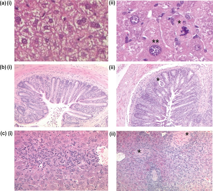
FIG 3 .
Representative hematoxylin- and eosin-stained sections from naive and infected F2 Giftm1a/tm1a and wild-type mice. (a) Liver sections from naive wild-type (i) and F2 Giftm1a/tm1a (ii) mice. A single asterisk indicates an abnormal mitotic figure, and double asterisks indicate enlarged cellular nuclei in an F2 Giftm1a/tm1a mouse (×400 magnification). (b) Colon sections obtained on day 14 after a C. rodentium challenge of wild-type (i) and F2 Giftm1a/tm1a (ii) mice. The asterisk indicates a crypt abscess in an F2 Giftm1a/tm1a mouse (×100 magnification). (c) Liver sections obtained on day 8 after an S. Typhimurium challenge of wild-type (i) and F2 Giftm1a/tm1a (ii) mice. Both images show inflammatory cellular infiltration and granuloma formation. Asterisks indicate the large necrotic regions seen in F2 Giftm1a/tm1a mice (× 200 magnification).

