Abstract
In this study, DNA arrays have been employed to monitor gene expression patterns in testis of mice exposed to tobacco smoke for 24 weeks and compared to control animals. The results of the analysis revealed significant changes in expression of several genes that may have a role in spermatogenesis. Cdk14 was chosen for further characterization because of a suggested role in the testis and in regulation of Wnt signaling. RT-PCR analysis confirmed down regulation of Cdk14 in mice exposed to cigarette smoke (CS). Cdk14 is expressed in all testicular cells; spermatogonia- and Sertoli-derived cell lines treated with cigarette smoke extract (CSE) in vitro showed down-regulation of CDK14 mRNA and protein levels as well as down-regulation of β-catenin levels. CS-induced down-regulation of CDK14 mRNA and protein levels was also observed in several lung epithelium-derived cell lines including primary normal human bronchial epithelial cells (NHBE), suggesting that the effect is not restricted to the testis. Similar to testicular cells, CS-induced down-regulation of CDK14 in lung cells correlated with decreased levels of β-catenin, a finding suggesting impaired Wnt signaling. In the lungs, CDK14 was localized to the alveolar and bronchial epithelium.
Keywords: Cigarette smoke, CDK14/PFTK1/PFTAIRE-1, Cyclin Y, β-Catenin, Spermatogenesis, Spermatogonia, DNA arrays, Lungs, Normal human bronchial cells
1. Introduction
It is generally agreed that tobacco smoke can be a cause of serious health-related problems (Witschi, 2001; Doll et al., 2004). Pathologies linked to smoking tobacco cigarettes include diseases of the cardiovascular system, in particular atherosclerosis, myocardial infarction and stroke, and diseases of the respiratory tract, such as emphysema, lung cancer and cancers of the larynx and mouth. These effects are thought to stem from long term exposure to more than 3000 chemicals found in tobacco smoke, such as nicotine, carbon monoxide, cyanide, and other compounds that are believed to be responsible for various forms of tissue damage. Exposure to cigarette smoke may be either active or passive. While the association between inhalation of mainstream smoke and diseases has been established for many years, the realization that exposure to second hand smoke adversely affects human health is more recent (Jinot and Bayard, 1994; Chidekel, 2000; Cook and Strachan, 1999; Strachan and Cook, 1997).
Adult spermatogenesis is a multiphase process that includes proliferation of spermatogonia, meiosis of spermatocytes, and post-meiotic maturation of spermatids (spermiogenesis). Spermatogenesis is regulated by controlled expression of specific genes at different stages of germ cell maturation. The process is supported by growth factors and hormones secreted by Sertoli and Leydig somatic cells. Successful progression through spermatogenesis is crucial for normal gamete formation and for transferring the genetic information to the next generations.
Several studies have shown that smoking might be associated with increased prevalence of abnormal sperm and changes in steroid hormone concentrations in humans (Attia et al., 1989; Chia et al., 1994; Field et al., 1994). A study in rats has shown a significant correlation between cigarette smoke exposure and impaired testicular histology; however, the mechanism underlying the adverse effect of tobacco on testicular cells has not been well-characterized. As recently shown by our group, exposure of mice to tobacco smoke in vivo causes oxidative stress and changes in posttranslational modifications of proteins in mouse testicular cells and in human sperm (Shrivastava et al., 2010; Vigodner et al., 2013). Another study has found a significant increase in germ-line mutation frequency in spermatogonial stem cells of mice exposed to tobacco (Yauk et al., 2007). It has also been demonstrated that CSE-treated spermatocytes show signs of oxidative damage and increased expression of several antioxidant genes (Esakky et al., 2012). However, tobacco-induced changes in gene expression in vivo remain largely uncharacterized. In this study, we employed DNA arrays to examine and compare gene expression patterns in testis of mice exposed to tobacco smoke for 24 weeks as compared to control animals. We observed significant changes in several genes with a putative role in spermatogenesis, and further analyzed the effects of cigarette smoke on the expression of CDK14 (cyclin-dependent kinase 14) in multiple cell lines in vitro.
2. Materials and methods
2.1. Tobacco exposure
Adult (6–8 week old) C57B16 male mice were obtained from the Jackson Laboratory (Bar Harbor, ME, USA). Animal Committee of Yeshiva University approved all animal protocols. The tobacco smoke exposure was performed in the laboratory of Dr. Jeanine D’Armiento, Division of Molecular Medicine, Columbia University, New York as previously described (Foronjy et al., 2006). After acclimatization for three days, five mice were exposed to cigarette smoke in a specially designed chamber for 6 h/day 5 days a week for 24 weeks at a total particulate matter (TPM) concentration of 250mg/m3. The TPM was determined by gravimetric analysis of filter samples taken during the exposure periods. Two 70mL puffs per minute from research cigarettes (Type 3RF; 3R4F: 10mg of tar and 0.8mg of nicotine, Tobacco Research, University of Kentucky, Lexington, KY, USA) were generated by a smoking machine (Teague Enterprises, Davis, CA) and then diluted with fresh air and delivered to whole body exposure chambers. Mice were provided with standard food and water ad libitum and maintained at room temperature in a 12-h dark/light cycle. Mice showed no signs of adverse effects or abnormal behavior during or after the smoke exposure. Five control mice were exposed to room air. The same exposure schedule has previously been used in several studies from D’Armiento’s laboratory and in a previously published study from our group showing an adverse effect of CS on testicular cells (Shrivastava et al., 2010).
2.2. Cell lines
GC1 spermatogonia and 15P1 Sertoli were obtained from American Type Culture Collection (ATCC). Cells were cultured in DMEM supplemented with 10% fetal bovine serum, incubated at 37 °C (5% CO2). Immortalized human bronchial epithelial cells (HBEC), a gift from Dr. Spivack (Albert Einstein College of Medicine), were cultured in keratinocyte serum-free medium (Life technologies, Cat# 17005-042) containing 50 mg/L bovine pituitary extract with 5μg/L epidermal growth factor (Tan et al., 2010). Human bronchial epithelial cells (BEAS-2B) were purchased from ATCC (Cat# CRL-9609™) and maintained in bronchial epithelial cell growth medium (BEGM, Cat# CC-3170) containing Clonetics® bronchial epithelial cell basal medium with supplements provided by Lonza. Normal human bronchial epithelial cells (NHBE) were purchased from Lonza (Cat# CC-2540) and maintained in BEGM produced by Lonza.
2.3. Preparation of cigarette smoke extract
CSEwas prepared as described previously (Calogero et al., 2009; Mercer et al., 2009; Lawson et al., 1998; Lemaitre et al., 2011). In brief, one research cigarette (3R4F) was attached to a tube connected to a Buchner flask containing 25mL PBS. The smoke derived from the cigarette was drawn into the flask under a vacuum generated by a nickel-platedwater aspirator. The pH of the solution was then adjusted to 7.2–7.4 with 1N HCl and filtered through a 0.22-μm pore filter to remove bacteria and large particles. The resulting 100% CSE was diluted with PBS to achieve 1–10% concentrations and used within 30 min of preparation. The concentrations of 1–10% CSE correspond to the nicotine concentrations in the extract which is similar to these measured in the blood of the smokers (Calogero et al., 2009; Lawson et al., 1998). This concentration range was also commonly used for cell treatment in other previously published studies (Mercer et al., 2009; Lemaitre et al., 2011). Treatments for the indicated time periods were followed by preparation of whole-cell protein lysates. Each experiment was repeated at least three times.
2.4. Gene array and statistical analysis
Testes of control and cigarette smoke-exposed mice were obtained, and RNAwas isolated from the samples using the RNeasy mini kit (Qiagen). RNA integrity was tested by microfluidic analysis using the Agilent 2100 BioAnalyzer. The microarray analysis was performed at the Albert Einstein College of Medicine of Yeshiva University Microarray Facility. For each sample, the Affymetrix whole-transcript protocol was used to amplify 300 ng RNA and hybridized to the Affymetrix Mouse Gene 1.0 ST Array. Three mice were used per condition (control and cigarette smoke) to provide biological triplicates. Images of each array have been captured using the Axon scanner, and the pixel intensities from each channel of individual features on the chip were determined using the Genepix 3.0 program.
The raw array data were imported into Expression Console v1.1 (Affymetrix), a software package that permits visualization, quality control and normalization of the data. Quality control (QC) results included array images, line graphs of labeling and hybridization controls, signal box plots and histograms before and after normalization for all hybridizations, as well as a heatmap for Spearman rank correlation between all pairs of hybridizations. The gene expression measures for all arrays that passed QC were then normalized by the robust multi-array analysis (RMA) approach in the Expression Console software package.
Ranking of genes by degree of differential expression was performed using Bioconductor package limma and in-house R code. Selection of significantly different gene expression profiles between the two experimental conditions was based on the empirical Bayes moderated t-statistic and Benjamini–Hochberg method was applied to correct for multiple testing. Significant genes were identified by adjusted-P value ≤0.05 and fold-difference in mean expression ≥|1.4|. All results are expressed as the mean±standard deviation (SD) unless otherwise stated.
2.5. Real time RT-PCR
Total RNA was extracted from testicular tissues using the RNeasy Mini Kit (Qiagen, Valencia, CA). The quality and concentration of RNA in samples was determined using Nanodrop 2000 (Thermo Fisher Scientific, West Palm Beach, FL). Equal amounts of RNA (500 ng) from different samples were used for cDNA synthesis using iScript cDNA synthesis kit (Bio-Rad Laboratories, Hercules, CA) according to the manufacturer’s instructions.
The primers used, based on the cDNA sequences were as follows:
Mouse primers:
Cdk14 forward: 5′-CTGCGAGGACTGTCTTACATC-3′;
Cdk14 reverse: 5′-TGGCTAGGGACGGATTTTG-3′
Gapdh forward: 5′-CTTTGTCAAGCTCATTTCCTGG-3′;
Gapdh reverse: 5′-TCTTGCTCAGTGTCCTTGC-3′
Human primers:
CDK14 forward: 5′-TGTCAGTACATGGACAAGCACCCT-3′;
CDK14 reverse: 5′-TGTAAGACAGACCTCGCAGCAACT-3′
GAPDH forward: 5′-GCAAGAGCACAAGAGGAAG-3′;
GAPDH reverse: 5′-TCTACATGGCAACTGTGAGG-3′
qPCR was performed in triplicate using iQ™ SYBR® green supermix and CFX96™ Real-time system (Bio-Rad Hercules, CA). Using the Bio-Rad CFX Manager analyzing program, the values of the relative quantities (RQs) of CDK14 were calculated from their quantification cycle (Cq) measurements and then normalized to the geometric mean of the GAPDH RQs to produce the normalized expression values. For cell line experiments, controls (untreated samples) were considered as 1 and other samples were normalized to the controls. To calculate the difference between samples, the experiments were performed at least three times for each condition. Student’s paired t-test was used. P values <0.05 were considered statistically significant.
2.6. Whole cell protein lysates, gel electrophoresis, western and oxyblot analysis
Whole cell protein lysateswas prepared as previously described (Shrivastava et al., 2010) using the whole cell extraction kit and protease inhibitor cocktail (Chemicon) according to the manufacturer’s instructions. Protein concentrations were determined via bicinchoninic acid (BCA) protein assay using bovine serum albumin (BSA) as the standard (Pierce, Rockford, IL, USA). Gel electrophoresis was performed under reducing conditions using NuPAGE 4–12% gradient bis-tris polyacrylamide gels and MOPS running buffer (Invitrogen, Carlsbad, CA, USA) at a constant voltage (200 V). After electrophoresis, proteins were transferred to a nitrocellulose membrane (0.45μm, Invitrogen, Carlsbad, CA, USA) using NuPAGE transfer buffer. Protein electrophoresis and transfer were performed using the Invitrogen XCell SureLock Mini-Cell electrophoresis system. Rabbit polyclonal antibody against CDK14 from Proteintech, (Chicago, IL, Cat# 21612-1-AP) and Abcam (Cambridge, MA, Cat# ab104150) were used at 1:750 and 1:100 dilution, respectively. Mouse monoclonal anti-β-catenin antibody (cell signaling, Beverly, MA, Cat# 104150) was used at 1:1000 dilution. Equal loading was ensured with monoclonal antiβ-tubulin antibody (Abcam, Cat# ab7291) at 1:2000 dilution or anti-β-actin antibody (Santz Cruz, Cat# sc-1615) at 1:1000. Western blot detection was performed using the ECL plus kit (GE Healthcare, Piscataway, NJ, USA), in accordance with the manufacturer’s instructions. Quantitative densitometry analyses were performed using the Quantity One software (Bio-Rad Laboratories, Hercules, CA, USA) and the density values were normalized to tubulin. In each experiment, controls (untreated samples) were considered as 1 and other samples were normalized to the controls. To calculate the difference between samples, Student’s paired t-test was used. P values <0.05 were considered statistically significant.
OxyBlot kit (Millipore, Billerica, MA, USA) was used to immunodetect carbonyl groups (which are introduced into protein side chains as a result of protein oxidation) according to the user’s manual and as previously described by our group (Shrivastava et al., 2010). Briefly, to derivatize the carbonyl groups to 2,4-dinitrophenylhydrazone (DNP-hydrazone), cell lysates (~10μg) were treated with DNP-hydrazine for 15 min at room temperature, and were then neutralized using the neutralization buffer provided with the kit. A negative control without DNP-hydrazone was used in each experiment. Proteins were then separated on 4–12% NuPAGE gels and transferred onto nitrocellulose membranes as described above. Western blotting was performed using an antibody specific to the DNP moiety of the proteins.
2.7. Germ cell separation by velocity sedimentation (STA-PUT)
Mouse testicular germ cells were separated using a STA-PUT velocity sedimentation cell separator (ProScience Inc., Scarborough, Ontario, Canada) as previously described (La Salle et al., 2009). Briefly,10–12 adult mice (45 days) were sacrificed, and their testes were isolated, decapsulated and enzymatically digested, first with collagenase (0.5 mg/mL) for 20 min and then with trypsin (0.5 mg/mL) and DNase I (0.5μg/mL) together for 13 min. Both digestions were performed with constant shaking at 225 rpm in a 32 °C water-bath. This cell suspension was then filtered and washed with 0.5% BSA in KRB media (120mM NaCl, 4.8mM KCl, 25.2mM NaHCO3, 1.2mM KH2PO4, 1.2mM MgSO4·7H2O, 1.3mM CaCl2, 1 × Pen/Strep/Glu (Life technologies), 1 × essential amino acids, 1 × non-essential amino acids (Lonza, Walkersville, MD, USA), and 11.1mM dextrose) and complemented with DNase I (0.4μg/mL). The cells were then resuspended in 25mL of 0.5% BSA in KRB media and loaded into the loading chamber of the STA-PUT apparatus. The cells were allowed to sediment for approximately 1 h 45 min, and 10mL fractions were collected. Every fifth fraction, starting from the 21st fraction, was examined using flow cytometry. The purity of the combined fractions was additionally confirmed microscopically.
2.8. Flow cytometry analysis
Selected fractions were centrifuged at 500 ×g for 7 min at 4 °C followed by aspiration of all but 1mL of supernatant. The pellets were then re-suspended with a brief, gentle vortex. From each resulting cell suspension, 150μl was individually mixed with propidium iodide (PI) staining solution (PBS complemented with 1% (v/v) RNase A, 10μg/mL PI, and 1% (v/v) Igepal CA-630) and incubated at 37 °C for 15 min in the dark. After incubation, the samples were filtered through a nylon mesh and subjected to a cell cycle assay. The flow-cytometry and the subsequent analyses were processed by CytoSoft software (Millipore, Billerica, MA, USA). Fractions containing primary spermatocytes (tetraploid cells) and spermatids (haploid cells) with a purity above 80% and 90%, respectively, were pooled together for cell slide preparation.
2.9. Isolation of spermatogonia and Sertoli cells
Five to ten 9-day old mice were sacrificed and their testes were isolated and decapsuled. The exposed seminiferous tubules were digested first by collagenase (1 mg/mL) for 4 min and then by a mixture of collagenase (1 mg/mL), trypsin (0.5 mg/mL), and hyaluronidase (1.5 mg/mL) for 8 min at 34 °C with shaking speed of 150 rpm. After that, tubules were subjected to pipetting up and down for 3 min on ice with a plastic pipette. The released cells were then filtered through a 70μm strainer, washed once with complete growth media, and seeded onto Matrigel™ pre-coated T-75 flasks (with a ration of 5 mice/per one flask). After an overnight incubation at 32 °C, 5% CO2, the floating cells (spermatogonia) were collected and used for preparation of slides. Adherent cells were grown in basal DMEM medium (with no serum) for 3 days to enrich for Sertoli cells. The cells were trypsinized and used for slide preparation.
2.10. Immunofluorescence
Cells were washed with PBS, attached to poly-L-lysine slides and fixed in 1% paraformaldehyde followed by washing twice in PBS. Fixed cells on slides were treated with 0.3% Igepal CA-630 for 10 min and blocked with Image-iT® FX Signal Enhancer (Life technologies, Cat# I36933) for 30 min. Cells were then rinsed with PBS and incubated for 2 h with the following primary antibodies diluted in PBS containing 1% BSA: anti-CDK14 (ProteinTech, at 1:150 dilution); anti-CDK14 (Abcam, at 1:50 dilution); anti-GATA4 (Affymetrix eBioscience, Cat# 14-9980-80, at 1:200 dilution); anti-TRA98 (Abcam Cat# ab82527 at 1:100 dilution). Following one wash with PBS, cells were incubated with Alexa Fluor 488- or 594-conjugated donkey anti-rabbit IgG, or Alexa Fluor 594-conjugated or 488-conjugated goat anti-rat IgG (Life technologies) at a 1:100 dilution in PBS containing 1% BSA for 1 h. Following two washes, the nuclei were stained for 5 min with 4μg/mL of 4,6-diamino-2-phenylindole (DAPI) (Sigma–Aldrich, Cat# D9542). Slides were rinsed and mounted with ProLong® Gold antifade reagent (Life technologies, Cat# 36930). Images were collected with a Nikon inverted fluorescence microscope using 60× and 100× objective lenses with DAPI, fluorescein isothiocyanate (FITC) and CY-5 filter sets. At least 50 cells were analyzed for each slide.
2.11. Immunohistochemistry
Lung tissues were fixed in 10% neutral buffered formalin for 24–36 h and routinely processed in a Leica ASP300 tissue processor for paraffin embedding. Samples were sectioned at 5μm, deparaffinized in xylene followed by graded alcohols. Antigen retrieval was performed in 10mM sodium citrate buffer at pH 6.0, heated to 96 °C for 20 min. Endogenous peroxidase activity was quenched using 3% hydrogen peroxide in PBS for 10 min. Blocking was performed by incubating sections in 5% normal donkey serum with 2% BSA for 30 min. Anti-PCNA primary antibody (Life technologies, Clone PC10, Cat# 180110) was used at 1:500 for 1 h at room temperature. Primary species (mouse IgG2a) was substituted for the primary antibody to serve as a negative control. Sections were stained by routine IHC methods, using HRP mouse polymer conjugate (Life technologies, SuperPicture HRP polymer conjugate mouse primary (DAB), Cat# 87-9163), for 15 min to localize the antibody bound to antigen with diaminobenzidine (DAB) as the final chromogen. After washing, sections were incubated with dual endogenous enzyme block (Dako, Cat# S2003) for 5 min to quench endogenous alkaline phosphatase. The primary antibody to CDK14 (ProteinTech) was used at 1:100 for 1 h. Rabbit IgG served as a negative control. Sections were then incubated with rabbit/mouse link (Dako, Envision G/2 system/AP, Rabbit/Mouse, Cat# K5355) for 30 min followed by a 30 min incubation with the AP enzyme enhancer (Dako, Envision G/2 system/AP) and developed with vector blue (Vector Labs, Alkaline Phosphatase Substrate kit III, Cat# SK-5300) for 15 min. After washing, sections were lightly counterstained with light green SF yellowish (Polyscientific, Cat# S232B) for 3 s. Sections were mounted in Vecta mount (Vector Labs, Cat# H-5000).
3. Results and discussion
3.1. CDK14 expression is down-regulated in testis of tobacco smoke-exposed mice
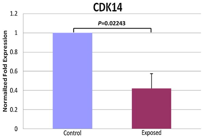
The microarray analyses of testes of mice exposed to tobacco smoke for 24 weeks revealed significant changes in expression of several genes that may be involved in spermatogenesis. The summary of the genes and their putative functions in testis are summarized in Table 1. The maximal changes were seen in the expression of Speer8 (a pseudogene) followed by Cdk14 which was down-regulated two-fold in the exposed mice. CDK14 (also known as PFTK1/PFTAIRE-1) is a novel member of the CDC2-like kinase family that is highly expressed in the testis and brain with lower expression level in other tissues (Besset et al., 1998; Shu et al., 2007). In somatic cells, CDK14 regulates cell cycle via interaction with Cyclin D3 (CCND3). However, in addition to this canonical mode of action, CDK14 also interacts with Cyclin Y (CCNY), the newest member of the cyclin family. The interaction with Cyclin Y enhances CDK14 kinase activity and recruits CDK14 to the plasma membrane, leading to activation of the Wnt signaling pathway through phosphorylation of LRP6 co-receptor (a key regulator of the Wnt pathway) (Davidson et al., 2009). CDK14 function in reproductive and other tissues is not yet fully understood. However, proper Wnt signaling and regulation of cyclin D are required for normal spermatogenesis (Kerr, 2014; Golestaneh et al., 2009; Zindy et al., 2001). Furthermore, mice with a deletion of the gene encoding CDK16 (another CDC2-like kinase that binds Cyclin Y), exhibit male-specific infertility (Mikolcevic et al., 2012). Therefore, CDK14 was chosen for further characterization. To confirm the microarray results, we performed RT-PCR analysis and observed significant down-regulation of CDK14 mRNA in CSE-exposed mice compared to controls (Fig. 1).
Table 1.
Top genes show statistically significant changes after tobacco exposure versus control at 24 weeks.
| Affyid | Symbol | Name | Entrez | LogFC | AveExpr | Adj.P. Val | B | Comments | |
|---|---|---|---|---|---|---|---|---|---|
| 4687 | 10390211 | Igf2bp1 | Insulin-like growth factor 2 mRNA binding protein 1 | 140486 | −0.62 | 7.69 | 0.02 | 5.41 | RNA-binding, IFG signaling |
| 19465 | 10527940 | Cdk14 | Cyclin-dependent kinase 14 | 18647 | −0.96 | 8.35 | 0.02 | 4.62 | Regulation of mitosis and meiosis |
| 9672 | 10435948 | Ccdc80 | Coiled-coil domain containing 80 | 67896 | −0.71 | 6.23 | 0.02 | 4.37 | Glucose, lipid metabolism; peroxisomal |
| 4647 | 10389759 | Ankfn1 | Ankyrin-repeat and fibronectin type III | 382543 | −0.56 | 5.52 | 0.02 | 4.22 | Membrane protein |
| 23915 | 10566201 | Olfr589 | Olfactory receptor (OR)589 | 259054 | −0.47 | 5.05 | 0.02 | 4.16 | OR receptors may function as a chemosensing receptors in mouse germ cells and sperm |
| 18835 | 10521498 | Crmp1 | Collapsin response mediator protein 1 | 12933 | −0.73 | 7.45 | 0.02 | 3.76 | Expressed in spermatids |
| 18692 | 10519811 | Speer8-ps1 | Spermatogenesis associated glutamate (E) | 74062 | −1.48 | 7.82 | 0.02 | 3.66 | A trascribed pseudogene; testis-specific. |
| 8111 | 10420286 | Gzmn | Granzyme N | 245839 | 0.4 | 9.77 | 0.02 | 3.3 | Expressed in spermatids and spermatocytes, may be involved in spermatogenesis |
| 4631 | 10389639 | Lpo | Lactoperoxidase | 76113 | −0.54 | 6.34 | 0.03 | 3.07 | May have a role in oxidative stress in testicular cell mitochnodria |
| 14980 | 10485372 | Rag1 | Recombination activating gene 1 | 19373 | −0.45 | 6.03 | 0.03 | 2.97 | Immunoglobulin; may also be produced by testicular cells |
LogFC: log2 fold change for that gene. A positive value indicates up-regulation of a gene, a negative value indicates down-regulation; aveExpr: average expression value for that gene across all the arrays; adj.P.Val: adjusted p-value (a p-value adjusted for multiple testing by Benjamini–Hochberg method); B: the log odds that the gene is differentially expressed. A B-statistic of zero corresponds to a 50–50 chance that the gene is differentially expressed.
Fig. 1.
Real time RT-PCR analysis of CDK14 expression levels in 3 tobacco-exposed and 3 control animals. qPCR was performed in triplicate for each sample. The values were normalized to the actin content of each sample. The results were normalized to control which was considered as 1. To calculate the difference between samples, Student’s paired t-test was used.
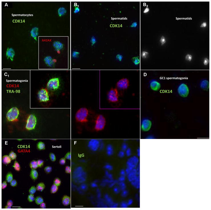
To examine the expression of Cdk14 in the testis, we performed immunofluorescent localization of CDK14 in different types of testicular cells. The spermatocytes and spermatids were separated with a STA-PUT procedure that utilizes differential sedimentation velocity at the unit gravity of different cell types (La Salle et al., 2009; Bellve et al., 1977). The contents of the fractions collected from the separation were examined using flow cytometry to identify tetraploid and haploid cells. The histogram of the whole testis single-cell suspension prior to the STA-PUT procedure and representative images of fractions enriched for tetraploid spermatocytes and haploid spermatids after the separation are shown in Supplementary Fig. 1. CDK14 was detected in the nuclear and perinuclear areas of spermatocytes (Fig. 2A, green; γH2AX (red) was used as a spermatocyte marker) and spermatids (Fig. 2B1 and B2; DAPI alone image is used to demonstrate nuclei of maturating spermatids found in the haploid fraction). Spermatogonia and Sertoli cells were isolated from testes of 9 day old mice. TRA-98 (Fig. 2C1, green) and GATA4 (Fig. 2E, red) were used as a marker of germ and Sertoli cells, respectively. In spermatogonia, CDK14 was localized to the nucleus and perinuclear regions of the cells (Fig. 2C1 and C2, red); similar localization pattern of CDK14 was observed in GC1 spermatogonia cell line (Fig. 2D and E, green). We detected a prominent CDK14 signal in both the cell nucleus and the cytoplasm of immature mouse Sertoli cells (green Fig. 2E). Together, the localization pattern of CDK14 is suggestive of its involvement in regulation of multiple aspects of spermatogenesis.
Fig. 2.
Immunofluorescent localization of CDK14 in testicular cells. Color-coding is indicated for each image. Scale bar is 10μm. Nuclei are stained by DAPI (blue). Different cell types have been separated from adult and pubertal testes as described in Section 2. (A) Spermatocytes; insert: double staining for CDK14 and γH2AX (a spermatocyte marker). (B1) Spermatids; (B2) DAPI alone panel of Fig. B1 to visualize different types of spermatid nuclei. (C1) TRA- and CDK14-positive spermatogonia; (C2) CDK14 staining alone of Fig. C1. (D) GC-1 spermatogonia. (E) Mouse immature Sertoli cells. (F) Negative control (rabbit IgG was used instead of primary antibody).
3.2. CDK14 expression is suppressed by cigarette smoke extract in testis-derived cell lines
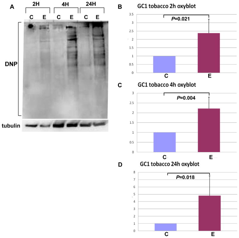
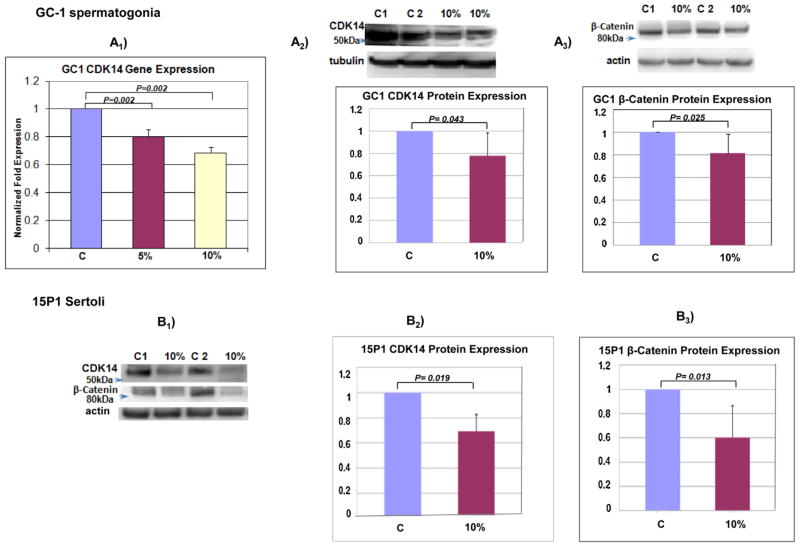
Because testicular tissue is composed of multiple cell types, it is important to determine a cell type-specific response to tobacco. These studies, however, can be affected by prolonged procedures of germ cell separation, which are performed after the cells are isolated from the exposed animals. Therefore, an in vitro CSE treatment of different cell types or cell lines can be a useful alternative. Proliferation in the testis is maintained by spermatogonia, which become spermatocytes after several mitotic divisions and differentiation. GC-1 cell line is derived from type B spermatogonia and expressseveral specific spermatogonia markers (Hofmann et al., 1992). Our previous study has shown a significant increase in oxidative stress and protein oxidation in the testes of mice exposed to tobacco smoke (Shrivastava et al., 2010). Similarly, treatment of GC-1 cells with CSE caused a significant increase in the amount of carbonyl groups (which are introduced into protein side chains as a result of protein oxidation), as depicted in Fig. 3. Treatment with CSE also resulted in a significant (p<0.05) down-regulation of CDK14 mRNA and protein levels in GC-1 cells (Fig. 4A1 and A2). This down-regulation, however, was less robust than the one obtained in the whole testis, suggesting that CDK14 expression in other testicular cells (e.g., spermatocytes, spermatids, Sertoli) can be affected more severely. Indeed, CSE treatment of mouse 15P-1 Sertoli cell line (which maintain several characteristics of Sertoli cells (Paquis-Flucklinger et al., 1993; Grandjean et al., 1997; Mather, 1980)) resulted in a more prominent down-regulation of CDK14 protein levels than the one obtained in the spermatogonia cell line (Fig. 4B1 and B2).
Fig. 3.
GC-1 spermatogonia cell line was treated with 5% CSE for 2, 4 and 24 h. Exposed cells (E) were compared to non-treated control (C) for each time point. (A) Protein samples were used for the Oxyblot analysis to detect the amount of carbonyl groups on cellular proteins which are introduced into protein side chains as a result of protein oxidation. Western blotting was performed using an antibody specific to the DNP moiety of the proteins. Equal loading was determined using anti-β-tubulin antibody. In each experiment, controls were considered as 1 and other samples – 2 (B), 4 (C) and 24 (D) h were normalized to the controls. To calculate the difference between samples, Student’s paired t-test was used.
Fig. 4.
(A1) Real time RT-PCR analysis of Cdk14 expression in GC1 spermatogonia cells treated with 5 and 10% CSE for 5 h. qPCRwas performed in triplicate and the results were normalized to Gapdh content of each sample. In each experiment, the results were normalized to control which was considered as 1. To calculate the difference between samples, Student’s paired t-test was used. (A2) A western blot analysis of CDK14 expression in GC1 cells treated with 10% CSE for 24 h. Densitometry data from six experiments are shown. Density values were normalized to tubulin. In each experiment, controls (untreated samples) were considered as 1 and other samples were normalized to the controls. (A3) A western blot analysis of β-catenin expression in GC1 cells treated with 10% CSE for 24 h. Densitometry data from four experiments are shown. Density values were normalized to actin. (B1) A representative western blot of CDK14 and β-catenin expression in 15P1 Sertoli cells treated with 10% CSE for 24 h. (B2) A western blot analysis of CDK14 expression in 15P1 Sertoli cells treated with 10% CSE for 24 h. Densitometry data from four experiments are shown. Density values were normalized to actin. (B3) A western blot analysis of β-catenin expression in 15P1 Sertoli cells treated with 10% CSE for 24 h. Densitometry data from four experiments are shown. Density values were normalized to actin.
Because Wnt signaling regulates gene expression through stabilization of β-catenin, we also tested the effect of CSE on the total levels of β-catenin. Treatment of GC-1 cells with CSE resulted in a small but significant (p<0.05) down-regulation of β-catenin protein levels in GC-1 cells (Fig. 4A3). Again, the β-catenin downregulation in Sertoli cells was more prominent than in spermatogonia (Fig. 4B3), suggesting that CSE preferentially affects Sertoli cell signaling.
The expression of β-catenin has previously been demonstrated in Sertoli and germ cells in the testis of adult rodents (Lombardi et al., 2013). β-catenin was localized to adherent junction formed between Sertoli cells, and between Sertoli and germ cells at the adluminal compartment of the seminiferous epithelium. Sporadic mutation inβ-catenin gene caused germ cell apoptosis and the loss of spermatocytes and spermatids (Kerr, 2014). Although this study suggests that postmitotic cells and not spermatogonia may be the major targets of active Wnt signaling, another study has shown activation of canonical Wnt signaling in spermatogonia during mouse development (Golestaneh et al., 2009). Spermatid-specific deletion of β-catenin resulted in significantly reduced sperm count, increased germ cell apoptosis, loss of Sertoli cell-germ cell adhesion and impaired fertility (Chang et al., 2011).
Both CDK14 and CDK16 activate Wnt signaling through interaction with cyclin Y, suggesting possible existence of other cyclin Y-interacting CDKs within the CDC2-lile kinase family; their overlapping and distinct functions as well as the role of cyclin Y during spermatogenesis should be further studied. It is also possible that CDK14 regulates mitosis and/or meiosis through interaction with cyclin D (as reported for CDK14 in somatic cells (Shu et al., 2007)). Three D-type cyclins have been implicated in regulation of spermatogenesis. In spermatogonia, cyclins D1 and D3 are implicated in cell cycle regulation, whereas cyclin D2 likely has a role in spermatogonial differentiation (Beumer et al., 2000). Cyclin D3 expression was also detected in Sertoli and Leydig cells. Male mice lacking both the Ink4c and Ink4d genes, which encode two inhibitors of D-type cyclin-dependent kinases (Cdks), are infertile (Zindy et al., 2001).
Taken together with previously published data from our laboratory (Shrivastava et al., 2010, 2014), the results presented herein for testicular cells in vivo and in vitro demonstrate that cigarette smoke causes oxidative damage and changes in post-translation modifications of proteins, altered gene expression and molecular signaling in testicular cells. Future studies will focus on understanding the cell type-specific functions of CDK14 and its interacting partners, and the consequences of their downregulation during normal spermatogenesis and under stress.
3.3. CDK14 expression is down-regulated by tobacco smoke in the lung epithelium-derived cells
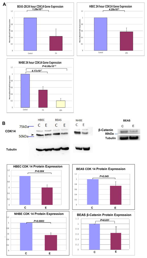
We next tested whether down-regulation of Cdk14 by tobacco is specific for testicular cells or may occur in other cell types. We focused on the lungs, which are the primary organ affected by tobacco exposure. Several lung epithelium-derived cell lines, including primary normal human bronchial cells (NHBE) were treated with CSE. Similar to the results observed in testicular cells, CSE treatment resulted in significant (p<0.05) down-regulation of CDK14 mRNA and protein levels in three different lung epithelial cell lines (Fig. 5A and B). The decrease in CDK14 correlated with a significant decrease in β-catenin level, suggesting that similar to the results in testicular cells, CSE can affect Wnt signaling in the lung through a decrease in CDK14 activity resulting in de-stabilization of β-catenin levels. Further studies will examine the effects of tobacco on nuclear accumulation of β-catenin and other Wnt pathway markers in vivo and in vitro.
Fig. 5.
(A) Real time RT-PCR analysis of CDK14 expression in three lung-derived epithelial cell lines (BEAS, HBEC and NHBE). Cells were treated with 5 or 10% CSE for 24 h. QPCR was performed in triplicates and the results were normalized to GAPDH content of each sample. In each experiment, the results were normalized to control which was considered as 1. To calculate the difference between samples, Student’s paired t-test was used. (B) A representative western blot analysis of CDK14 expression for each lung epithelial cell line and β-catenin for BEAS cell line treated with 10% CSE is shown.
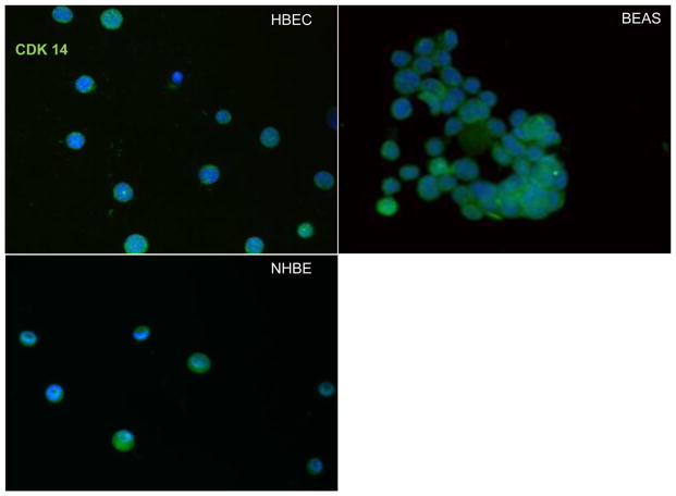
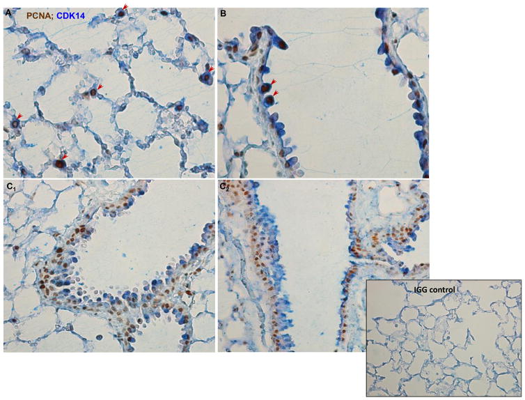
CDK14 was found to be localized to the nucleus and cytoplasm of lung-derived cell lines including NHBE (Fig. 6, green). Notably, similar to the cell lines, CDK14 was also detected in the nucleus and cytoplasm of mouse bronchial and alveolar epithelium (Fig. 7, blue; PCNA staining (brown) was used to detect proliferative cells). Interestingly, in the alveoli and small bronchiole, CDK14 expression was often observed in proliferative (PCNA-positive) cells (Fig. 7, A and B, arrow heads). In contrast, in the bronchial epithelium, mostly non-proliferative and more differentiated cells were positively stained with the CDK14 antibody (Fig. 7C1 and C2). The results presented herein may serve as a foundation for further studies of the function of CDK14 in the lung and lung-related diseases caused by tobacco. Interestingly, both cell cycle arrest and misregulation of the Wnt pathway was implicated in the development of several smoking-related airway disorders such as COPD. A decrease in Wnt target genes and an increase in Wnt inhibitors were reported in bronchial epithelial cells of smokers and COPD patients, and in experimental mouse models of emphysema (Wang et al., 2011; Kneidinger et al., 2011). Notably, preventive and therapeutic Wnt/β-catenin activation led to a significant reduction of experimental emphysema (Kneidinger et al., 2011). Therefore, CS-induced down-regulation of CDK14 can contribute to de-stabilization of β-catenin and misregulation of Wnt signaling in lung epithelium. Both Cyclin D and Wnt signaling were implicated in regulation of cell cycle. Furthermore, in other cell types, genetically-mediated reduction of CDK14 expression caused a cell cycle arrest (Shu et al., 2007). Thus, CSE-mediated down regulation of CDK14 may decrease the robustness of Wnt signaling and cell cycle progression in lung epithelium, potentially leading to progression to disease. Further studies analyzing long-term cigarette smoke exposure of mice with down-regulated level and/or activity of CDK14 and its interacting partners will address this hypothesis.
Fig. 6.
Immunofluorescent localization of CDK14 (green) in several lung-derived epithelial cell lines. Nuclei are stained by DAPI (blue).
Fig. 7.
Immunocytochemical localization of CDK14 (blue) and PCNA (brown) in alveoli (A), bronchioli (B) and bronchi (C1 and C2). Arrowheads indicate PCNA/CDK14-positive cells. Insert: negative isotype control.
Acknowledgments
Funding
This study was supported by a grant from the Flight Attendant Medical Research Institute (to MV).
The authors would like to thank Dr. Rani Sellers and Dr. Barbara Cannella from histotechnology and comparative pathology facility (AECOM) for help with performing immunohistochemistry procedures on lung tissue sections. We also thank Dr. David Reynolds and Wen Tran, from the AECOM Affymetrix Facility for help with gene array analysis.
Appendix A. Supplementary data
Supplementary data associated with this article can be found, in the online version, at http://dx.doi.org/10.1016/j.toxlet.2015.02.006.
Footnotes
Conflict of interest
The authors declare that there are no conflicts of interest.
Transparency document
The Transparency document associated with this article can be found in the online version.
References
- Attia AM, et al. Cigarette smoking and male reproduction. Arch Androl. 1989;23(1):45–49. doi: 10.3109/01485018908986788. [DOI] [PubMed] [Google Scholar]
- Bellve AR, et al. Spermatogenic cells of the prepuberal mouse: isolation and morphological characterization. J Cell Biol. 1977;74(1):68–85. doi: 10.1083/jcb.74.1.68. [DOI] [PMC free article] [PubMed] [Google Scholar]
- Besset V, Rhee K, Wolgemuth DJ. The identification and characterization of expression of Pftaire-1: a novel Cdk family member, suggest its function in the mouse testis and nervous system. Mol Reprod Dev. 1998;50(1):18–29. doi: 10.1002/(SICI)1098-2795(199805)50:1<18::AID-MRD3>3.0.CO;2-#. [DOI] [PubMed] [Google Scholar]
- Beumer TL, et al. Involvement of the D-type cyclins in germ cell proliferation and differentiation in the mouse. Biol Reprod. 2000;63(6):1893–1898. doi: 10.1095/biolreprod63.6.1893. [DOI] [PubMed] [Google Scholar]
- Calogero A, et al. Cigarette smoke extract immobilizes human spermatozoa and induces sperm apoptosis. Reprod Biomed Online. 2009;19(4):564–571. doi: 10.1016/j.rbmo.2009.05.004. [DOI] [PubMed] [Google Scholar]
- Chang YF, et al. Role of beta-catenin in post-meiotic male germ cell differentiation. PLoS One. 2011;6(11):e28039. doi: 10.1371/journal.pone.0028039. [DOI] [PMC free article] [PubMed] [Google Scholar]
- Chia SE, Ong CN, Tsakok FM. Effects of cigarette smoking on human semen quality. Arch Androl. 1994;33(3):163–168. doi: 10.3109/01485019408987820. [DOI] [PubMed] [Google Scholar]
- Chidekel AS. The respiratory health effects of passive smoking on children. Del Med J. 2000;72(5):203–207. [PubMed] [Google Scholar]
- Cook DG, Strachan DP. Health effects of passive smoking-10: summary of effects of parental smoking on the respiratory health of children and implications for research. Thorax. 1999;54(4):357–366. doi: 10.1136/thx.54.4.357. [DOI] [PMC free article] [PubMed] [Google Scholar]
- Davidson G, et al. Cell cycle control of wnt receptor activation. Dev Cell. 2009;17(6):788–799. doi: 10.1016/j.devcel.2009.11.006. [DOI] [PubMed] [Google Scholar]
- Doll R, et al. Mortality in relation to smoking: 50 years’ observations on male British doctors. BMJ. 2004;328(7455):1519. doi: 10.1136/bmj.38142.554479.AE. [DOI] [PMC free article] [PubMed] [Google Scholar]
- Esakky P, et al. Cigarette smoke condensate induces aryl hydrocarbon receptor-dependent changes in gene expression in spermatocytes. Reprod Toxicol. 2012;34(4):665–676. doi: 10.1016/j.reprotox.2012.10.005. [DOI] [PubMed] [Google Scholar]
- Field AE, et al. The relation of smoking: age, relative weight, and dietary intake to serum adrenal steroids, sex hormones, and sex hormone-binding globulin in middle-aged men. J Clin Endocrinol Metab. 1994;79(5):1310–1316. doi: 10.1210/jcem.79.5.7962322. [DOI] [PubMed] [Google Scholar]
- Foronjy RF, et al. Superoxide dismutase expression attenuates cigarette smoke- or elastase-generated emphysema in mice. Am J Respir Crit Care Med. 2006;173(6):623–631. doi: 10.1164/rccm.200506-850OC. [DOI] [PMC free article] [PubMed] [Google Scholar]
- Golestaneh N, et al. Wnt signaling promotes proliferation and stemness regulation of spermatogonial stem/progenitor cells. Reproduction. 2009;138(1):151–162. doi: 10.1530/REP-08-0510. [DOI] [PubMed] [Google Scholar]
- Grandjean V, et al. Stage-specific signals in germ line differentiation: control of Sertoli cell phagocytic activity by spermatogenic cells. Dev Biol. 1997;184(1):165–174. doi: 10.1006/dbio.1997.8518. [DOI] [PubMed] [Google Scholar]
- Hofmann MC, et al. Immortalization of germ cells and somatic testicular cells using the SV40 large T antigen. Exp Cell Res. 1992;201(2):417–435. doi: 10.1016/0014-4827(92)90291-f. [DOI] [PubMed] [Google Scholar]
- Jinot J, Bayard S. Respiratory health effects of passive smoking: EPA’s weight-of-evidence analysis. J Clin Epidemiol. 1994;47(4):339–349. doi: 10.1016/0895-4356(94)90154-6. (discussion 351-3) [DOI] [PubMed] [Google Scholar]
- Kerr GE, et al. Regulated Wnt/beta-catenin signaling sustains adult spermatogenesis in mice. Biol Reprod. 2014;90(1):3. doi: 10.1095/biolreprod.112.105809. [DOI] [PubMed] [Google Scholar]
- Kneidinger N, et al. Activation of the WNT/beta-catenin pathway attenuates experimental emphysema. Am J Respir Crit Care Med. 2011;183(6):723–733. doi: 10.1164/rccm.200910-1560OC. [DOI] [PubMed] [Google Scholar]
- La Salle S, Sun F, Handel MA. Isolation and short-term culture of mouse spermatocytes for analysis of meiosis. Methods Mol Biol. 2009;558:279–297. doi: 10.1007/978-1-60761-103-5_17. [DOI] [PubMed] [Google Scholar]
- Lawson GM, et al. Application of serum nicotine and plasma cotinine concentrations to assessment of nicotine replacement in light: moderate, and heavy smokers undergoing transdermal therapy. J Clin Pharmacol. 1998;38(6):502–509. doi: 10.1002/j.1552-4604.1998.tb05787.x. [DOI] [PubMed] [Google Scholar]
- Lemaitre V, Dabo AJ, D’Armiento J. Cigarette smoke components induce matrix metalloproteinase-1 in aortic endothelial cells through inhibition of mTOR signaling. Toxicol Sci. 2011;123(2):542–549. doi: 10.1093/toxsci/kfr181. [DOI] [PMC free article] [PubMed] [Google Scholar]
- Lombardi AP, et al. Physiopathological aspects of the Wnt/beta-catenin signaling pathway in the male reproductive system. Spermatogenesis. 2013;3(1):e23181. doi: 10.4161/spmg.23181. [DOI] [PMC free article] [PubMed] [Google Scholar]
- Mather JP. Establishment and characterization of two distinct mouse testicular epithelial cell lines. Biol Reprod. 1980;23(1):243–252. doi: 10.1095/biolreprod23.1.243. [DOI] [PubMed] [Google Scholar]
- Mercer BA, et al. Identification of a cigarette smoke-responsive region in the distal MMP-1 promoter. Am J Respir Cell Mol Biol. 2009;40(1):4–12. doi: 10.1165/rcmb.2007-0310OC. [DOI] [PMC free article] [PubMed] [Google Scholar]
- Mikolcevic P, et al. Cyclin-dependent kinase 16/PCTAIRE kinase 1 is activated by cyclin Y and is essential for spermatogenesis. Mol Cell Biol. 2012;32(4):868–879. doi: 10.1128/MCB.06261-11. [DOI] [PMC free article] [PubMed] [Google Scholar]
- Paquis-Flucklinger V, et al. Expression in transgenic mice of the large T antigen of polyomavirus induces Sertoli cell tumours and allows the establishment of differentiated cell lines. Oncogene. 1993;8(8):2087–2094. [PubMed] [Google Scholar]
- Shrivastava V, et al. SUMO proteins are involved in the stress response during spermatogenesis and are localized to DNA double-strand breaks in germ cells. Reproduction. 2010;139(6):999–1010. doi: 10.1530/REP-09-0492. [DOI] [PubMed] [Google Scholar]
- Shrivastava V, et al. Cigarette smoke affects posttranslational modifications and inhibits capacitation-induced changes in human sperm proteins. Reprod Toxicol. 2014;43:125–129. doi: 10.1016/j.reprotox.2013.12.001. [DOI] [PubMed] [Google Scholar]
- Shu F, et al. Functional characterization of human PFTK1 as a cyclin-dependent kinase. Proc Natl Acad Sci U S A. 2007;104(22):9248–9253. doi: 10.1073/pnas.0703327104. [DOI] [PMC free article] [PubMed] [Google Scholar]
- Strachan DP, Cook DG. Health effects of passive smoking. 1 Parental smoking and lower respiratory illness in infancy and early childhood. Thorax. 1997;52(10):905–914. doi: 10.1136/thx.52.10.905. [DOI] [PMC free article] [PubMed] [Google Scholar]
- Tan XL, et al. Candidate dietary phytochemicals modulate expression of phase II enzymes GSTP1 and NQO1 in human lung cells. J Nutr. 2010;140(8):1404–1410. doi: 10.3945/jn.110.121905. [DOI] [PMC free article] [PubMed] [Google Scholar]
- Vigodner M, et al. Localization and identification of sumoylated proteins in human sperm: excessive sumoylation is a marker of defective spermatozoa. Hum Reprod. 2013;28(1):210–223. doi: 10.1093/humrep/des317. [DOI] [PMC free article] [PubMed] [Google Scholar]
- Wang R, et al. Down-regulation of the canonical Wnt beta-catenin pathway in the airway epithelium of healthy smokers and smokers with COPD. PLoS One. 2011;6(4):e14793. doi: 10.1371/journal.pone.0014793. [DOI] [PMC free article] [PubMed] [Google Scholar]
- Witschi H. A short history of lung cancer. Toxicol Sci. 2001;64(1):4–6. doi: 10.1093/toxsci/64.1.4. [DOI] [PubMed] [Google Scholar]
- Yauk CL, et al. Mainstream tobacco smoke causes paternal germ-line DNA mutation. Cancer Res. 2007;67(11):5103–5106. doi: 10.1158/0008-5472.CAN-07-0279. [DOI] [PubMed] [Google Scholar]
- Zindy F, et al. Control of spermatogenesis in mice by the cyclin D-dependent kinase inhibitors p18(Ink4c) and p19(Ink4d) Mol Cell Biol. 2001;21(9):3244–3255. doi: 10.1128/MCB.21.9.3244-3255.2001. [DOI] [PMC free article] [PubMed] [Google Scholar]


