Figure 1.
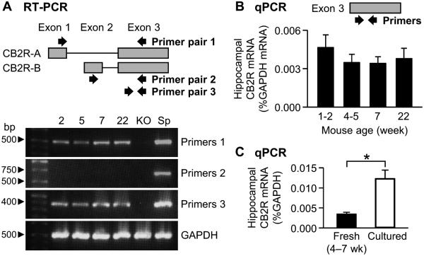
Quantification of CB2R mRNAs in the mouse hippocampus. A. RT-PCR with mRNAs extracted from the hippocampi of C57BL/6J and CB2R KO mice. A schematic diagram of the structure of mouse CB2R genome is illustrated on top. Approximate locations of the primers used for RT-PCR are indicated by arrows. Ages of C57BL/6J mice (2–22 weeks) were indicated above the images. KO, the hippocampus of CB2R KO mice. Sp, the spleen of C57BL/6J mice. B. qPCR with mRNAs from the hippocampus of C57BL/6J mice. The primers for qPCR targeted the exon 3 of Cnr2. The amount of CB2R mRNA was normalized to that of GAPDH mRNA. C. qPCR with mRNAs extracted from cultured hippocampal slices of C57BL/6J mice or freshly fixed hippocampus of C57BL/6J mice. The data of fresh tissue were from Figure 1B (age 4–7 weeks). *, P = 0.00003, t-test. Error bars indicate SEM.

