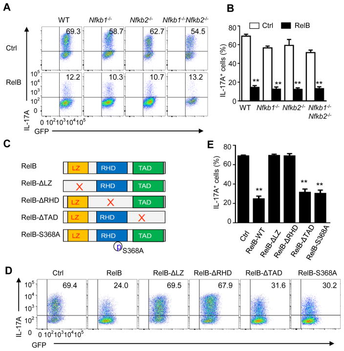
Figure 4. RelB inhibits Th17 cells independent of p50, p52, and its transactivation domain.
(A and B) Induction of Th17 cells from WT B6, Nfkb1−/−, Nfkb2−/−, −/−, Nfkb1−/−Nfkb2−/− DKO CD4+ T cells transduced with empty vector (Ctrl) or with retrovirus expressing RelB and cultured for 3 days under Th17-polarizing conditions (A). Graphs in (B) depict Mean ± SEM of 5 experiments. ** p <0.01.
(C) Schematic representation of RelB structure and RelB mutants showing deleted leucine zipper domain (LZ), Rel homology domain (RHD), transactivation domain (TAD), respectively, as well as a serine 368 to alanine point mutation (RelB-S368A).
(D and E) Induction of Th17 cells from WT CD4+ T cells transduced with empty vector (Ctrl) or with retroviruses expressing full length RelB (WT-RelB) or RelB mutants (as in C) and cultured for 3 d under Th17-polarizing conditions (D). Graphs in (E) depict Mean ± SEM of 3 independent experiments with triplicate cultures. ** p <0.01.

