Abstract
The regulators GadE, GadW and GadX (which we refer to as GadEWX) play a critical role in the transcriptional regulation of the glutamate-dependent acid resistance (GDAR) system in Escherichia coli K-12 MG1655. However, the genome-wide regulatory role of GadEWX is still unknown. Here we comprehensively reconstruct the genome-wide GadEWX transcriptional regulatory network and RpoS involvement in E. coli K-12 MG1655 under acidic stress. Integrative data analysis reveals that GadEWX regulons consist of 45 genes in 31 transcription units and 28 of these genes were associated with RpoS-binding sites. We demonstrate that GadEWX directly and coherently regulate several proton-generating/consuming enzymes with pairs of negative-feedback loops for pH homeostasis. In addition, GadEWX regulate genes with assorted functions, including molecular chaperones, acid resistance, stress response and other regulatory activities. These results show how GadEWX simultaneously coordinate many cellular processes to produce the overall response of E. coli to acid stress.
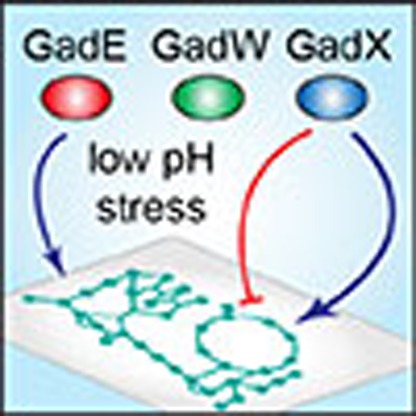
 GadEWX regulons play a critical role in transcription regulation in response to acid stress. By reconstructing genome-wide GadEWX transcriptional network, here the authors show how GadEWX simultaneously coordinates many other cellular processes to produce the overall response of E. coli to acid stress.
Bacteria depend on maintaining pH homeostasis, as most proteins have optimal ranges of pH where they function1. Diverse strategies for pH sensing and homeostasis allow bacteria to tolerate or grow at acidic external pH values. One of the most well-studied model organisms, Escherichia coli K-12 MG1655, has sophisticated acid resistance (AR) systems to survive in acidic pH environments. The entire AR system of E. coli and the complexity of its regulation have been extensively studied and well described in recent reviews2,3,4,5. Briefly, of the four clearly defined AR systems, AR1 is a RpoS-dependent oxidative (glucose-repressed) AR system that requires F1F0-ATPase, while its detailed molecular mechanism remains elusive6,7,8. The other two AR systems require amino acid decarboxylation9. The AR2 is a glutamate-dependent AR (GDAR) system that converts glutamate to γ-aminobutyric acid with decarboxylases (GadA and GadB)10,11,12 and imports glutamate in exchange for the γ-aminobutyric acid by selecting among protonated substrates using a charge-based mechanism of anti-porter (GadC)13. Similarly, the AR3 and AR4 are composed of arginine decarboxylase (AdiA) with the arginine/agmatine anti-porter (AdiC)14 and lysine decarboxylase (CadA) with lysine/cadaverine anti-porter (CadB)15, respectively. Consequently, this cycle pumps out cytoplasmic protons to the extracellular environment and reverses electrical membrane potential to slow proton movement into the cell, resulting in an increase of the intracellular pH to survive acidic stress. Of them, the AR2 is known to be the most effective system to relieve the acid stress12.
As mentioned above, the three genes (gadA, gadB and gadC) as well as several other genes in acid fitness islands (AFI) are mainly involved in the AR2 (GDAR) system; however, it is also under multiple levels of control. The GadE, GadW and GadX transcription factors (TFs) are at the first level of this regulatory cascade for direct regulation of these genes16,17,18. In addition, many other regulators such as PhoP19, YdeO20, EvgA20, TrmE21, RpoS22, RcsB23,24, Crp25, H-NS26,27 and TorR28 have been reported to affect induction of this AR system by forming a complex hierarchical regulatory cascade in response to different environmental signals. Although the general role of GadEWX in the regulation of the GDAR system and other genes in AFI has been extensively investigated with in vitro DNA-binding experiments, mutation analysis and comparative transcriptomics16,17,18,27,29,30,31,32,33,34,35,36,37, little is known about genome-scale in vivo GadEWX-binding events and the broader regulatory networks they comprise. A complete reconstruction of the GadEWX transcriptional regulatory networks under acidic stress would reveal the detailed mechanism of GadEWX regulation and its role in coordinating all cellular functions in response to acid stress.
In this study, we apply a systems biology approach to decode the GadEWX regulatory networks under acidic stress. We integrate genome-scale data from chromatin immunoprecipitation (ChIP) with lambda exonuclease digestion followed by high-throughput sequencing (ChIP-exo) for GadE, GadW, GadX and RpoS with those from strand-specific massively parallel cDNA sequencing (RNA-seq). To fully reconstruct the GadEWX regulons and RpoS involvement, we first examine the GadEWX- and RpoS-binding sites on the E. coli K-12 MG1655 genome. Then, we measure transcription levels of genes in the wild-type strain and knockout mutants of each TF on a genome scale to identify causal transcriptional regulatory relationships. From these data, we identify that the GadEWX regulatory networks maintain intracellular proton concentration by regulating proton-efflux/influx system and proton-generating/consuming metabolic enzymes and interconnecting the tree of them with pairs of negative-feedback loops. Reconstruction of the GadEWX regulatory networks provides a comprehensive view of the coordinative genome-wide regulatory roles of these TFs under acidic stress.
Results
Genome-wide binding profiles of GadEWX
Previously, several binding sites of GadEWX have been characterized in E. coli by in vitro DNA-binding experiments and mutation analysis16,17,18,27,29,30,31,32,33,34,35,36,37. However, direct measurement in vivo of GadEWX binding has not been achieved. Therefore, we employed a recently developed high-resolution ChIP-exo method to determine the in vivo genome-wide binding profiles of these three TFs with near 1-bp resolution in E. coli under acidic stress (pH 5.5).
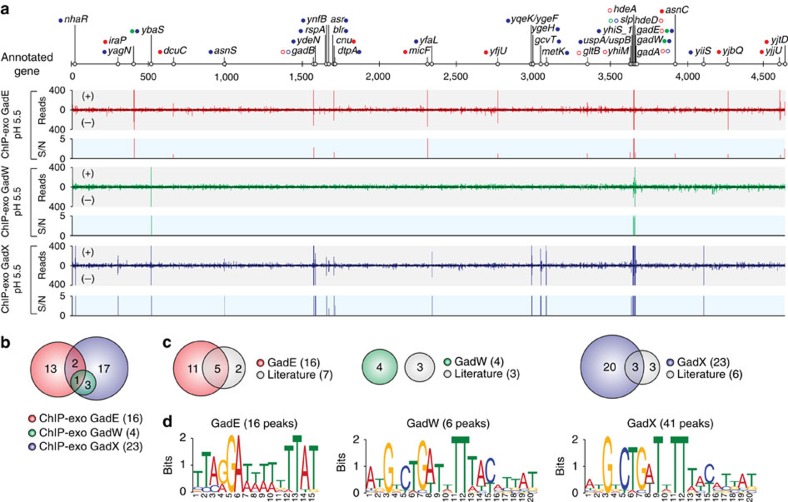
Using a peak finding algorithm (MACE program), 16, 6 and 41 reproducible binding peaks were identified for GadE, GadW and GadX, respectively, under acidic stress (Fig. 1a and Supplementary Tables 1–3). The number of binding sites of GadEWX was much lower than that of global TFs, suggesting that GadEWX may act more like local TFs to relieve acidic stress. In total, 16, 4 and 23 binding sites with multiple peaks were identified for GadE, GadW and GadX, respectively (Fig. 1b). We also found that the in vivo GadX binding sites include all of GadW binding sites as suggested in the previous comparative transcriptome study17. These numbers of binding sites and their overlap suggest that GadE and GadX play broader roles than GadW and that they have distinct regulatory networks from each other32. Before this study, 16 binding sites had been identified in vitro for GadEWX with strong experimental evidence, 50% (8 out of 16) of which were also detected in this study (Fig. 1c and Supplementary Table 4). It is unclear why these eight binding sites are missing from the data set obtained here. One possibility is that they are detectible only with in vitro methods and may not occur in vivo because of the interference by other regulators. Collectively, the identification of a total of 35 new binding sites was made possible by the new ChIP-exo method deployed here, significantly expanding the current knowledge of the scope of the GadEWX regulatory network.
Figure 1. Genome-wide landscape of GadEWX-binding sites.
(a) An overview of GadEWX-binding profiles across the E. coli genome (KiloBase) at mid-exponential growth phase (OD600=0.3) under acidic stress (pH 5.5). Open and closed dots indicate previously known and newly found GadEWX-binding sites, respectively. S/N denotes signal-to-noise ratio. (+) and (−) indicate reads mapped on forward and reverse strands, respectively. Binding peaks that overlap with Mock-IP signal were eliminated. Red, GadE; Green, GadW; Blue, GadX. (b) Overlaps between GadEWX-binding sites under the low-pH condition. (c) Comparison of the GadEWX binding sites obtained from this study (ChIP-exo) with the known binding sites from the literature. (d) Sequence logo representations of the GadEWX-DNA-binding motifs.
We next assessed the widths and the genomic locations of the GadEWX-binding sites using currently available genome annotation. The widths of binding sites of each TF were 29±3.6, 30±7.6 and 28.7±8.0, respectively (Supplementary Fig. 1). The majority of GadEWX-binding sites (88%, or 38 out of 43) were observed within regulatory regions (that is, upstream of promoters, promoters and 5'-proximal to coding regions). The remaining 12% were found in intragenic regions or between two coding regions of convergent genes (Supplementary Tables 1–3). These results show a strong preference of GadEWX-binding site location being within noncoding intergenic regions with relatively fixed binding widths. To identify DNA sequence motifs of these newly identified GadEWX-binding sites, we used the sequence of each binding peak in the motif search procedure. The identified sequence motif of GadE from 16 binding peaks (TTARGAWWWWAAATA) was mostly consistent with the previously characterized asymmetric GadE-binding site (TTAGGAttTTgTTATTTAAa) (Fig. 1d)16. The motif search of GadW and GadX from 6 and 41 binding peaks, respectively, also yielded a sequence (AKGKCTGWTWTTWWYMYVAK), which resembled the previously reported binding motif (AtGtcTGATtTttatattat)17,31. This same motif is due to the binding regions of GadW being overlapped with those of GadX as revealed in the previous study32.
Identification of GadEWX regulons and causal relationships
Currently, a total of 16 genes in 10 transcription units (TUs) (GadE: 12 genes in 7 TUs; GadW: 6 genes in 3 TUs; and GadX: 10 genes in 6 TUs) have been characterized with strong evidence as members of GadEWX regulons in E. coli15,16,17,26,28,29,30,31,32,33,34,35,36. Based on our ChIP-exo data sets, we can significantly expand the size of the GadEWX regulons to comprise 45 target genes in 31 TUs (GadE: 20 genes in 15 TUs; GadW: 6 genes in 4 TUs; GadX: 29 genes in 19 TUs, several genes are co-regulated) (Supplementary Tables 1–3). Functional analysis with clusters of orthologous groups (COG) categories was performed to look for any functional enrichment of GadEWX regulon genes (Supplementary Fig. 2). The COG category for amino acid metabolism and transport (E) was found to be statistically overrepresented (hypergeometric test P-value=4 × 10⁻⁶) and it includes gadA, gadBC, gltBD and ybaST, whose reactions are closely related with glutamate metabolism. Besides, a diverse range of COG functional categories indicate that GadEWX may play complicated roles beyond regulating genes in AFI to coordinate associated cellular processes in E. coli K-12 MG1655 as GadE has multiple roles in other pathogenic E. coli strains38,39,40. We in addition analysed what other TFs are known to regulate these 45 genes based on the strong evidence in RegulonDB41 and found that 21 of them are regulated by 22 other TFs (Supplementary Table 5). Interestingly, the core genes in the GDAR system (gadA and gadBC) were regulated by three additional TFs (H-NS, FliZ and AdiY)42,43. This result represents the importance of complicated regulation of these genes according to the changes of the environments. The remaining 24 genes were not directly regulated by other TFs, meaning that GadEWX exclusively regulates these genes in response to acid stress.
To determine the causal relationships between the binding of GadEWX and changes in RNA transcript levels of genes in the GadEWX regulons, we compared transcript levels between the wild-type strain and that of each deletion mutant (ΔgadE, ΔgadW and ΔgadX) grown under acidic stress conditions. Overall, a total of 351 genes were differentially expressed in at least one mutant. Genes with expression changes with log2 fold change≥0.5 and false discovery rate <0.01 were defined as differentially expressed. Only 25 out of 351 were differentially expressed in all mutants (Supplementary Fig. 3 and Supplementary Data 1–3). This unexpectedly small overlap in the transcriptional response suggests that each TF has a specific transcriptional response and may have a distinct role in regulating the AR system.
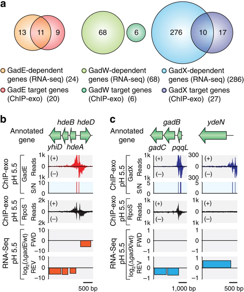
RpoS, one of the alternative sigma factors in E. coli, is known to be crucial for expression of GadEWX and GDAR as well as performing general stress management in association with EvgS/EvgA and PhoQ/PhoP44,45,46. To investigate the involvement of RpoS in regulating GadEWX regulons, we additionally performed ChIP-exo to determine the in vivo binding profile of RpoS under acid stress. Combining our GadEWX- and RpoS-binding maps with a GadEWX-dependent transcriptome, we could determine the causal relationships between the binding of GadEWX and the changes in transcript levels of genes in GadEWX regulons under acidic stress as well as RpoS involvement for expression (Fig. 2). Among 45 target genes identified from ChIP-exo analysis of GadEWX, we determined that 19 genes in 13 TUs (GadE, 11 genes in 7 TUs; GadX, 10 genes in 7 TUs; gadBC are overlapped) (43%) were directly regulated by GadEWX under acidic stress (Fig. 2a, Supplementary Fig. 4 and Supplementary Tables 1–3). In addition, the expression of 62% of GadEWX regulon members (28 out of 45) was involved in the association of RpoS (Supplementary Fig. 4 and Supplementary Tables 1–3). Among the binding events of RpoS on GadEWX regulon members, 64% of them (18 out of 28) were novel (Supplementary Fig. 4 and Supplementary Tables 1–3). This result confirms the importance of RpoS recruitment for acid-stress response and expands the current knowledge of RpoS involvement in E. coli.
Figure 2. Genome-wide identification of GadEWX regulons and RpoS involvement.
(a) Comparison of ChIP-exo results and gene expression profiles to define direct GadEWX regulons. Examples of (b) GadE regulon (hdeAB-yhiD and hdeD with divergent promoters) and (c) GadX regulon (gadBC and ydeN). S/N denotes signal-to-noise ratio. (+) and (−) in ChIP-exo data indicate reads mapped on forward and reverse strands, respectively.
For example, the divergent promoters upstream of hdeA and hdeD TUs were extensively occupied by GadE and RpoS, and this GadE binding significantly increased transcript level of downstream genes under the acidic condition (Fig. 2b). Likewise, GadE acted as an activator on other promoters (Supplementary Table 1). The calculation of relative distance from GadE bindings to transcription start site41,47 showed that GadE binding sites are localized to either further upstream or downstream of the promoter region (−35 and −10 boxes) (Supplementary Table 1). This observation suggests that GadE may prevent binding of other repressors or directly activate transcription through another mechanism, such as recruiting RNAP to activate transcription32. However, unlike GadE, GadX acted as a dual regulator either to activate or repress the transcription of target genes. For instance, the association of GadX and RpoS on gadB promoter increased transcript level under acidic stress (Fig. 2c). However, the binding of GadX on ydeN promoter decreased transcript level and RpoS association was not observed (Fig. 2d). Although GadW bound to the promoter regions (Supplementary Table 2), we were not able to observe significant changes in transcript levels upon gadW knockout as shown in the previous transcriptome study17. Also, the remaining 59% of the genes (including GadW target genes) lacked significant changes in transcript levels despite the binding of corresponding TFs. These results indicate that the changes in their transcript levels may require additional regulatory signals such as association or dissociation of other TFs.
Genome-wide roles of GadEWX regulons
To determine how GadEWX coherently regulate gene expression in response to acidic stress, we reconstructed the genome-wide GadEWX regulatory network in E. coli. First, we looked into the regulatory circuit that GadEWX comprises. Although we detected bindings of each TF on either itself or other TFs as known previously2, we were not able to observe changes of transcript levels by association of these TFs except for the repression of gadW by GadX (Fig. 3a). Since we compared transcript levels between wild-type and each of deletion mutant (ΔgadE, ΔgadW or ΔgadX) to determine causal relationships, we are not able to examine the autoregulation of TFs. A previous study showed that the transcription of gadE is activated by GadW and GadX when cells were grown to stationary phase at pH 5.5 in LB glucose media18. However, a different growth phase or growth media could have contributed to different transcriptional regulation on gadE. (Fig. 3a and Supplementary Tables 2 and 3). We believe that multiple reiterative-control circuits consisting of other regulators still activate GadE, which is crucial in the GDAR system for AR, even if GadW or GadX is deleted48.
Figure 3. Genome-wide roles of the GadEWX regulons in E. coli.
Regulation of (a) GadEWX regulatory circuits and (b) core genes in the glutamate-dependent acid resistance (GDAR) system. Closed and open circles represent whether target genes are bound by TF or not. Triangle, inverted triangle, and dash indicate whether the expression levels of target genes are activated, repressed, or not changed upon TF knockout. Red, GadE; Green, GadW; Blue, GadX. (c) Schematic diagram for the reconstructed GadEWX regulatory motif for acidic pH homeostasis. The GadEWX repressed proton-influx system (TI) and proton-generating metabolic enzymes (MG) while activated proton-consuming metabolic enzymes (MC). We were not able to observe regulation of proton-efflux system (TE) by GadEWX. IM, inner membrane; OM, outer membrane. (d) GadEWX regulatory networks regulate diverse cellular functions. Arrows with dotted lines indicate that the regulation is not observed in this study.
A recent kinetic study of promoters and regulators in the AR2 system claimed that GadX (gadXW double mutation) did not affect the kinetics of the promoters of core genes (gadA and gadBC) in the GDAR system48. However, another comparative transcriptome analysis suggested that these promoters are significantly activated by GadX17. Interestingly, our study observed that both GadE and GadX activate transcription of these core genes, but with a different activation ratio (over 50-fold activation by GadE but less than 2-fold by GadX; Supplementary Data 1 and 3). These results demonstrate that GadE is a much stronger activator than GadX and consequently GadX cannot fully compensate for the loss of activation of the GDAR system caused by gadE deletion. Similarly, physiological studies showing that the gadE mutant was more sensitive to acid challenge than the gadX mutant also supported this observation16,17,49.
The comprehensive genome-wide reconstruction of the GadEWX regulatory network in E. coli allowed us to extend the scope of its roles under acidic stress. After functional classification of 43 genes out of 45 GadEWX regulons excluding gadEW, we observed that the functions of 23% (10) of those genes were mainly involved in proton consumption, generation and transport. Under acidic stress, only GadX-repressed genes involved in proton generation and influx such as speG (spermidine acetyltransferase) and dtpA (proton-dependent oligopeptide transporter) (Fig. 3c). On the other hand, both GadE and GadX activated several genes, such as gadA and gadB that encode enzyme consuming protons. We could not observe any proton-efflux systems regulated by GadEWX in E. coli K-12 MG1655. From this analysis, we believe that GadEWX connect proton-efflux/influx and generating/consuming enzymes with negative-feedback loop pairs to maintain intracellular proton concentration for pH homeostasis (Fig. 3c).
In addition to the control of proton flow, GadE directly regulated the expression of chaperone-encoding genes (hdeA and hdeB)5. Both GadE and GadX participated in controlling the expression of other acid-resistance related genes (asr, slp, yhiM, yhiD and hdeD) known to enhance the survival of cells under acid stress17,49,50 (Fig. 3d and Supplementary Fig. 4). Based on genome-wide binding properties, GadX was likely to participate in the regulation of general stress response (uspB, uspA and uspD). Interestingly, GadEWX were coordinated with other regulatory networks (iraP, cnu, micF, nhaR, asnC and dctR) indicating that GadEWX may play more complex genome-wide roles in association with other TFs. Several genes with predicted or unknown functions (yjbQ, yjbR, yjjU, yjtD, yiiS, ynfB, yfaL, yqeK, ygeF and ygeH) were also regulated by GadE and GadX (Fig. 3d). Recent studies revealed that expression of genes of the locus of enterocyte effacement (LEE) for pathogenesis of E. coli O157:H7 was indirectly regulated by GadE and GadX39,40. In E. coli K-12 MG1655, we found GadX binding peaks (yqeK, ygeF and ygeH) within a remnant of a pathogenicity island (Supplementary Table 3). Since it was found that GadE and GadX indirectly regulate LEE in E. coli O157:H7 (ref. 39), investigation of genome-wide in vivo bindings of these regulators in pathogenic strains might be able to reveal regulatory cascades in LEE that cause pathogenicity. To further examine the physiological roles of GadEWX regulon members with predicted or unknown functions under acid stress, we chose five genes (yjbQ, yjbR, yjjU, yjtD and yiiS) that either were activated or showed no causal relationship upon single TF knockout. Acid-stress resistance assays of knockout strains of these genes revealed that they were not directly related to GDAR, but three of them (yjjU, yjtD and yiiS) were implicated in general AR (Supplementary Fig. 5). This result indicates that GadEWX contribute to generate more global acid-resistance responses in E. coli beyond the regulation of the GDAR system.
Evolutionary aspects of GadEWX regulons
A recent survey on conservation analysis of the gadBC system in mostly enteric bacteria suggested that the presence and expression of this operon in pathogenic bacteria has been strongly connected to the requirement to survive under extremely acidic conditions but not necessarily in other non-pathogenic bacteria12. We further expanded our investigation of how E. coli GadEWX and their entire regulons have been evolved in the proteobacteria phylum including 134 γ-proteobacteria, 40 β-proteobacteria and 58 α-proteobacteria. We first compared the conservation of the genes encoding the regulators (gadEWX). They were almost conserved in Escherichia and Shigella, but not in Salmonella, Yersinia and even further distant enterobacteria (Fig. 4). As stated in the previous studies12,51, the presence of the GDAR system (gadA, gadB and gadC) in these bacteria would allow them to survive in an extreme acidic environment (pH 2) in the presence of glutamate. Interestingly, gadX as well as gadAB are conserved in some of β- and α-proteobacteria whereas gadE and gadW are not. Probably, GadX alone would be able to regulate the GDAR system in part when GadE is absent in these classes. Genes related to the proton consumption/generation (gltBD, gcvHTP, ybaS and so on) exhibited higher conservation ratio across proteobacteria even in the species that do not have the GDAR system (Fig. 4). These genes may be regulated by other TFs and participate in controlling intracellular proton flow.
Figure 4. Evolution of GadEWX regulons.
Conservation rates of GadEWX regulons across γ-, β-, α-proteobacteria are presented based on the ortholog calculation. The genes are subdivided by their functional categories.
By maintaining pH homeostasis under alkaline condition, the Na+/H+ anti-porter plays a crucial role in adaptation under alkaline challenge4. It should be noted that nhaR encoding Na+/H+ anti-porter activator was strongly bound by GadX and RpoS under acidic stress (Supplementary Table 3). Since the deletion of GadX did not significantly affect the transcription level of nhaR, additional signal and/or TF associations may be required in regulation of this activator under acidic condition so that it can be activated only under alkaline condition. Unlike the GDAR system, it seems like the involvement of cation–proton anti-porter is a commonly used strategy for alkaline challenge across proteobacteria given the high conservation rate of nhaR (Fig. 4).
Discussion
We comprehensively reconstructed the GadEWX regulons at the genome scale in E. coli by combining genome-wide GadEWX-binding maps and GadEWX-dependent transcriptome daunder acidic stress. We identified (i) a total of 45 genes in 31 TUs that belong to GadEWX regulons and associations of RpoS for 28 of those genes; (ii) 19 genes in 13 TUs showing causal relationships upon single TF knockout; (iii) the negative-feedback loop pairs of proton transporters and utilization enzymes by GadEWX for pH homeostasis; and (iv) the additional roles of GadEWX regulatory networks beyond the GDAR system.
Using active proton transports4 is a major strategy for bacterial pH homeostasis. They include the proton-pumping respiratory chain complexes such as cytochrome bo terminal oxidase (cbo) and NADH:ubiquinone oxidoreductase (ndh). Under conditions of acid challenge, E. coli increases expression of those complexes that pump protons out of the cell. Our recently developed genome-scale model of metabolism and gene expression (ME-Model) in E. coli52 predicted that these proton-pumping complexes are more efficient for relieving the acid stress than other amino acid decarboxylase-anti-porter pairs (AR2 and AR3) (Supplementary Fig. 6). Nevertheless, AR2 and AR3 are certainly required by cells for survival when passing through extreme acidic environments such as the stomach as they secrete cytoplasmic protons to the extracellular environment53,54.
Recently, two studies showed interesting observations on AR in E. coli. One study revealed that polyamines such as spermidine, putrescine and cadaverine induce multiple components in GDAR and they are important for protection against acidic stress55. Surprisingly, we also found that GadX represses RpoS-involved transcription of speG (spermidine acetyltransferase), which consequently controls the polyamine concentration by degradation (Fig. 3c and Supplementary Fig. 4). This reaction also generates protons during acetylation of spermidine which agrees with our hypothesis that proton-generating enzymes will be repressed for the control of proton flow. By doing so, cells can regulate the level of intracellular proton and polyamine concentration, so that they can survive under acidic stress. The other study reported that enzymatic release of ammonia by ybaS encoding glutaminase neutralizes protons, resulting in elevated intracellular pH under acidic environment53. We identified that GadW, GadX and RpoS bound to the promoter region of ybaS while we could not observe significant changes in transcript level upon single TF knockout (Supplementary Fig. 4). However, a previous microarray-based study showed that the double knockout strain (ΔgadXW) showed significant changes in transcript level of ybaS compared to wild type17, indicating that transcription of ybaS can be activated in the presence of either GadW or GadX. In addition, the promoter region of gcvHTP encoding glycine cleavage system, which is known to release ammonia similar to ybaS, was also occupied by GadX (Supplementary Table 3). Given the properties of the ybaS system, it is plausible that this system may also contribute to AR by releasing ammonia. In summary, we have described the complex roles of the GadEWX regulatory network in E. coli under acidic stress by a systems approach that integrates various types of cutting-edge genome-scale experimental data. Future efforts on revealing the precise molecular role of small RNAs in GadEWX transcriptional regulatory networks by recently developed techniques such as ribosome profiling56 would expand acid-response regulatory networks including the post-transcription level. Understanding AR in microbes has important implications for prevention and clinical treatment as well as health-care applications such as probiotics57 and even for engineering microbes to produce organic acids58. Further, similar to how the genome-scale model of E. coli metabolism with protein structures (GEM-PRO)59 successfully predicted thermostability of proteomes, the inclusion of structural information under acidic stress with these comprehensive operon structures into the current model52 would expand the predictive capability of the model on future complex phenotypes.
Methods
Bacterial strains and growth conditions
All strains used are E. coli K-12 MG1655 and its derivatives. The E. coli strains harbouring GadE-8myc, GadW-8myc and GadX-8myc were generated by a λ Red-mediated site-specific recombination system targeting C-terminal region of each gene60. Deletion mutants (ΔgadX, ΔgadW, ΔgadX and other y-genes) were also constructed by a λ Red-mediated site-specific recombination system61. Glycerol stocks of E. coli strains were inoculated into fresh 70 ml of M9 minimal medium (47.8 mM Na2HPO4, 22 mM KH2PO4, 8.6 mM NaCl, 18.7 mM NH4Cl, 2 mM MgSO4 and 0.1 mM CaCl2) supplemented with 0.2% (w/v) glucose in 500 ml flask and cultured overnight at 37 oC with 250 r.p.m. For acidic stress, the overnight cultures were inoculated into the fresh 70 ml of M9 minimal medium at pH 5.5 (adjusted with HCl) in 500 ml flask and continued to culture at 37oC with 250 r.p.m. to OD600=0.3±0.02. At the sampling point, the pH was measured to be 5.28±0.03.
ChIP-exo
To identify GadEWX- and RpoS-binding maps in vivo, we isolated the DNA bound to each protein from formaldehyde cross-linked E. coli cells by ChIP with the specific antibodies that specifically recognizes myc tag (9E10, Santa Cruz Biotechnology) and RpoS (1RS1, Neoclone) with 1:50 dilutions, and Dynabeads Pan Mouse IgG magnetic beads (Invitrogen) were also used to capture specific antibody followed by stringent washings62. With ChIP materials (chromatin-beads), we performed on-bead enzymatic reactions of the original ChIP-exo method63 with following modifications as shown in our previous study64. Briefly, the sheared DNA of chromatin-beads was end-repaired, dA-tailed and ligated with the first adaptor (5'-phosphorylated). After that, nick sites of each fragment were repaired before Lambda exonuclease and RecJf exonuclease treatment. The protein-DNA crosslink was reversed by overnight incubation at 65 °C with RNAs and proteins removal. DNA samples were used to perform primer extension and second adaptor ligation. The size-selected DNA sample was amplified and quantified. Prepared DNA library was sequenced using MiSeq (Illumina) in accordance with the manufacturer's instructions. ChIP-exo experiments were performed in biological duplicate. Sequence reads generated from ChIP-exo were mapped onto the reference genome (NC_000913.2) using bowtie65 with default options to generate SAM output files. MACE program ( https://code.google.com/p/chip-exo/)66-based in-house script was used to define peak candidates from biological duplicates with sequence depth normalization. To reduce false-positive peaks, peaks with signal-to-noise ratio less than 1.0 and same signal with Mock-IP were removed as in our previous study64.
RNA-seq expression profiling
Total RNAs including small RNAs were isolated using the cells treated with RNAprotect Bacteria Reagent (Qiagen) at OD600=0.3±0.02 followed by purification steps using Qiagen RNeasy Plus Mini Kit (Qiagen) in accordance to the manufacturer's instruction to collect both of them. Strand-specific RNA-seq library was prepared using the dUTP method67 with the following modifications as shown in our previous study64. Briefly, the rRNA-removed RNA sample was fragmented, and then first and second strands were synthesized. The samples were sequenced using MiSeq (Illumina) in accordance with the manufacturer's instructions. RNA-seq experiments were performed in biological duplicate. Sequence reads generated from RNA-seq were mapped onto the reference genome (NC_000913.2) using bowtie65 with the maximum insert size of 1000, bp, and 2 maximum mismatches after trimming 3 bp at 3' ends. These files were used for Cufflinks ( http://cufflinks.cbcb.umd.edu/)68 and Cuffdiff to calculate fragments per kilobase of exon per million fragments and differential expression, respectively. Cufflinks and Cuffdiff were run with default options with library type of dUTP RNA-seq. The RNA-seq data generated by our study were reproducible when comparing fragments per kilobase of exon per million fragments values of all genes and sRNAs between replicates (Supplementary Fig. 7). From cuffdiff output, genes with differential expression with log2 fold change ≥0.5 and a false discovery rate value<0.01 were considered as differentially expressed genes.
Motif search and analysis
The GadEWX-binding motif analyses were completed using the MEME tool from the MEME software suite with default settings69. We extended the sequence of each binding site by 10–30 bp at each end to allow for adjacent sequences to be included in the analysis.
COG functional enrichment
The GadEWX regulons were categorized according to their annotated COG category. Functional enrichment of COG categories was determined by performing one-tailed Fisher's exact test (Hypergeometric test), and P-value<0.05 was considered significant.
Acid resistance assays
AR assays followed the procedures of previous study with modification of usage of growth media49. Cells grown for overnight in M9 minimal media (pH 7.0) were diluted into fresh M9 media at pH 5.5 and cultured to OD600=0.3. These adapted cell cultures were inoculated into M9 minimal media at pH 2.5 without or with 1.5 mM sodium glutamate. The initial cell density inoculated for acid challenge was between 1 × 10⁶ and 3 × 10⁶ colony-forming unit (CFU) per ml. The pH 2.5 cultures were then incubated at 37 °C without shaking, and samples were collected after 2 h. Aliquots were serially diluted, and triplicates were plated onto LB agar plates. Colonies were counted after 24 h. Percent survival was calculated as follows49: ((CFU per ml at time 2 h)/(CFU per ml at time zero)) × 100. The results presented are averages of triplicate experiments and include the standard deviations.
Conservation analysis of GadEWX regulons
Gene annotation of strains and species were obtained from the SEED server ( http://theseed.org) and ortholog calculation to E. coli K-12 MG1655 was also performed on RAST (Rapid Annotation using Subsystem Technology) server70. Conservation level of gadEWX and genes in GadEWX regulons were calculated from orthologs retained from RAST output.
Additional information
Accession codes: All raw data for ChIP-exo and RNA-seq has been deposited into the Gene Expression Omnibus at the National Center for Biotechnology Information under the accession code GSE66482.
How to cite this article: Seo, S.W. et al. Decoding genome-wide GadEWX-transcriptional regulatory networks reveals multifaceted cellular responses to acid stress in Escherichia coli. Nat. Commun. 6:7970 doi: 10.1038/ncomms8970 (2015).
Supplementary Material
Supplementary Figures 1-7 and Supplementary Tables 1-5
Differentially expressed genes (DEGs) due to gadE deletion under acidic stress
Differentially expressed genes (DEGs) due to gadW deletion under acidic stress
Differentially expressed genes (DEGs) due to gadX deletion under acidic stress
Acknowledgments
This work was supported by The Novo Nordisk Foundation (NNF) Center for Biosustainability at the Technical University of Denmark.
Footnotes
Author contributions S.W.S., D.K. and B.O.P. designed research. S.W.S and D.K. constructed myc-tagged and knockout strains. S.W.S. performed ChIP-exo and RNA-seq; R.S. ran MiSeq; S.W.S., D.K., E.J.O. and B.O.P. analysed data; and S.W.S., D.K. and B.O.P. wrote and edited the manuscript.
References
- Talley K. & Alexov E. On the pH-optimum of activity and stability of proteins. Proteins 78, 2699–2706 (2010). [DOI] [PMC free article] [PubMed] [Google Scholar]
- Foster J. W. Escherichia coli acid resistance: tales of an amateur acidophile. Nat. Rev. Microbiol. 2, 898–907 (2004). [DOI] [PubMed] [Google Scholar]
- Lund P., Tramonti A. & De Biase D. Coping with low pH: molecular strategies in neutralophilic bacteria. FEMS Microbiol. Rev. 38, 1091–1125 (2014). [DOI] [PubMed] [Google Scholar]
- Krulwich T. A., Sachs G. & Padan E. Molecular aspects of bacterial pH sensing and homeostasis. Nat. Rev. Microbiol. 9, 330–343 (2011). [DOI] [PMC free article] [PubMed] [Google Scholar]
- Hong W., Wu Y. E., Fu X. & Chang Z. Chaperone-dependent mechanisms for acid resistance in enteric bacteria. Trends. Microbiol. 20, 328–335 (2012). [DOI] [PubMed] [Google Scholar]
- Castanie-Cornet M. P. et al. Control of acid resistance in Escherichia coli. J. Bacteriol. 181, 3525–3535 (1999). [DOI] [PMC free article] [PubMed] [Google Scholar]
- Lin J. et al. Comparative analysis of extreme acid survival in Salmonella typhimurium, Shigella flexneri, and Escherichia coli. J. Bacteriol. 177, 4097–4104 (1995). [DOI] [PMC free article] [PubMed] [Google Scholar]
- Sun Y., Fukamachi T., Saito H. & Kobayashi H. Respiration and the F(1)Fo-ATPase enhance survival under acidic conditions in Escherichia coli. PLoS ONE 7, e52577 (2012). [DOI] [PMC free article] [PubMed] [Google Scholar]
- Diez-Gonzalez F. & Karaibrahimoglu Y. Comparison of the glutamate-, arginine- and lysine-dependent acid resistance systems in Escherichia coli O157:H7. J. Appl. Microbiol. 96, 1237–1244 (2004). [DOI] [PubMed] [Google Scholar]
- Hersh B. M. et al. A glutamate-dependent acid resistance gene in Escherichia coli. J. Bacteriol. 178, 3978–3981 (1996). [DOI] [PMC free article] [PubMed] [Google Scholar]
- Lin J. et al. Mechanisms of acid resistance in enterohemorrhagic Escherichia coli. Appl. Environ. Microbiol. 62, 3094–3100 (1996). [DOI] [PMC free article] [PubMed] [Google Scholar]
- De Biase D. & Pennacchietti E. Glutamate decarboxylase-dependent acid resistance in orally acquired bacteria: function, distribution and biomedical implications of the gadBC operon. Mol. Microbiol. 86, 770–786 (2012). [DOI] [PubMed] [Google Scholar]
- Tsai M. F., McCarthy P. & Miller C. Substrate selectivity in glutamate-dependent acid resistance in enteric bacteria. Proc. Natl Acad. Sci. USA 110, 5898–5902 (2013). [DOI] [PMC free article] [PubMed] [Google Scholar]
- Fang Y., Kolmakova-Partensky L. & Miller C. A bacterial arginine-agmatine exchange transporter involved in extreme acid resistance. J. Biol. Chem. 282, 176–182 (2007). [DOI] [PubMed] [Google Scholar]
- Meng S. Y. & Bennett G. N. Nucleotide sequence of the Escherichia coli cad operon: a system for neutralization of low extracellular pH. J. Bacteriol. 174, 2659–2669 (1992). [DOI] [PMC free article] [PubMed] [Google Scholar]
- Ma Z. et al. GadE (YhiE) activates glutamate decarboxylase-dependent acid resistance in Escherichia coli K-12. Mol. Microbiol. 49, 1309–1320 (2003). [DOI] [PubMed] [Google Scholar]
- Tucker D. L. et al. Genes of the GadX-GadW regulon in Escherichia coli. J. Bacteriol. 185, 3190–3201 (2003). [DOI] [PMC free article] [PubMed] [Google Scholar]
- Sayed A. K., Odom C. & Foster J. W. The Escherichia coli AraC-family regulators GadX and GadW activate gadE, the central activator of glutamate-dependent acid resistance. Microbiology 153, 2584–2592 (2007). [DOI] [PubMed] [Google Scholar]
- Zwir I. et al. Dissecting the PhoP regulatory network of Escherichia coli and Salmonella enterica. Proc. Natl Acad. Sci. USA 102, 2862–2867 (2005). [DOI] [PMC free article] [PubMed] [Google Scholar]
- Masuda N. & Church G. M. Regulatory network of acid resistance genes in Escherichia coli. Mol. Microbiol. 48, 699–712 (2003). [DOI] [PubMed] [Google Scholar]
- Cabedo H. et al. The Escherichia coli trmE (mnmE) gene, involved in tRNA modification, codes for an evolutionarily conserved GTPase with unusual biochemical properties. EMBO J. 18, 7063–7076 (1999). [DOI] [PMC free article] [PubMed] [Google Scholar]
- De Biase D., Tramonti A., Bossa F. & Visca P. The response to stationary-phase stress conditions in Escherichia coli: role and regulation of the glutamic acid decarboxylase system. Mol. Microbiol. 32, 1198–1211 (1999). [DOI] [PubMed] [Google Scholar]
- Johnson M. D. et al. RcsB is required for inducible acid resistance in Escherichia coli and acts at gadE-dependent and -independent promoters. J. Bacteriol. 193, 3653–3656 (2011). [DOI] [PMC free article] [PubMed] [Google Scholar]
- Castanie-Cornet M. P. et al. The glutamate-dependent acid resistance system in Escherichia coli: essential and dual role of the His-Asp phosphorelay RcsCDB/AF. Microbiology. 153, 238–246 (2007). [DOI] [PubMed] [Google Scholar]
- Ma Z., Richard H. & Foster J. W. pH-Dependent modulation of cyclic AMP levels and GadW-dependent repression of RpoS affect synthesis of the GadX regulator and Escherichia coli acid resistance. J. Bacteriol. 185, 6852–6859 (2003). [DOI] [PMC free article] [PubMed] [Google Scholar]
- De Biase D., Tramonti A., Bossa F. & Visca P. The response to stationary-phase stress conditions in Escherichia coli: role and regulation of the glutamic acid decarboxylase system. Mol. Microbiol. 32, 1198–1211 (1999). [DOI] [PubMed] [Google Scholar]
- Tramonti A. et al. Functional characterization and regulation of gadX, a gene encoding an AraC/XylS-like transcriptional activator of the Escherichia coli glutamic acid decarboxylase system. J. Bacteriol. 184, 2603–2613 (2002). [DOI] [PMC free article] [PubMed] [Google Scholar]
- Bordi C., Theraulaz L., Mejean V. & Jourlin-Castelli C. Anticipating an alkaline stress through the Tor phosphorelay system in Escherichia coli. Mol. Microbiol. 48, 211–223 (2003). [DOI] [PubMed] [Google Scholar]
- Ma Z. et al. Collaborative regulation of Escherichia coli glutamate-dependent acid resistance by two AraC-like regulators, GadX and GadW (YhiW). J. Bacteriol. 184, 7001–7012 (2002). [DOI] [PMC free article] [PubMed] [Google Scholar]
- Sayed A. K. & Foster J. W. A 750bp sensory integration region directs global control of the Escherichia coli GadE acid resistance regulator. Mol. Microbiol. 71, 1435–1450 (2009). [DOI] [PubMed] [Google Scholar]
- Tramonti A., De Canio M. & De Biase D. GadX/GadW-dependent regulation of the Escherichia coli acid fitness island: transcriptional control at the gadY-gadW divergent promoters and identification of four novel 42bp GadX/GadW-specific binding sites. Mol. Microbiol. 70, 965–982 (2008). [DOI] [PubMed] [Google Scholar]
- Tramonti A. et al. Mechanisms of transcription activation exerted by GadX and GadW at the gadA and gadBC gene promoters of the glutamate-based acid resistance system in Escherichia coli. J. Bacteriol. 188, 8118–8127 (2006). [DOI] [PMC free article] [PubMed] [Google Scholar]
- Giangrossi M. et al. Antagonistic role of H-NS and GadX in the regulation of the glutamate decarboxylase-dependent acid resistance system in Escherichia coli. J. Biol. Chem. 280, 21498–21505 (2005). [DOI] [PubMed] [Google Scholar]
- Opdyke J. A., Kang J. G. & Storz G. GadY, a small-RNA regulator of acid response genes in Escherichia coli. J. Bacteriol. 186, 6698–6705 (2004). [DOI] [PMC free article] [PubMed] [Google Scholar]
- Hommais F. et al. GadE (YhiE): a novel activator involved in the response to acid environment in Escherichia coli. Microbiology. 150, 61–72 (2004). [DOI] [PubMed] [Google Scholar]
- Tramonti A., De Canio M., Bossa F. & De Biase D. Stability and oligomerization of recombinant GadX, a transcriptional activator of the Escherichia coli glutamate decarboxylase system. Biochim. Biophys. Acta 1647, 376–380 (2003). [DOI] [PubMed] [Google Scholar]
- Castanie-Cornet M. P. & Foster J. W. Escherichia coli acid resistance: cAMP receptor protein and a 20bp cis-acting sequence control pH and stationary phase expression of the gadA and gadBC glutamate decarboxylase genes. Microbiology 147, 709–715 (2001). [DOI] [PubMed] [Google Scholar]
- Tree J. J. et al. Transcriptional regulators of the GAD acid stress island are carried by effector protein-encoding prophages and indirectly control type III secretion in enterohemorrhagic Escherichia coli O157:H7. Mol. Microbiol. 80, 1349–1365 (2011). [DOI] [PMC free article] [PubMed] [Google Scholar]
- Branchu P. et al. NsrR, GadE, and GadX interplay in repressing expression of the Escherichia coli O157:H7 LEE pathogenicity island in response to nitric oxide. PLoS. Pathog. 10, e1003874 (2014). [DOI] [PMC free article] [PubMed] [Google Scholar]
- Kailasan Vanaja S., Bergholz T. M. & Whittam T. S. Characterization of the Escherichia coli O157:H7 Sakai GadE regulon. J. Bacteriol. 191, 1868–1877 (2009). [DOI] [PMC free article] [PubMed] [Google Scholar]
- Salgado H. et al. RegulonDB v8.0: omics data sets, evolutionary conservation, regulatory phrases, cross-validated gold standards and more. Nucleic Acids Res. 41, D203–D213 (2013). [DOI] [PMC free article] [PubMed] [Google Scholar]
- Yoshida T., Yamashino T., Ueguchi C. & Mizuno T. Expression of the Escherichia coli dimorphic glutamic acid decarboxylases is regulated by the nucleoid protein H-NS. Biosci. Biotechnol. Biochem. 57, 1568–1569 (1993). [DOI] [PubMed] [Google Scholar]
- Krin E., Danchin A. & Soutourina O. Decrypting the H-NS-dependent regulatory cascade of acid stress resistance in Escherichia coli. BMC Microbiol. 10, 273 (2010). [DOI] [PMC free article] [PubMed] [Google Scholar]
- Gaida S. M. et al. Synthetic tolerance: three noncoding small RNAs, DsrA, ArcZ and RprA, acting supra-additively against acid stress. Nucleic Acids Res. 41, 8726–8737 (2013). [DOI] [PMC free article] [PubMed] [Google Scholar]
- Bak G., Han K., Kim D. & Lee Y. Roles of rpoS-activating small RNAs in pathways leading to acid resistance of Escherichia coli. Microbiologyopen 3, 15–28 (2014). [DOI] [PMC free article] [PubMed] [Google Scholar]
- Eguchi Y., Ishii E., Hata K. & Utsumi R. Regulation of acid resistance by connectors of two-component signal transduction systems in Escherichia coli. J. Bacteriol. 193, 1222–1228 (2011). [DOI] [PMC free article] [PubMed] [Google Scholar]
- Kim D. et al. Comparative analysis of regulatory elements between Escherichia coli and Klebsiella pneumoniae by genome-wide transcription start site profiling. PLoS Genet. 8, e1002867 (2012). [DOI] [PMC free article] [PubMed] [Google Scholar]
- Burton N. A. et al. Novel aspects of the acid response network of E. coli K-12 are revealed by a study of transcriptional dynamics. J. Mol. Biol. 401, 726–742 (2010). [DOI] [PubMed] [Google Scholar]
- Mates A. K., Sayed A. K. & Foster J. W. Products of the Escherichia coli acid fitness island attenuate metabolite stress at extremely low pH and mediate a cell density-dependent acid resistance. J. Bacteriol. 189, 2759–2768 (2007). [DOI] [PMC free article] [PubMed] [Google Scholar]
- Seputiene V. et al. Molecular characterization of the acid-inducible asr gene of Escherichia coli and its role in acid stress response. J. Bacteriol. 185, 2475–2484 (2003). [DOI] [PMC free article] [PubMed] [Google Scholar]
- Zhao B. & Houry W. A. Acid stress response in enteropathogenic gammaproteobacteria: an aptitude for survival. Biochem. Cell. Biol. 88, 301–314 (2010). [DOI] [PubMed] [Google Scholar]
- O'Brien E. J. et al. Genome-scale models of metabolism and gene expression extend and refine growth phenotype prediction. Mol. Syst. Biol. 9, 693 (2013). [DOI] [PMC free article] [PubMed] [Google Scholar]
- Lu P. et al. L-glutamine provides acid resistance for Escherichia coli through enzymatic release of ammonia. Cell Res. 23, 635–644 (2013). [DOI] [PMC free article] [PubMed] [Google Scholar]
- Richard H. & Foster J. W. Escherichia coli glutamate- and arginine-dependent acid resistance systems increase internal pH and reverse transmembrane potential. J. Bacteriol. 186, 6032–6041 (2004). [DOI] [PMC free article] [PubMed] [Google Scholar]
- Chattopadhyay M. K. & Tabor H. Polyamines are critical for the induction of the glutamate decarboxylase-dependent acid resistance system in Escherichia coli. J. Biol. Chem. 288, 33559–33570 (2013). [DOI] [PMC free article] [PubMed] [Google Scholar]
- Bazzini A. A., Lee M. T. & Giraldez A. J. Ribosome profiling shows that miR-430 reduces translation before causing mRNA decay in zebrafish. Science 336, 233–237 (2012). [DOI] [PMC free article] [PubMed] [Google Scholar]
- Ruder W. C., Lu T. & Collins J. J. Synthetic biology moving into the clinic. Science 333, 1248–1252 (2011). [DOI] [PubMed] [Google Scholar]
- Warnecke T. & Gill R. T. Organic acid toxicity, tolerance, and production in Escherichia coli biorefining applications. Microb. Cell Fact. 4, 25 (2005). [DOI] [PMC free article] [PubMed] [Google Scholar]
- Chang R. L. et al. Structural systems biology evaluation of metabolic thermotolerance in Escherichia coli. Science 340, 1220–1223 (2013). [DOI] [PMC free article] [PubMed] [Google Scholar]
- Cho B. K., Knight E. M. & Palsson B. O. PCR-based tandem epitope tagging system for Escherichia coli genome engineering. Biotechniques 40, 67–72 (2006). [DOI] [PubMed] [Google Scholar]
- Datta S., Costantino N. & Court D. L. A set of recombineering plasmids for gram-negative bacteria. Gene 379, 109–115 (2006). [DOI] [PubMed] [Google Scholar]
- Cho B. K. et al. Genome-scale reconstruction of the Lrp regulatory network in Escherichia coli. Proc. Natl Acad. Sci. USA 105, 19462–19467 (2008). [DOI] [PMC free article] [PubMed] [Google Scholar]
- Rhee H. S. & Pugh B. F. ChIP-exo method for identifying genomic location of DNA-binding proteins with near-single-nucleotide accuracy. Curr. Protoc. Mol. Biol. Chapter 21, 21–24 (2012). [DOI] [PMC free article] [PubMed] [Google Scholar]
- Seo S. W. et al. Deciphering Fur transcriptional regulatory network highlights its complex role beyond iron metabolism in Escherichia coli. Nat. Commun. 5, 4910 (2014). [DOI] [PMC free article] [PubMed] [Google Scholar]
- Langmead B., Trapnell C., Pop M. & Salzberg S. L. Ultrafast and memory-efficient alignment of short DNA sequences to the human genome. Genome. Biol. 10, R25 (2009). [DOI] [PMC free article] [PubMed] [Google Scholar]
- Wang L. et al. MACE: model based analysis of ChIP-exo. Nucleic Acids Res. 42, e156 (2014). [DOI] [PMC free article] [PubMed] [Google Scholar]
- Levin J. Z. et al. Comprehensive comparative analysis of strand-specific RNA sequencing methods. Nat. Methods 7, 709–715 (2010). [DOI] [PMC free article] [PubMed] [Google Scholar]
- Trapnell C. et al. Transcript assembly and quantification by RNA-Seq reveals unannotated transcripts and isoform switching during cell differentiation. Nat. Biotechnol. 28, 511–515 (2010). [DOI] [PMC free article] [PubMed] [Google Scholar]
- Bailey T. L. et al. MEME SUITE: tools for motif discovery and searching. Nucleic Acids Res. 37, W202–W208 (2009). [DOI] [PMC free article] [PubMed] [Google Scholar]
- Aziz R. K. et al. The RAST Server: rapid annotations using subsystems technology. BMC Genomics 9, 75 (2008). [DOI] [PMC free article] [PubMed] [Google Scholar]
Associated Data
This section collects any data citations, data availability statements, or supplementary materials included in this article.
Supplementary Materials
Supplementary Figures 1-7 and Supplementary Tables 1-5
Differentially expressed genes (DEGs) due to gadE deletion under acidic stress
Differentially expressed genes (DEGs) due to gadW deletion under acidic stress
Differentially expressed genes (DEGs) due to gadX deletion under acidic stress


