

Abstract
Background
This study explored the relationship between foot types and corresponding variations in shock attenuating properties of the heel.
Methods
Thirty matched participants were assigned to 3 groups: pronated, neutral, and supinated. A low-mass accelerometer was mounted to the calcaneus of the right leg of each participant.
Results
Acceleration at heel strike for supinators was significantly higher than that in individuals with pronated and neutrally aligned feet. No significant difference was found in mean and maximum acceleration at heel strike between pronators and neutrals.
Conclusion
Cavus feet undergo significantly higher mean and maximum acceleration forces than neutrally aligned and pronated feet.
Keywords: Heel strike, Acceleration, Shock absorption, Gait
1. Introduction
The impact of the foot with the ground at heel strike generates repetitive impulsive forces which are transmitted throughout the whole body, mostly along the tibia, knees, femur, hip, lower back, and up to the skull. As these forces travel up along the lower extremities, they may be detrimental to the musculoskeletal system. During this process, the lower limbs are responsible for absorbing shock and decreasing impact forces,1 by the action of the fibro-fatty tissue beneath the calcaneum which is the first line of defense against these acceleration force, since this is the first anatomical region of the foot that normally hits the ground. This is then followed by rapid plantarflexion in the first phase of initial contact, controlled by eccentric deceleration by the ankle dorsiflexors, coupled later on by knee flexion during the initial period of the stance phase. Indeed, literature suggests that walking with reduced knee flexion torque at heel contact results in a reduced heel deceleration.2
There is increasing evidence supporting the link between heel impact forces, once excessive, and the development of a number of pathological conditions, including degenerative joint disease such as osteoarthritis,3 plantar fasciitis, headaches, prosthetic joint loosening, muscle tears, and lower back pain.4
There is a surprising paucity of information, however, regarding the repetitive heel impact forces during normal walking, and any relationship that there might be with foot posture. In an investigation of the highest force recorded in the Fz (vertical force) direction during landing, Hargrave et al. (2003) de-emphasized the importance of anatomical foot alignment on impact forces and absorption during a single-leg drop landing, concluding that further investigations are needed to fully elucidate the role of subtalar pronation in force dissipation during dynamic functional activities.5
On the other hand, during running, the repetitive loading together with the impact shocks cause microtrauma to underlying tissues, which may cause damage and impair function.6
Acceleration at the calcaneus can be quantified by an accelerometer, which is an inexpensive and reliable tool that can measure certain gait parameters, shock attenuation, and body segment accelerations during gait.7, 8 Notwithstanding the ready availability of these devices, only few studies have investigated calcaneal acceleration in relation to foot types. Ledoux et al. (2001) reported that acceleration of the calcaneus at heel strike was not sensitive for foot type, for neutrally aligned, and pes planus feet; however, the authors did not include pes cavus feet in their study.9
A pes cavus, or supinated, foot is assumed to be relatively rigid with reduced shock attenuation abilities while a flat, or pronated, foot is more flexible and able to attenuate the transient forces more efficiently.10 However, to date, no evidence supports this assumption about the relationship between supinated feet and related reduced shock attenuating properties. Literature has demonstrated a lack of congruency regarding the relation between foot structure and lower limb mechanics such as shock attenuation. Despite this general assumption that individuals with a supinated foot type have diminished shock attenuation properties, there is a considerable lack of scientific evidence supporting this theory. This study aimed to explore possible calcaneal acceleration variation during heel strike in the different foot types and consequently this paper outlines the various shock-attenuation characteristics of the main 3 foot types during initial heel strike in healthy adults with controlled cadence.
2. Materials and methods
This study adopted a non-experimental quantitative and comparative design. Thirty healthy participants were recruited via a convenience sampling method, from a university student population following approval by the University Research Ethics Committee. Participants were excluded from the study if they were over 60 years of age, body mass index exceeded 29 kg/m2, and if they presented with any orthopedic pathologies, including limb length discrepancy of more than 2 cm and ankle equinus. Neurological disorders, which could potentially prevent normal heel strike during stance phases including peripheral nerve injury, diabetic peripheral neuropathy, and Charcot joint disease, were also excluded.
Prior to commencing data collection, all participants, who signed informed consent, underwent a thorough lower limb examination by an experienced clinician. During this screening process, the feet of each participant were examined for ranges of motion, muscle strength, and quality of the heel fat pad. The subjects were then assigned to three groups of ten participants each, whose foot type according to the Foot Posture Index (FPI-6) determined the group. The FPI is a validated tool which classifies foot posture in a neutral, pronated, or supinated position according to 6 criteria11, 12 and has been used extensively in various studies where classification of foot type was evaluated.13, 14
The three groups were matched for gender, age, and weight. The first group included participants having an FPI-6 value ranging from −1 to −12 (supinated feet), the second group comprised of subjects with values of 0 to +5 (neutrally aligned feet) while the subjects in the third group had an FPI-6 value between +6 and +12 (pronated feet).
A low-mass (12 g) triaxial accelerometer (x-IMU; 33 × 42 × 10 mm; 512 Hz; range up to ±8 g) was then adhered to the subjects while standing barefoot in an upright position on even ground. The accelerometer was directly mounted on the posterior aspect of the medial surface of the calcaneus of the right leg of each participant (posterior to and inferior to the medial malleolus) using adhesive taping and reinforced with an elastic strap, in order to preload the accelerometer and to prevent movement of skin15 (Fig. 1). Before commencing trials, while each subject was standing still in quiet stance, the active axis of the accelerometer was aligned to the long axis of the tibia, so the acceleration due to gravity could be measured. During eventual data calculation, this was then subtracted from the overall calcaneal acceleration value. The participants then walked barefooted on a treadmill at a fixed speed of 3.5 km/h for 5 min. Five sets of 5 stride cycles were collected at random intervals without the participants’ knowledge, effectively blinding the participants as they were unaware of the exact moment data was being collected.
Fig. 1.
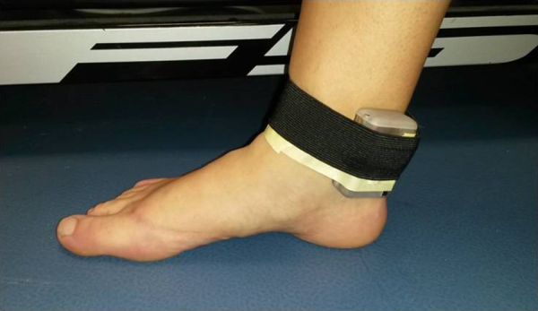
Accelerometer loaded on the leg.
Data was collected using x-IMU GUI software (version 13.0.4911.40774; X-IO Technologies Limited, UK) which logged and converted raw data (in g's) which was streamed via a Bluetooth connection from the unit to a nearby laptop. This raw data was then exported to a Microsoft Excel spreadsheet (Fig. 2). For each participant, the average acceleration and maximum acceleration at heel strike of the 5 stride cycles was calculated. Statistical analysis was preceded by the use of Kolmogorov–Smirnov test to check for distribution of data. Since the data was found to be normally distributed, the One-Way ANOVA test was subsequently used to compare the average and maximum acceleration at heel strike between pronated, neutral, and supinated feet.
Fig. 2.

Typical calcaneal acceleration versus time graph.
3. Results
Twenty-one female and 9 male participants, with a mean age of 21 years (SD 2.5) and mean weight of 65 kg (SD 10.3), were recruited.
3.1. Mean acceleration at heel strike
The mean acceleration at heel strike for supinated feet (2.127 g) was significantly higher than in pronated (1.344 g) and neutral feet (1.317 g), (p = 0.002). The Dunnett T3 post-hoc test revealed comparable average acceleration at heel strike for the pronated and neutrally aligned feet (p = 0.994). On the other hand, the mean acceleration at heel strike for supinated feet was significantly higher than the average acceleration for pronated and neutral foot types (p = 0.039 and p = 0.031 respectively).
3.2. Maximum acceleration at heel strike
The same results were obtained when analyzing maximum acceleration at heel strike. The one-way ANOVA test revealed an average maximum acceleration which was significantly higher in supinated feet (2.709 g) as opposed to pronated (1.733 g) and neutral (1.665 g) foot types (p = 0.002).
When values for pronated and neutral feet were analyzed, the maximum acceleration at heel strike was not significantly different (p = 0.968). However, when comparing supinated feet to the pronated and neutral feet, a significant difference (p = 0.040 and p = 0.025 respectively) was found between foot types.
4. Discussion
This study aimed to explore possible calcaneal acceleration variation during heel strike in the different foot types. Results confirm that supinated feet had significantly higher mean and higher maximum acceleration at the calcaneus during heel strike than the neutral and pronated types, i.e. this foot type resulted in reduced shock attenuation. Although this has for long been suspected, the scientific evidence is conspicuously lacking.
The results of this study also revealed no significant difference for the average acceleration and the maximum acceleration at heel strike between pronated and neutral foot types. This supports results of the study by Ledoux and Hillstrom (2001), who also report no differences in subjects with pes planus and neutrally aligned feet.9 Despite the fact that supinated feet were not concluded in their study, the authors had concluded that this lack of significant difference for peak acceleration was attributed to the fact that acceleration impulse occurs too fast (over a span of approximately 5 ms) for changes in foot structure to affect foot function. Since acceleration at the calcaneus during walking in healthy subjects has not been extensively studied, direct comparison was only possible with the above-mentioned study,9 despite some variations in the methodology between the two studies.
Pronation is thought to act as a mechanism that dampens impact forces from the foot up the lower limb and to the rest of the musculoskeletal system.16 Although the higher acceleration at heel strike in supinators may be attributed to the shortening of the invertor group of muscles of the foot in these individuals and thus to their diminished capability to pronate adequately, it is unlikely that pronation-dependent mechanisms of shock-absorption may have come into play at the instant of heel strike.
Although supinated feet are not as common as pronated or neutral feet, the implications of these findings for clinical practice should be considered. Accelerometers are cheaply-available nowadays and the testing of patients in clinic is easily possible. This would enable the clinician to quantify calcaneal acceleration and, where appropriate, to advice the necessary management strategies in order to reduce the detrimental effects of this increased acceleration at heel strike through the use of adequate footwear and possible heel inserts that would absorb or dissipate these heel impact forces. Although investigating running and not normal walking, O’Leary et al. (2008) have shown that the use of cushioned insoles significantly reduced the mean vertical ground reaction force peak impact by 6.8% (p = 0.004) when compared with the shoe-only condition.17 The use of viscoelastic insoles as protection against running overuse injuries has already been suggested.18 Thus, these could also possibly be effective during normal walking.
As already pointed out, since there is evidence supporting the link between excessive heel impact forces and the development of degenerative joint disease such as osteoarthritis,3 the cavus foot may be particularly susceptible to arthritis, an important inference with significant clinical implications.
In-depth knowledge of calcaneal acceleration could affect the choice of foot orthoses material for those patients with cavus feet, which material could differ as a consequence of these testing carried out in-clinic. This could possibly necessitate the wider utilization of shock-absorbing materials in those foot orthotic devices prescribed once supinated, or cavus, feet are diagnosed. A further possibility is for the use of this shock absorbing material to be strategically placed within the orthotic device so that it actively replaces the shock-absorbing mechanism within the foot at the appropriate time during the stance phase of gait.
An important limitation of this study is the fact that, in order to control cadence which is known to affect calcaneal acceleration, all participants underwent treadmill walking at a constant speed. As yet, one cannot accurately extrapolate these results onto walking on uneven surfaces at different speeds, and certainly further studies are warranted.
Certainly further research is necessary into what magnitude of acceleration forces constitute pathological levels that require management. Furthermore, this testing modality opens up new opportunities for research into those patient populations who suffer from low back pain and a number of musculoskeletal conditions, who could possibly benefit from these quantifications, since the relationship between shock and these conditions is well documented.
5. Conclusion
This study demonstrates that cavus, or supinated, feet, undergo significantly higher mean and maximum acceleration forces when compared to neutrally aligned and pronated feet. Keeping this in mind, especially in the clinical field, could be beneficial in order to prevent and reduce the prevalence of musculoskeletal problems caused by excess acceleration, such as degenerative joint disease, low back pain, muscle tears, headaches, and Achilles tendon pathologies.
Conflicts of interest
None.
References
- 1.Williams D.S., McClay I.S., Hamill J., Buchanan T.S. Lower extremity kinematic and kinetic differences in runners with high and low arches. J Appl Biomech. 2001;17(2):153–163. [Google Scholar]
- 2.Beschorner K., Cham R. Impact of joint torques on heel acceleration at heel contact, a contributor to slips and falls. Ergonomics. 2008;51(12):1799–1813. doi: 10.1080/00140130802136479. Available from: http://www.ncbi.nlm.nih.gov/pubmed/18937108. [DOI] [PubMed] [Google Scholar]
- 3.Collins J.J., Whittle M.W. Impulsive forces during walking and their clinical implications. Clin Biomech. 1989;4(3):179–187. doi: 10.1016/0268-0033(89)90023-5. [DOI] [PubMed] [Google Scholar]
- 4.Pratt D.J. Mechanisms of shock attenuation via the lower extremity during running. Clin Biomech. 1989;4:51–57. doi: 10.1016/0268-0033(89)90068-5. [DOI] [PubMed] [Google Scholar]
- 5.Hargrave M.D., Carcia C.R., Gansneder B.M., Shultz S.J. Subtalar pronation does not influence impact forces or rate of loading during a single-leg landing. J Athl Train. 2003;38(1):18–23. [PMC free article] [PubMed] [Google Scholar]
- 6.Milner C.E., Ferber R., Pollard C.D., Hamill J., Davis I.S. Biomechanical factors associated with tibial stress fracture in female runners. Med Sci Sports Exerc. 2006;38(2):323–328. doi: 10.1249/01.mss.0000183477.75808.92. [DOI] [PubMed] [Google Scholar]
- 7.Mayagoitia R.E., Nene A.V., Veltink P.H. Accelerometer and rate gyroscope measurement of kinematics: an inexpensive alternative to optical motion analysis systems. J Biomech. 2002;35(4):537–542. doi: 10.1016/s0021-9290(01)00231-7. [DOI] [PubMed] [Google Scholar]
- 8.Kavanagh J.J., Menz H.B. Accelerometry: a technique for quantifying movement patterns during walking. Gait Posture. 2008;28(1):1–15. doi: 10.1016/j.gaitpost.2007.10.010. [DOI] [PubMed] [Google Scholar]
- 9.Ledoux W.R., Hillstrom H.J. Acceleration of the calcaneus at heel strike in neutrally aligned and pes planus feet. Clin Biomech (Bristol, Avon) 2001;16(7):608–613. doi: 10.1016/s0268-0033(01)00041-9. Available from: http://www.ncbi.nlm.nih.gov/pubmed/11470303. [DOI] [PubMed] [Google Scholar]
- 10.Warden S.J., Burr D.B., Brukner P.D. Stress fractures: pathophysiology, epidemiology, and risk factors. Curr Osteoporos Rep. 2006:103–109. doi: 10.1007/s11914-996-0029-y. [DOI] [PubMed] [Google Scholar]
- 11.Redmond A.C., Crosbie J., Ouvrier R.A. Development and validation of a novel rating system for scoring standing foot posture: the foot posture index. Clin Biomech. 2006;21(1):89–98. doi: 10.1016/j.clinbiomech.2005.08.002. [DOI] [PubMed] [Google Scholar]
- 12.Keenan A.M., Redmond A.C., Horton M., Conaghan P.G., Tennant A. The foot posture index: Rasch analysis of a novel. Foot-specific outcome measure. Arch Phys Med Rehabil. 2007;88(January):88–93. doi: 10.1016/j.apmr.2006.10.005. [DOI] [PubMed] [Google Scholar]
- 13.Cowley E., Marsden J. The effects of prolonged running on foot posture: a repeated measures study of half marathon runners using the foot posture index and navicular height. J Foot Ankle Res. 2013;6(1):20. doi: 10.1186/1757-1146-6-20. Available from: http://www.pubmedcentral.nih.gov/articlerender.fcgi?artid=3668212&tool=pmcentrez&rendertype=Abstract. [DOI] [PMC free article] [PubMed] [Google Scholar]
- 14.Evans A.M., Rome K., Peet L. The foot posture index, ankle lunge test, Beighton scale and the lower limb assessment score in healthy children: a reliability study. J Foot Ankle Res. 2012;5(1):1. doi: 10.1186/1757-1146-5-1. Available from: http://www.jfootankleres.com/content/5/1/1. [DOI] [PMC free article] [PubMed] [Google Scholar]
- 15.Wakeling J.M., Nigg B.M. Modification of soft tissue vibrations in the leg by muscular activity. J Appl Physiol. 2001;90(2):412–420. doi: 10.1152/jappl.2001.90.2.412. Available from: http://jap.physiology.org/content/90/2/412. [DOI] [PubMed] [Google Scholar]
- 16.Nachbauer W., Nigg B. Effects of arch height of the foot on ground reaction forces in running. Med Sci Sports Exerc. 1992;24(11):1264–1269. [PubMed] [Google Scholar]
- 17.O’Leary K., Vorpahl K.A., Heiderscheit B. Effect of cushioned insoles on impact forces during running. J Am Podiatr Med Assoc. 2008;98(1):36–41. doi: 10.7547/0980036. [DOI] [PubMed] [Google Scholar]
- 18.Nigg B.M., Herzog W., Read L.J. Effect of viscoelastic shoe insoles on vertical impact forces in heel-toe running. Am J Sports Med. 1988;16(1):70–76. doi: 10.1177/036354658801600113. [DOI] [PubMed] [Google Scholar]