Abstract
PURPOSE
We assessed the repeatability and reproducibility of abutment teeth dental impressions, digitized with a blue light scanner, by comparing the discrepancies in repeatability and reproducibility values for different types of abutment teeth.
MATERIALS AND METHODS
To evaluate repeatability, impressions of the canine, first premolar, and first molar, prepared for ceramic crowns, were repeatedly scanned to acquire 5 sets of 3-dimensional data via stereolithography (STL) files. Point clouds were compared and the error sizes were measured (n=10, per type). To evaluate reproducibility, the impressions were rotated by 10-20° on the table and scanned. These data were compared to the first STL data and the error sizes were measured (n=5, per type). One-way analysis of variance was used to assess the repeatability and reproducibility of the 3 types of teeth, and Tukey honest significant differences (HSD) multiple comparison test was used for post hoc comparisons (α=.05).
RESULTS
The differences with regard to repeatability were 4.5, 2.7, and 3.1 µm for the canine, premolar, and molar, indicating the poorest repeatability for the canine (P<.001). For reproducibility, the differences were 6.6, 5.8, and 11.0 µm indicating the poorest reproducibility for the molar (P=.007).
CONCLUSION
Our results indicated that impressions of individual abutment teeth, digitized with a blue light scanner, had good repeatability and reproducibility.
Keywords: Blue light scanner, Repeatability, Reproducibility, Color-difference-map
INTRODUCTION
Scanners are increasingly being utilized in the field of dentistry, and the evaluation of their repeatability and reproducibility is necessary to validate their clinical use.1,2,3 Common prosthesis fabrication includes model fabrication by pouring dental stone into impressions of the patient's teeth. Also, assessment of the repeatability and reproducibility of the process is determined by comparing 3-D scan data digitized with the stone models. However, this fabrication process is time-consuming and expensive.2,3,4 To overcome these disadvantages, a prosthesis fabrication method using intraoral scanners was developed; however, it did not demonstrate a high degree of repeatability and reproducibility.1,5,6,7,8 Therefore, methods to provide three-dimensional data by directly scanning dental impressions with an extraoral scanner have been developed.3,5,6,7,8,9
Previous clinical studies evaluating the repeatability and reproducibility of impressions digitized using an extraoral scanner (e.g., touch probe scanner, laser scanner, white light scanner) have not demonstrated any evidence that supports its clinical application.1,4,10 Recently, to compensate for the disadvantages of this type of scanners, blue light scanners that can scan impressions with high repeatability and reproducibility (even when scanning narrow and deep forms) and high scanning speed were developed.11 But, no study has assessed the repeatability and reproducibility of impressions digitized using blue light scanners or assess them for different types of abutment teeth. The ISO-12836 standard specifies repeatability and reproducibility as the factors that need to be established for scanners.12 Generally, reference data is usually referenced to the data scanned in the reference scanner of engineering (concept of trueness by ISO-12836). However, in this study, the repeatability and reproducibility of blue light scanner was assessed through the comparison of each other's data. Comparison of errors in data obtained from repeated scanning was used to verify repeatability,5,13 while reproducibility was established through comparison of errors in data obtained from scanning of rotated models.3,14,15,16
The purpose of this study was assessed the repeatability and reproducibility of impressions digitized using a blue light scanner for the canine, premolar, and molar. In addition, reference values for potential clinical applications were obtained.
MATERIALS AND METHODS
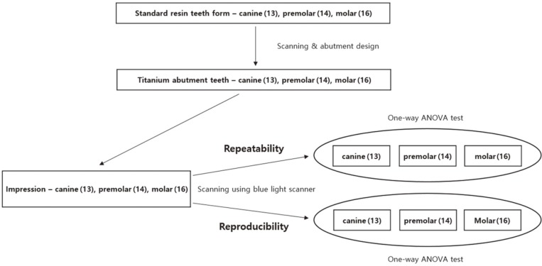
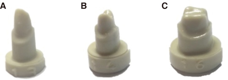
Maxillary molars and adjoining anterior teeth were chosen for scanning. Specifically, artificial resin models with conventional tooth forms (AG-3, Frasaco, GmbH, Tettang, Germany) of the canine, first premolar (stated below as "premolar"), and first molar (stated below as "molar") were selected. Next, the tooth models were prepared to have the shape of the abutments for all-ceramic crowns. The characters of the artificial resin tooth abutments are explained below (Fig. 1).
Fig. 1. Scheme showing the experimental design.
The canine model was the longest and sharpest tooth. The length of the premolar was similar to that of the molar, but the occlusal dimensions of the molar were greater than that of the premolar. The premolar's axial wall was almost vertical. Deep chamfer margins were placed in the cervical third of the buccal/labial, lingual, mesial, and distal surfaces. For preparation, 1-1.5 mm cuts were placed on the labial/buccal and lingual axial walls, while 1.5-2 mm cuts were placed on the incisal edges or occlusal surfaces. The labial/buccal, lingual, mesial, and distal surfaces were prepared, maintaining an angle of 6° with the axial wall. The points and margin angles were rounded off and smoothened (Fig. 2).3,5,6
Fig. 2. Resin tooth abutments. (A) Canine (#13), (B) Premolar (#14), (C) Molar (#16).
After tooth preparation, the CAM system (DT400, Doosan Infracore Co., Ltd., Seoul, South Korea) was used to fabricate titanium overlays to minimize abrasion of the abutment tooth surfaces during impression taking (titanium exhibits better wear resistance) and increase the repeatability and reproducibility of the impression. Next, the extra-light body vinyl polysiloxane impression material (Aquasil Ultra, Dentsply, St. York, PA, USA), which is indicated for scanning purposes, was used to record the impressions of the titanium tooth abutments.3,5,6
A blue light scanner (Identica Blue, Medit, Seoul, Korea) was utilized to scan these impressions to evaluate repeatability and reproducibility. Repeatability was evaluated by first fixing the maxillary canine impression onto the scanner table and collecting 5 sets of 3-dimensional shape data with steriolithography (STL) by repeated scanning (C_repeat1 - C_repeat5). A similar procedure was used to collect 5 sets of STL files each for the impressions of the premolar (P_repeat1 - P_repeat5) and molar (M_repeat1 - M_repeat5).
To verify reproducibility, the maxillary canine impression was scanned after rotating the table by 10-20° from the initial position, and 5 sets of STL files (C_repro1 - C_repro5) were obtained. Following the same procedure, data were obtained for the premolar (P_repro1 - P_repro5) and molar impressions (M_repro1 - M_repro5). Unnecessary regions under the margins on each scan were eliminated from the STL file for the impressions.3,4,5,6,8
After converting the STL files into point clouds (ASCII files) with CopyCAD7.350 SP3 software (Delcam plc., Birmingham, UK), a color-difference map and a report were generated with PowerInspect 2012 software (Delcam plc., Birmingham, UK) from the point cloud ASCII files.
Repeatability was confirmed by mutually superimposing the point clouds for the 5 scans of abutment teeth on the STL files and determining the best fit alignment (n = 10, per tooth type).
Reproducibility was confirmed by superimposing the scans of the rotated impressions onto the first scans from repeatability evaluation and determining the best fit alignment (n = 5, per tooth type).
The repeatability and reproducibility values for the canine, premolar, and molar were assessed using parametric one-way analysis of variance (ANOVA), and Tukey honest significant test (HSD) was used for performing post hoc comparisons. An alpha value of 0.05 was set as the significance level for each experiment. The above-mentioned statistical analyses were performed with SPSS v. 21.0 software (IBM SPSS Inc., Chicago, IL, USA).
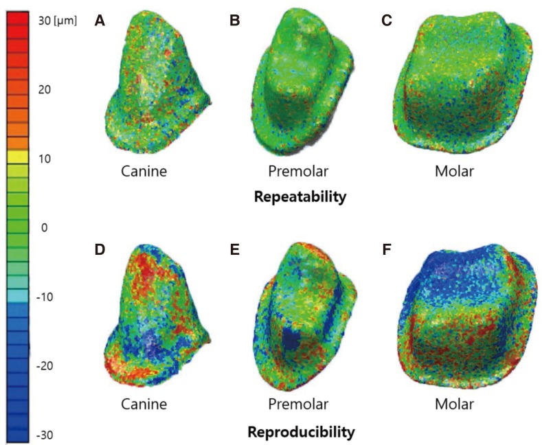
Color-difference maps were generated after superimposition of 3-dimensional data showing the repeatability and reproducibility of digitized impressions for canine, premolar, and molar. Yellow or red represents positive error, turquoise to blue represents negative error, and green represents good fit.
RESULTS
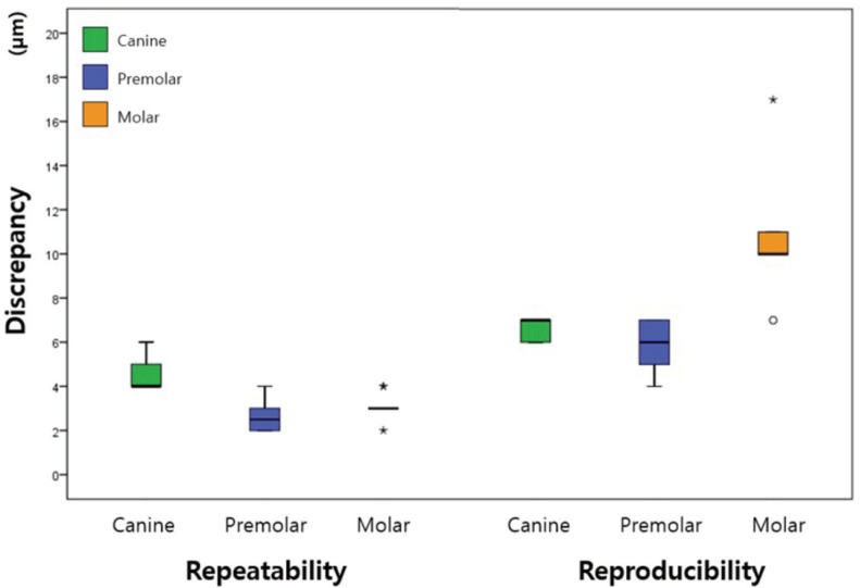
A color-difference map (qualitative data, Fig. 3) and a report (quantitative data, Fig. 4) of the projections were obtained for each tooth type.
Fig. 3. Evaluation of the repeatability and reproducibility of digitized impressions obtained with a blue light scanner for three types of abutment teeth.
Fig. 4. Box plot showing the repeatability and reproducibility of impressions digitized with a blue light scanner for three types of abutment teeth; repeatability (n = 10 per type), reproducibility (n = 5 per type).
Figure 3 shows the qualitative repeatability and reproducibility data for each tooth type. For repeatability, the canine primarily exhibited green color, indicating no significant error. Smaller red and blue areas indicating small positive and negative errors were observed along the longitudinal axis. For reproducibility, the canine primarily exhibited red and blue areas, representing positive and negative errors.
The premolar was also predominantly green for repeatability; however, it showed negative (blue) errors for reproducibility and a larger green area compared with the molar. The molar was primarily green for repeatability, but it showed negative (blue) errors on the occlusal surface and positive and negative (red and blue) errors along the longitudinal axis. For reproducibility, it mostly showed larger positive and negative (red and blue) errors compared with the other teeth.
Figure 4 shows the quantitative repeatability and reproducibility data for each tooth type. Repeatability discrepancies for the canine were significantly larger than those for the other teeth (P < .001). There was a significantly larger reproducibility discrepancy for the molar as compared to the other teeth (P = .007).
Discrepancies with regard to repeatability were significantly larger for the canine than for the other teeth (Table 1); this can be ascribed to the morphologic features of the canine, a narrow and deep shape that results in more shadow compared with the premolar and molar (Fig. 3).2,3,5
Table 1. Repeatability and reproducibility of digital impressions obtained with a blue light scanner for three types of abutment teeth.
| Mean (95% CI) | P value | ||
|---|---|---|---|
| Repeatability | Canine | 4.5 (4.0-5.0)a* | |
| Premolar | 2.7 (2.1-3.3)b | < .001 | |
| Molar | 3.1 (2.7-3.5)b | ||
| Reproducibility | Canine | 6.6 (5.9-7.3)a | |
| Premolar | 5.8 (4.2-7.4)a | .007 | |
| Molar | 11.0 (6.4-15.6)b |
Unit: µm; CI: confidence interval
*Different letters indicate significant differences (P < .05).
DISCUSSION
The purpose of this study was to compare the repeatability and reproducibility of impressions digitized using the recently developed blue light scanner for abutment tooth types. The repeatability and reproducibility data was shown using color-difference maps (qualitative data, Fig. 3) and box plots (quantitative data, Fig. 4).
Discrepancies for reproducibility were significantly larger for the molars than for the other teeth (Table 1). This indicates that occlusal line angular area is the largest in the molar and that is the highest lots of points in the steep parts of the axial walls compared with the canine and premolar.3,5,6,10 Previous studies have shown an average ofapproximately 10 µm error in impression scanning, which is considered to be within a reliable level.2,3,5,6,17 A greater level of repeatability and reproducibility was observed in the results of the current study, with errors less than 10 µm. The reason for this finding is that a blue light scanner uses a light source with a shorter wavelength, resulting in smaller scanning errors for the variables affecting the color and shape of the object being scanned.11,17,18
In this study, an impression material made of vinyl polysiloxane specially formulated for optical scanning was used to measure the repeatability and reproducibility of the scanned impressions.5 The use of this material improves the validity of repeatability and reproducibility evaluations of impressions digitized using blue light scanners.19
ISO-12836 introduced the concept of repeatability and reproducibility. Its goal was to ensure the validity of measurements in compliance with the experimental process and design.12
This study has a few limitations. First, we adapted measurement process on 3-dimensional superimposed data to confirm repeatability and reproducibility. It is difficult to explain the errors in the best fit alignment, confirming errors among data,3,17,20,21 and investigating whether the error in the process of best fit alignment occurs during the verification or scanning processes.3,5,10 In addition, we could not include the control group scanners and the trueness assessment with reference dataset was omitted. Future researches are required to get over these limitations, to find out means of reducing errors, and to improve the quality of impressions digitized using a blue light scanner.
CONCLUSION
This study shows that the digitization of impressions of individual abutment teeth using a blue light scanner provides good repeatability and reproducibility, indicating a possible clinical advantage for blue light scanners for digitizing impressions of single abutments.
References
- 1.Nedelcu RG, Persson AS. Scanning accuracy and precision in 4 intraoral scanners: an in vitro comparison based on 3-dimensional analysis. J Prosthet Dent. 2014;112:1461–1471. doi: 10.1016/j.prosdent.2014.05.027. [DOI] [PubMed] [Google Scholar]
- 2.Persson A, Andersson M, Oden A, Sandborgh-Englund G. A three-dimensional evaluation of a laser scanner and a touch-probe scanner. J Prosthet Dent. 2006;95:194–200. doi: 10.1016/j.prosdent.2006.01.003. [DOI] [PubMed] [Google Scholar]
- 3.Jeon JH, Kim HY, Kim JH, Kim WC. Accuracy of 3D white light scanning of abutment teeth impressions: evaluation of trueness and precision. J Adv Prosthodont. 2014;6:468–473. doi: 10.4047/jap.2014.6.6.468. [DOI] [PMC free article] [PubMed] [Google Scholar]
- 4.Quaas S, Rudolph H, Luthardt RG. Direct mechanical data acquisition of dental impressions for the manufacturing of CAD/CAM restorations. J Dent. 2007;35:903–908. doi: 10.1016/j.jdent.2007.08.008. [DOI] [PubMed] [Google Scholar]
- 5.Jeon JH, Lee KT, Kim HY, Kim JH, Kim WC. White light scanner-based repeatability of 3-dimensional digitizing of silicon rubber abutment teeth impressions. J Adv Prosthodont. 2013;5:452–456. doi: 10.4047/jap.2013.5.4.452. [DOI] [PMC free article] [PubMed] [Google Scholar]
- 6.Jeon JH, Choi BY, Kim CM, Kim JH, Kim HY, Kim WC. Three-dimensional evaluation of the repeatability of scanned conventional impressions of prepared teeth generated with white- and blue-light scanners. J Prosthet Dent. 2015;114:549–553. doi: 10.1016/j.prosdent.2015.04.019. [DOI] [PubMed] [Google Scholar]
- 7.Jeon JH, Jung ID, Kim JH, Kim HY, Kim WC. Three-dimensional evaluation of the repeatability of scans of stone models and impressions using a blue LED scanner. Dent Mater J. 2015;34:686–691. doi: 10.4012/dmj.2014-347. [DOI] [PubMed] [Google Scholar]
- 8.Luthardt RG, Loos R, Quaas S. Accuracy of intraoral data acquisition in comparison to the conventional impression. Int J Comput Dent. 2005;8:283–294. [PubMed] [Google Scholar]
- 9.Ender A, Mehl A. Accuracy of complete-arch dental impressions: a new method of measuring trueness and precision. J Prosthet Dent. 2013;109:121–128. doi: 10.1016/S0022-3913(13)60028-1. [DOI] [PubMed] [Google Scholar]
- 10.Persson AS, Andersson M, Odén A, Sandborgh-Englund G. Computer aided analysis of digitized dental stone replicas by dental CAD/CAM technology. Dent Mater. 2008;24:1123–1130. doi: 10.1016/j.dental.2008.01.008. [DOI] [PubMed] [Google Scholar]
- 11.Bernal C, de Agustina B, Marin MM, Camacho AM. Performance evaluation of optical scanner based on blue LED structured light. Procedia Eng. 2013;63:591–598. [Google Scholar]
- 12.ISO-12836. Dentistry - Digitizing devices for CAD/CAM systems for indirect dental restorations-test methods for assessing accuracy. Geneva; Switzerland: ISO; 2015. [Accessed March 2, 2016]. Available from: http://www.iso.org/iso/store.html. [Google Scholar]
- 13.Hoyos A, Soderholm KJ. Influence of tray rigidity and impression technique on accuracy of polyvinyl siloxane impressions. Int J Prosthodont. 2011;24:49–54. [PubMed] [Google Scholar]
- 14.Wöstmann B, Rehmann P, Balkenhol M. Accuracy of impressions obtained with dual-arch trays. Int J Prosthodont. 2009;22:158–160. [PubMed] [Google Scholar]
- 15.Ziegler M. Digital impression taking with reproducibly high precision. Int J Comput Dent. 2009;12:159–163. [PubMed] [Google Scholar]
- 16.Chandran DT, Jagger DC, Jagger RG, Barbour ME. Two- and three-dimensional accuracy of dental impression materials: effects of storage time and moisture contamination. Biomed Mater Eng. 2010;20:243–249. doi: 10.3233/BME-2010-0638. [DOI] [PubMed] [Google Scholar]
- 17.Logozzo S, Zanetti EM, Franceschini G, Kilpelä A, Mäkynen A. Recent advances in dental optics – Part I: 3D intraoral scanners for restorative dentistry. Opt Lasers Eng. 2014;54:203–221. [Google Scholar]
- 18.Martorelli M, Ausiello P, Morrone R. A new method to assess the accuracy of a Cone Beam Computed Tomography scanner by using a non-contact reverse engineering technique. J Dent. 2014;42:460–465. doi: 10.1016/j.jdent.2013.12.018. [DOI] [PubMed] [Google Scholar]
- 19.Persson M, Andersson M, Bergman B. The accuracy of a high-precision digitizer for CAD/CAM of crowns. J Prosthet Dent. 1995;74:223–229. doi: 10.1016/s0022-3913(05)80127-1. [DOI] [PubMed] [Google Scholar]
- 20.Naidu D, Freer TJ. Validity, reliability, and reproducibility of the iOC intraoral scanner: a comparison of tooth widths and Bolton ratios. Am J Orthod Dentofacial Orthop. 2013;144:304–310. doi: 10.1016/j.ajodo.2013.04.011. [DOI] [PubMed] [Google Scholar]
- 21.Flügge TV, Schlager S, Nelson K, Nahles S, Metzger MC. Precision of intraoral digital dental impressions with iTero and extraoral digitization with the iTero and a model scanner. Am J Orthod Dentofacial Orthop. 2013;144:471–478. doi: 10.1016/j.ajodo.2013.04.017. [DOI] [PubMed] [Google Scholar]


