

INTRODUCTION
Macrodystrophia lipomatosa (ML) is a rare non-hereditary congenital developmental anomaly causing disproportionate overgrowth of one or several digits. It may cause a localised gigantism or involve the entire limb. It is characterised by progressive hamartomatous enlargement of the fibro-fatty tissue and should be differentiated from macromelia or hemihypertrophy. The imaging is important in making the diagnosis.
CASE REPORT
A 18-year-old male presented to our institute with a history of enlargement of right great toe since childhood (Figure 1). He did not offer any other complaints. There was no history of similar complaints or neurofibromatosis in his family members suggestive of non-hereditary nature of the lesion. There were no obvious cutaneous vascular lesions. There was no neurological deficit. The other digits and contralateral foot were normal. Radiographs of the right foot showed gross increase in the soft tissue of great toe as compared to the contralateral side. The phalanges and metatarsals were enlarged as compared to contralateral side. There was an accessory bone present adjacent to the distal phalanx on its medial aspect (Figure 2). There were no features of degenerative change. Ultrasonography and colour Doppler using high-resolution probe revealed abundant increase in the fat tissue with no evidence of any vascular malformation. The patient underwent surgery for cosmetic reasons and (to achieve medical fitness prior to recruitment for defence services) as a prerequisite to apply for a job in the defence forces. Histopathology showed presence of adipose tissue in the excised specimen scattered in a fine mesh-like fibrous tissue extending to involve the periosteum (Figure 3). A diagnosis of ML was offered.
Figure 1.
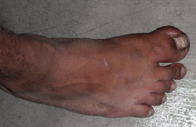
Photograph of the enlarged right toe.
Figure 2.
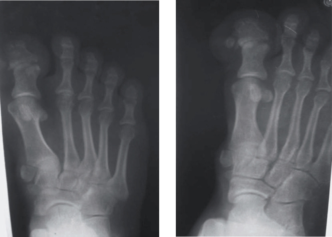
Radiograph antero-posterior view of right toe showing gross increase in the soft tissue of great toe with enlarged phalanges and metatarsals. There is an additional bone present medial to the distal phalanx.
Figure 3.

Photomicrograph of the operated specimen showing abundant fatty tissue within mesh-like fibrous mesh work.
DISCUSSION
Macrodystrophia lipomatosa is a rare form of localised gigantism characterised by progressive overgrowth of all the mesenchymal elements with a disproportionate increase in the fibroadipose tissues. This congenital abnormality occurs most frequently in the distribution of the median nerve in the upper extremity and in the distribution of the plantar nerves in the lower extremity. In 1925, Feriz was the first person to use this term for localised gigantism of the lower extremity. Barsky on the other hand gave a more detailed description of the disease involving local gigantism of digits. He described two forms of true macrodactyly. In the first or the static form, the size of the enlarged digits increases proportionally in relation to the rest of body. In the second or the progressive form which is less common, the growth rate of the enlarged digits is disproportionate to the growth of rest of the body. The progressive form may be associated with fatty overgrowth, and it is similar to what Feriz described.1 Our case would fit into the first category.
This disease is non-hereditary in nature and has a slight male preponderance as in our case. The lower limb is involved more commonly. The region supplied by medial plantar nerve, that is, first to third toes, is frequently involved and the fifth digit is the rarest to be involved. ML commonly involves unilateral digits but rarely, a bilateral involvement has been described in literature. It is usually recognised in the neonatal period but starts to cause problems as the child grows. There may be difficulty in walking though, more often than not, a surgical consultation is sought for cosmetic rather than mechanical reasons. As the child grows there may be degenerative changes of small joints and compression of the neurovascular structure.2 Patient may present with features of carpal tunnel syndrome due to involvement of median nerve. The growth of bones normally ceases by puberty. Histopathology shows abundant increase in the adipose tissue scattered in a fine mesh-like fibrous tissue. Besides affecting subcutaneous tissue it can involve underlying nerve sheaths, muscles, periosteum, and bone marrow. Exact aetiopathogenesis is obscure. The proposed mechanisms include lipomatous degeneration, disturbed foetal circulation, disturbances of growth inhibiting factors, an error in segmentation, and trophic influence of tumefied nerve. The pathogenesis of bony enlargement is thought to be because of endosteal and periosteal deposition of bone.2
The imaging plays an important role in the diagnosis of this condition by characterising the nature of the hypertrophied tissue. Plain radiographs may demonstrate abnormalities in both the soft tissues and bony elements. The soft tissue overgrowth is most marked on the volar aspect. This volar overgrowth results in dorsal deviation of the digits.2 The affected phalanges and metatarsal bones show an increase in width and length, and often splayed at their distal ends. The articular surfaces may slant and in late childhood severe secondary degenerative changes may affect the joints. All these features were present in our case. Characteristic radiolucencies seen are indicative of the fatty nature of the soft tissue.3 Presence of syndactyly, polydactyly, and clinodactyly has been reported. Computed tomography of the involved extremities shows excessive fatty proliferation. Fatty infiltrations of the involved muscles with separation of the muscle fibres are also often evident.4 Magnetic resonance imaging characteristically shows abundant adipose tissues in the involved areas. This fat has the same signal intensity as the normal subcutaneous fat. But contrary to lipoma, the abnormal fat in this disorder is not encapsulated. Linear hypointense bands may be noted within this redundant adipose tissue representing fibrous strands. Fatty infiltration of the involved muscles as well as, the bony overgrowth and cortical thickening of the enlarged bones can be well demonstrated on the MR imaging.4 Histopathology in our case proved the diagnosis.
There is a plethora of differential diagnosis to localised gigantism of the digit. These include congenital localised gigantism such as fibrolipomatous hamartoma of a nerve, neurofibromatosis, haemangiomatosis, lymphangiomatosis, Proteus syndrome and Klippel-Trenaunay-Weber syndrome that includes macrodactyly with haemangiomas. But in the absence of family history and any cutaneous or systemic manifestation these diagnoses are unlikely.5 In this situation an ultrasound and Doppler using a high-resolution linear probe can distinguish vascular or lymphatic component. In addition to this presence of well-defined isoechoic to hypoechoic lesions along the nerve sheath will help in differentiating ML from lesions of nerve sheath. It is often difficult to distinguish this lesion from plexiform neurofibromatosis. The involvement in plexiform neurofibromatosis is often bilateral and growth of bones continues beyond puberty. In contradistinction to ML the distal phalanx is not predominantly involved.
Patients seek medical help more often for cosmetic reasons rather than for mechanical difficulties which is normally late in life. Surgical correction is the treatment of choice for ML. Some authors have advocated a conservative approach in case of involvement of plantar or median nerve. Our patient sought surgical intervention as he believed this disability would have made him unfit for recruitment in the defence forces. The main surgical principle in treating this lesion is to improve cosmetic appearance and preserve neurologic function as far as possible and this is usually done after puberty when growth ceases. Judicious and planned use of multiple debulking procedures, epiphysiodesis, and various osteotomies are advised to achieve the best results.6 Complications associated with overzealous debulking procedure lead to nerve injury with an incidence reported as high as 30–50%. A localised recurrence rate of 33–60% makes the management of ML more demanding.7
In conclusion, ML is a rare form of congenital localised gigantism, and surgical consultation is often sought for cosmetic reasons. Imaging provides vital clues to an accurate diagnosis which is confirmed by histopathology. Surgery can be offered to patients after puberty for cosmetic as well as functional problems.
CONFLICTS OF INTEREST
None.
REFERENCES
- 1.Boren WL, Henry REC, Wintch K. MR diagnosis of fibrolipomatous hamartoma of nerve: association with nerve territory-oriented macrodactyly (macrodystrophia lipomatosa) Skeletal Radiol. 1995;24:296–297. doi: 10.1007/BF00198419. [DOI] [PubMed] [Google Scholar]
- 2.Gupta SK, Sharma OP, Sharma SV, Sood B, Gupta S. Macrodystrophia lipomatosa: radiographic observations. BJR. 1992;65:769–773. doi: 10.1259/0007-1285-65-777-769. [DOI] [PubMed] [Google Scholar]
- 3.Jain R, Sawhney S, Berry M. CT diagnosis of macrodystrophia lipomatosa. Acta Radiol. 1992;33:554–555. [PubMed] [Google Scholar]
- 4.Sone M, Ehara S, Tamakawa Y, Nishida JN, Honjoh S. Macrodystrophia lipomatosa: CT and MR findings. Radiation Med. 2000;18:129–132. [PubMed] [Google Scholar]
- 5.Baruchin AM, Herold ZH, Shmueli G. Macrodystrophia lipomatosa of the foot. J Pediatr Surg. 1988;23:192–194. doi: 10.1016/s0022-3468(88)80157-x. [DOI] [PubMed] [Google Scholar]
- 6.Wu KK. Macrodactylia lipomatosis of the foot. J Foot Surg. 1991;30:402–405. [PubMed] [Google Scholar]
- 7.Brodwater BK, Major NM, Goldner RD, Layfield LJ. Macrodystrophia lipomatosa with associated fibrolipomatous hamartoma of the median nerve. Pediatr Surg Int. 2000;16:216–218. doi: 10.1007/s003830050728. [DOI] [PubMed] [Google Scholar]