

Abstract
Background
Radical Neck Dissection done in cases of squamous carcinoma of the head and neck with cervical metastases leads to significant morbidity due to the ‘shoulder syndrome’ arising from denervation and atrophy of the trapezius muscle supplied by the Spinal Accessory Nerve (SAN). Preservation of the SAN (described as Modified RND Type 1) has been described by various workers.
Methods
We present our experience with 24 patients of squamous carcinoma of the head and neck with cervical metastases. MRND Type 1 was performed in those cases where the SAN was not involved by metastatic disease.
Results
The SAN could be preserved in all the patients who were cN0, cN1 and cN2. Only one patient who was cN3 required sacrifice of SAN as the nerve was infiltrated by metastatic disease. Shoulder function was good where SAN was preserved. Conclusion : This procedure is oncologically safe and morbidity associated with the “Shoulder Syndrome” was prevented.
Key Words: Spinal accessory nerve, Modified neck dissection
Introduction
It was Crile in 1906 [1] who first described the technique of en bloc removal of the cervical lymphatics in the treatment of head and neck cancer. This was popularized by Martin [2], and remained essentially unchanged for several decades. At that time, the emphasis was primarily on optimal cancer control by radical surgery and sacrifice of function was accepted. In neck dissections the most crippling complication is the “Shoulder Syndrome” arising from denervation and atrophy of the trapezius muscle due to sacrifice of the spinal accessory nerve (SAN)[3]. Various modifications to the original classic RND have come to be practiced to reduce post operative morbidity. Of these the modified RND Type 1 is the most commonly performed procedure in the clinically positive neck[4]. We present our experience with this procedure at our centre.
Material and Methods
Patients who presented to the Dept of ENT- Head &Neck Surgery, associated with the Malignant Diseases Treatment Centre, of Command Hospital (Eastern Command) Kolkata from Jan 2002 to May 2004 with squamous cell carcinoma of the oral cavity, oropharynx, hypopharynx, larynx or squamous metastases from an unknown primary site in the upper aero - digestive tract were included in the study. The patients were assessed for cervical metastases by clinical examination, Ultrasonography, FNAC and CT scan of the neck. Patients with a clinically positive neck underwent comprehensive neck dissection and those with clinically negative neck (N0) were candidates for an elective prophylactic neck dissection if the probability of occult disease was 20% or greater. In this category, patients with a primary in the oral cavity were taken up for a Supra Omohyoid Neck Dissection (SOHND) and if the frozen section revealed squamous metastases, a comprehensive neck dissection was completed. None of the patients had any previous neck surgery or radiotherapy. The dissection was always started with the intention of preserving the SAN.
Site of lesion is given in Table 1. The clinical N stage and pathological N stage are given in Table 2.
Table 1.
Site of primary lesions
| Oral cavity | 12 |
| Oropharynx | 02 |
| Hypopharynx | 04 |
| Larynx | 06 |
| Unknown | 03 |
Table 2.
Clinical and Pathological N stages of the patients
| Stage | Clinical | Pathological |
|---|---|---|
| N0 | 04 | 03 |
| N1 | 03 | 04 |
| N2 | 09 | 08 |
| N3 | 03 | 04 |
Post operatively, the patients were advised shoulder exercises. The patients were given post operative radiotherapy where indicated. The indications were: multiple positive lymph nodes in the neck, extra capsular extension by metastatic disease, perivascular or perineural invasion by tumour and gross residual disease following neck dissection.
The technique of surgery was to identify the SAN during dissection at level 2 and then dissect out the entire nerve till the anterior border of the trapezius muscle. The method of identifying the nerve first at the anterior border of the trapezius muscle was avoided as it sometimes leads to injury to the nerve on account of it lying superficially. Fig 1 shows the nerve dissected out and skeletonised throughout its course in the neck from the jugular foramen to its entry into the trapezius muscle.
Fig. 1.

Dissection showing the skeletonised SAN
Results
The SAN could be preserved in all the patients who were cN0, cN1 and cN2. Only one patient who was cN3 required sacrifice of the SAN, as the nerve was infiltrated by the metastatic disease and could not be dissected free.
Of the 18 patients in whom the SAN could be preserved, all had preservation of normal contour of trapezius muscle (Fig 2). Shoulder function is preserved, and the patient was able to completely abduct his arm to 180o (Fig 3). Patient is alive and free of disease on follow up as shown in Table 3.
Fig. 2.

Preservation of contour of trapezius muscle
Fig. 3.
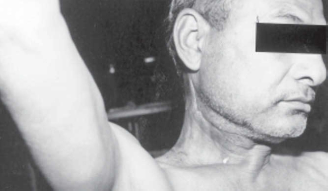
Preservation of shoulder function
Table 3.
Patient free of disease on last follow-up
| Site of primary | No. of patients |
|---|---|
| Oral cavity | 09 |
| Oropharynx | 01 |
| Hypopharynx | 03 |
| Larynx | 05 |
| Unknown | 02 |
Discussion
Historically the classical RND was described for treatment of the clinically positive neck in squamous cancer of the head and neck. Loss of trapezius muscle function represents the single most important source of long term morbidity from a radical neck dissection. Apart from the aesthetic deformity due to loss of the sternomastoid muscle, there is imbalance of the shoulder musculature due to the paralyzed trapezius muscle causing drooping of the shoulder and functional disability due to inability to abduct the shoulder beyond 90 degrees cephalad. Thus, the “Shoulder Syndrome” is characterized by pronounced deformity of the neck, long standing pain in the shoulder and decreased range of shoulder movements.
Neck dissections, performed either alone or in conjunction with surgery for the primary lesion constitute the commonest surgical procedure in a head & neck oncology centre. In order to reduce the morbidity due to neck dissections various modifications are practiced. The currently accepted classification of these procedures as standardized by Robbins et al [5] is as follows:
-
1.
RND
-
2.
Modified RND
-
3.
Selective neck dissection
-
a)
Supraomohyoid neck dissection
-
b)
Posterolateral neck dissection
-
c)
Lateral neck dissection
-
d)
Anterior compartment neck dissection
-
4.
Extended RND
Modified Radical Neck Dissection refers to the excision of all lymph nodes routinely removed by radical neck dissection with preservation of one or more non lymphatic structures ie spinal accessory nerve, internal jugular vein and sternomastoid muscle. In modified RND type 1 only the SAN is preserved.
In the presence of clinically positive nodal metastasis, the benefit of preserving the SAN has to be weighted against the possible risk of increased failure in the neck. The oncological safety of this procedure has been demonstrated by various studies. Anderson et al established that the SAN can be preserved in the clinically positive neck if the nerve is not involved or enveloped by metastatic disease [6]. This procedure avoids the “Shoulder Syndrome”. The contour of the shoulder is preserved. Shoulder function is preserved and the patient is able to abduct his arm fully at the shoulder joint. Postoperative exercises are mandatory to prevent the shoulder syndrome.
Doubts have been expressed by some workers whether maintaining physical integrity of the SAN actually leads to preservation of its function [7]. However reports continue to be published stating the favourable outcome of SAN preservation [8,9,10]. Our cases have been operated upon and followed up by a single surgeon, under service conditions, using standard techniques and the results support the usefulness of this technique.
Conflicts of Interest
None identified
References
- 1.Crile G. Excision of cancer of the head and neck, with special reference to the plan of dissection based on one hundred thirty two operations. JAM. 1906;47:1780–1785. doi: 10.1001/jama.258.22.3286. [DOI] [PubMed] [Google Scholar]
- 2.Martin H, Del Valle B, Ehrlich H, Cahan W. Neck dissection. Cancer. 1950;4:441–499. doi: 10.1002/1097-0142(195105)4:3<441::aid-cncr2820040303>3.0.co;2-o. [DOI] [PubMed] [Google Scholar]
- 3.Nahum A, Mullally W, Marmor L. A syndrome resulting from radical neck dissection. Arch Otolaryngol Head Neck Surg. 1961;74:82–86. doi: 10.1001/archotol.1961.00740030433011. [DOI] [PubMed] [Google Scholar]
- 4.Ballantyne AJ, Guinn GA. Reduction of shoulder disability after neck dissection. Am J Surg. 1996;112:662–667. doi: 10.1016/0002-9610(66)90100-0. [DOI] [PubMed] [Google Scholar]
- 5.Robbins KT, Medina JE, Wolfe GT. Standardising neck dissection terminology. Arch Otolaryngol Head Neck Surg. 1991;117:601–605. doi: 10.1001/archotol.1991.01870180037007. [DOI] [PubMed] [Google Scholar]
- 6.Andersen PE, Shah JP. The role of comprehensive neck dissection with preservation of the spinal accessory nerve in the clinically positive neck. Am J Surg. 1994;168:499–502. doi: 10.1016/s0002-9610(05)80110-2. [DOI] [PubMed] [Google Scholar]
- 7.Erisen L. Shoulder function after accessory nerve sparing neck dissection. Head Neck. 2004 doi: 10.1002/hed.20095. Sep 30 [Epub ahead of print] [DOI] [PubMed] [Google Scholar]
- 8.Caversaccio M, Negri S, Nolte LP, Zbaren P. Neck dissection shoulder syndrome: quantification and three dimensional evaluation with an optoelectronic tracking system. Ann Otol Rhinol Laryngol. 2003;112(11):939–946. doi: 10.1177/000348940311201105. [DOI] [PubMed] [Google Scholar]
- 9.El Ghani F, Van Den Brekel MW, De Gode CJ, Kuik J, Leemans CR, Smeele LE. Shoulder function and patient well being after various types of neck dissection. Clin Otolaryngol. 2002;27(5):403–408. doi: 10.1046/j.1365-2273.2002.00604.x. [DOI] [PubMed] [Google Scholar]
- Cheng PT, Hao SP, Lin YH, Yeh AR. Ann Otol Rhinol Laaryngol. 2000;109(8 pt 1):761–766. doi: 10.1177/000348940010900811. [DOI] [PubMed] [Google Scholar]