Abstract
Treatment of head and neck cancers (HNCs) involves radiotherapy. Patients undergoing radiotherapy for HNCs are prone to dental complications. Radiotherapy to the head and neck region causes xerostomia and salivary gland dysfunction which dramatically increases the risk of dental caries and its sequelae. Radiation therapy (RT) also affects the dental hard tissues increasing their susceptibility to demineralization following RT. Postradiation caries is a rapidly progressing and highly destructive type of dental caries. Radiation-related caries and other dental hard tissue changes can appear within the first 3 months following RT. Hence, every effort should be focused on prevention to manage patients with severe caries. This can be accomplished through good preoperative dental treatment, frequent dental evaluation and treatment after RT (with the exception of extractions), and consistent home care that includes self-applied fluoride. Restorative management of radiation caries can be challenging. The restorative dentist must consider the altered dental substrate and a hostile oral environment when selecting restorative materials. Radiation-induced changes in enamel and dentine may compromise bonding of adhesive materials. Consequently, glass ionomer cements have proved to be a better alternative to composite resins in irradiated patients. Counseling of patients before and after radiotherapy can be done to make them aware of the complications of radiotherapy and thus can help in preventing them.
Keywords: Cancer, oral complications and dental caries, radiotherapy
INTRODUCTION
Head and neck squamous cell carcinoma is the sixth most common form of cancer worldwide and represents approximately 5% of all cancers diagnosed annually in the United States.[1] India continues to report the highest prevalence of oral cancers with 75,000–80,000 new cases of such cancers reported every year. There are about 700,000 new cases of cancers every year in India out of which tobacco-related cancers are 300,000. According to WHO 8.2 million people worldwide died from cancer in 2012, 60% of world's total new annual cases occur in Africa, Asia, and Central and South America.
Head and neck cancers (HNCs) are often treated with radiation therapy (RT), a technique that utilizes ionizing radiation and semi-selectively damages the genetic material of vulnerable malignant cells, directly or through the production of free radicals, leading to cell death. Beech et al. mentioned in a study that RT damages normal cells also, especially those that are rapidly dividing, by the same mechanism thus producing RT-induced adverse effects.[2] The oral cavity is a common site for radiation-induced adverse effects. The adverse effects can be due to high oral mucosal cells turnover rates, a diverse and complex microflora, and trauma to oral tissues during normal function.[3]
Radiation-related adverse effects can be both direct and indirect on oral structures, and they may be acute or chronic. These adverse effects include mucositis, xerostomia, loss of taste, dental caries, infection, trismus, and osteoradionecrosis.[4] Fattore et al. said one of the earliest problems after RT is the development of abnormal caries.[5] Irradiated patients are at increased risk for the development of a rapid, rampant carious process known as radiation caries.[6] Caries frequently becomes severe in the cervical and incisal edges of teeth and, if left untreated, can progress rapidly to involve the pulp.[5] Dentists play an important role in the prevention of the condition via comprehensive oral healthcare before, during, and after the active cancer therapy.
The aim of this article is to review the mechanisms underlying the development of radiation-induced caries including its prevention and clinical management.
MATERIALS AND METHODS
Literature was selected through a search of PubMed and MEDLINE electronic databases for the following keywords: Cancer, radiotherapy, oral complications, and dental caries. The research was restricted from 1939 to May 2015. Fifty-seven articles met the inclusion criteria and were included in the study.
Etiological aspects of radiation caries
RT to the head and neck causes salivary gland dysfunction and xerostomia which increases the risk of dental caries and its sequelae. RT also affects the dental hard tissues increasing their susceptibility to demineralization.[7]
Thus, the problem of tooth decay in irradiated patients of HNC is caused by radiation to the salivary glands and due to radiation to teeth that weakens the dentin-enamel bonds and results in shear fracturing.
Radiation-induced xerostomia
Irradiation may irreversibly affect the production and quality of saliva in the major and minor salivary glands. Even a low dose of 20 Gy can result in changes in the amount of saliva and its consistency. Saliva can become sparse, thick and ropy after just 4–5 fractions.[4] According to Epstein et al. whole stimulated and resting saliva productions are decreased by 36.67% and 47.9%, respectively, by the end of 1 week of RT.[8]
The incidence is also related to the tumor location and the technique used to deliver radiotherapy. Newer radiotherapy techniques such as intensity-modulated radiotherapy (IMRT) avoid larger radiation doses to the glands and retain greater function.[2] Recovery of saliva output and the grade of xerostomia post-IMRT are much better in patients whose contralateral gland is spared.[9] Studies have shown that the gland volume remains unchanged (as there is no change in a number of cells), only the excretory function is decreased or lost.[9]
The patient can become uncomfortable as salivary lubrication is lost leading to sticky mucosa, difficulty in swallowing (dysphagia) and food sticking to the teeth. Individuals also complain of burning sensation on eating spicy food. This results in decreased nutritional intake and weight loss. Dry mucosa is also more prone to bleeding, resulting in bleeding gums.[10]
Radiotherapy also results in an alteration in the composition of saliva, increase in viscosity, reduced buffering capacity, changes in salivary electrolyte concentrations, and changed antibacterial system responsible for immunity. According to Kielbassa et al., on an average, pH after radiation falls from 7·0 to 5·0, which is cariogenic. As the pH and buffering capacity of saliva are low, the minerals of enamel and dentin dissolve easily. Thus, the process of remineralization of the dental hard tissue does not occur in the oral environment of HNC patients after radiotherapy is prone to demineralization. Consequently, remineralization capacity of saliva is hampered.[11]
Accompanied by the reduced oral clearance, these effects result in tremendous changes of the oral flora in patients treated with radiotherapy, with an increase in acidogenic, and cariogenic microorganisms (Streptococcus mutans, Lactobacillus, and Candida species).[8,12] These changes occur from the onset of radiotherapy to 3 months after completion and remain more or less constant thereafter. Undoubtedly, the shift in oral microflora toward cariogenic bacteria, the reduced salivary flow (oral clearance), and the altered saliva composition (buffer capacity, pH, immunoproteins, and oral clearance) clearly result in an enormous increase of caries risk, along with a raised risk for periodontal infections.[11]
Direct effect on hard tissues
Springer et al. concluded in a study that irradiation is thought to have a direct destructive effect on dental hard tissue, especially at the dentinoenamel junction (DEJ).[13] Besides destruction at the DEJ, significant micromorphometric differences in the demineralized nature of irradiated enamel occur, suggesting that enamel is less resistant to acid attack after irradiation.[13] When teeth are located in the irradiation field, hypovascularity results in a decrease in the circulation through pulpal tissue.[13] The effect of radiation on vascular flow to the dentition as a whole also plays a role in this multifaceted caries-promoting cycle.[14]
In the study done by Springer et al., a significant increase of the collagen cross-links hydroxylysylpyridinoline and lysylpyridinoline in dialyzed and ultrafiltrated probes of pulpal tissue of irradiated as compared to nonirradiated teeth, indicated a significant increase in amounts of collagen fragments by direct radiogenic destruction.[13] It was suggested that radiogenic destruction of collagen within the dental pulp may contribute to secondary fibrosis and decreased vascularity, thereby impairing the odontoblastic metabolism.
The degeneration of the odontoblast processes leading to obliteration of dentinal tubules was found due to direct radiogenic cell damage with hampered vascularization and metabolism particularly in the area of the terminations of the odontoblast processes.[15] A deficit in metabolism combined with a latent damage of the parenchyma ultimately resulted in functional symptoms such as subsurface caries.[15] Subsurface caries is the main factor contributing to the atypical and comparatively rapid progress of irradiation caries which may not be explained by hyposalivation alone.[16]
An increase in the stiffness of enamel and dentine near the DEJ was observed. The increased stiffness is hypothesized due to a radiation-induced decrease in the protein content, with a much greater reduction in the enamel sites as compared to dentin. These changes in mechanical properties and chemical composition can contribute to DEJ biomechanical failure and enamel delamination that occurs postradiotherapy.[17] It was observed that minimal tooth damage occurs below 30 Gy; there was a 2–3 times increased risk of tooth breakdown between 30 Gy and 60 Gy likely related to salivary gland impact; and a 10 times increased risk of tooth damage when the tooth-level dose is above 60 Gy indicating radiation-induced damage to the tooth in addition to salivary gland damage. These findings suggest a direct effect of radiation on tooth structure with increasing radiation dose to the tooth.[18]
Thus, radiogenic dental damage is the result of reduced salivary flow, as well as possible direct radiogenic damage.
CLINICAL PICTURE
Clinically, radiation caries begin on the labial surface at the cervical areas of the teeth, and the caries affect smooth surfaces including mandibular anterior teeth which are unexpected since these areas are the most resistant to caries in nonradiated populations.[19,20] This effect is thought to be due to mechanical cleansing of these surfaces by the continuous flow of saliva that is severely impeded in radiation-induced hyposalivation. Lesions progress and encircle the cervical areas of the tooth, indicating that this region seems to be especially prone to caries.[21] Subsequently, changes in translucency and color (brown-black discoloration of the entire tooth crowns), leading to increased friability and breakdown (accompanied by wear of the incisal and occlusal surfaces) of the tooth can follow, and complete amputation of the crown can be seen.
Clinically, three different patterns have been identified.
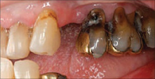
Type 1 - Most common pattern seen. It affects the cervical aspect of the teeth [Figure 1] and extends to the cementoenamel junction. A circumferential decay develops, and crown amputation often occurs
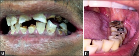
Type 2 - Appears as areas of demineralization on all dental surfaces. Generalized erosions and worn out occlusal and incisal surfaces are seen [Figure 2]
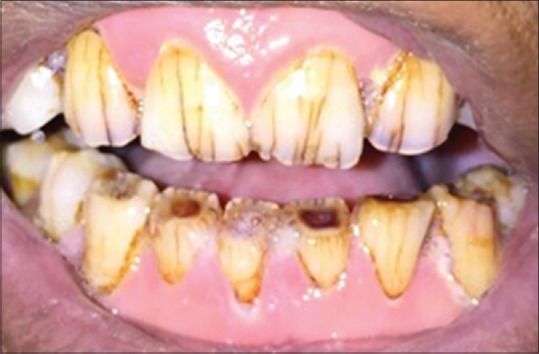
Type 3 - Least common pattern. Seen as color changes in the dentin. The crown becomes dark brown-black and occlusal and incisal wear [Figure 3] can be seen.[22,23,24,25]
Figure 1.

Type 1 are lesions affecting the cervical aspect of the teeth and extending along the cementoenamel junction
Figure 2.

(a) Type 2 presents with demineralized and worn occlusal surfaces. (b) Type 2 presents with demineralized and worn occlusal surfaces
Figure 3.

Type 3 lesions present as color changes in the dentin. The crown is dark brown-black, along with occlusal wear
Until now, no microscopic differences between initial radiation carious lesions and healthy incipient lesions have been recorded. This similarity is true for histological features for both enamel[26,27] and dentin,[28] and for initial reactions regarding clinical demineralization[29,30] and remineralization.[31]
MANAGEMENT OF RADIATION CARIES
Management of radiation caries includes management of xerostomia and that of radiation-induced dental caries.
Preventive measures prior to radiation therapy
A complete dental examination (clinical examination and full mouth radiographs), diagnosis, and treatment should be done before the start of radiotherapy. A complete examination of the mucosa, dentition, and periodontium should be done. Teeth vitality should be assessed. Restoration of carious lesion, endodontic therapy, and recontouring of restorations should be done prior to initiation of radiotherapy to prevent any future complications. Teeth showing severe pulpal or periodontal infection should be extracted in the preradiation phase so as to reduce the risk of osteoradionecrosis. A thorough dental prophylaxis should be performed.[32]
The patient should be given preventive home care instructions that include rigorous oral hygiene (including interdental techniques such as flossing), daily self-application of topical fluoride, restricted intake of cariogenic foods, and remineralizing mouth rinse solutions or artificial saliva preparations. Daily topical 1.0% sodium fluoride gel application by means of custom-made fluoride carriers is recommended for reducing caries occurrence.[33,34] The classic study by Dreizen et al.[35] showed an application of a 1% neutral sodium fluoride gel applied daily in custom trays could significantly reduce caries in irradiated patients. Neutral fluoride containing mouth rinses have also proved to be beneficial in preventing caries occurrence.
Besides fluorides, other alternatives have been studied. A clinical trial compared the caries preventive efficacy of a mouth rinse solution containing casein derivatives coupled with calcium phosphate (CD-CP) with a 0.05% sodium fluoride mouth rinse. It was found CD-CP preparations hold promise as caries preventive agents for individuals with dry mouth. Similarly, the efficacy of remineralizing toothpastes (which also deliver soluble calcium and phosphate ions) was recently investigated, and it was concluded that it may prevent root caries in irradiated patients.[36,37]
Prevention of xerostomia
Salivary gland sparing RT, cytoprotective agents, preservation by stimulation with cholinergic muscarinic agonists (pilocarpine, cevimeline), and the surgical transfer of submandibular glands according to Management Guidelines and Quality of Life Recommendations according to (the American Society of Clinical Oncology) clinical practice guidelines were presented by Brennan et al. (Oral Care Study Group 2010).[38]
Salivary-sparing radiation technique
Salivary output has been shown to increase overtime in patients receiving parotid-sparing IMRT instead of conventional radiotherapy technique as IMRT restricts radiation exposure to healthy structures adjacent to the radiation targets.[39]
Cytoprotective drugs
Radioprotection can be achieved by the use of certain drugs (Amifostine [WR-2721, EthyolR]) that accumulates in salivary gland tissue and makes it less sensitive to radiation damage. The drug enters the bloodstream, where it is rapidly hydrolyzed by endothelial alkaline phosphatase and is converted to its active form WR-1065. The drug then enters cells and nuclei and acts as a scavenger against free radicals and prevents radiation damage to DNA.[40] It was seen in a clinical study that incidence of grade >2 acute xerostomia significantly reduced from 78% to 51% and chronic xerostomia from 57% to 34% after administration of 200 mg/m2 of the drug daily before each fraction.[41] Amifostine administration produces mild to severe adverse toxicity events including nausea, vomiting (generally mild), and transient hypotension. Some other issues associated are the costs of the therapy and logistic problems as the drug needs to administered immediately prior to each RT session.
Submandibular gland transfer
The usual radiation portals in the treatment of HNC deliver 60–65 Gy to the major salivary glands. However, the submental region receives only scatter radiation amounting to 5% of the total dose. Surgical techniques were introduced in the early 1980s to spare salivary glands from head and neck radiotherapy.[42] The procedure involves the transfer of a single submandibular salivary gland into the submental space, while pedicled on the facial artery, facial vein, and submandibular ganglion.[43] It can be done only in patients with clinically negative cervical lymph nodes using the gland on the contralateral side of the primary tumor and is, therefore, not appropriate for all patients.
Management during and after radiotherapy
A good oral hygiene should be maintained throughout the treatment. It includes brushing 2–4 times daily with a soft-bristled toothbrush, daily flossing. To control for plaque accumulation, chlorhexidine mouthwashes should be continued in conjunction with and after normal daily tooth brushing. Fluoride prophylaxis with custom-made carriers and high concentrated fluorides (5000 ppm) should be maintained.[11]
Salivary substitutes to relieve symptoms and sialogogic agents to stimulate saliva can be used.[44] Sialogogues: Unstimulated whole salivary flow rates significantly increased over 3 and 6 months in patients who received pilocarpine (dose more than 2.5 mg 3 times daily for 8–12 weeks),[45] although there were no significant differences in xerostomia.[46] A study indicated that the efficacy of oral pilocarpine was dependent on the dose distributed to the gland.[47] Contraindications include asthma, iritis, and glaucoma. Caution is advised in patients with chronic obstructive pulmonary disease and cardiovascular disease. A newer muscarinic agonist, cevimeline, when administered 30–45 mg 3 times daily for 52 weeks produced very few adverse effects, increased unstimulated but not stimulated saliva.[48] Lemon candy can be sucked to increase the amount of whole saliva secretion and hence improve oral dryness. Sugar-free gums containing xylitol may stimulate salivary flow, buffering, sugar clearance, and can prevent dental decay.[49]
Oral mucosal lubricants/saliva substitutes are the treatment of choice for patients who do not respond to pharmacological gustatory or masticatory stimulation. Saliva substitutes are based on different substances, including animal mucin, carboxymethyl-cellulose, xanthan gum, and aloe vera. All may relieve xerostomia, but a common major disadvantage is the, generally, short duration of relief they provide.
Manual acupuncture using auricular points and, in some cases, supplemented with electrostimulation is the most well-described method for providing relief from xerostomia.
It is administered twice weekly for 6 weeks, xerostomia problems significantly improved, and unstimulated whole saliva flow rates increased.[50]
Stem cell replacement therapy may be a good option to treat radiotherapy-induced hyposalivation, but a better understanding of the mechanism is still needed.[51]
After completion of radiotherapy, frequent follow-up appointments should be scheduled for patients. Scaling and root planning are done under proper antibiotic coverage if proper oral hygiene is not maintained by the patient. Carious lesions are restored immediately. Dental extractions after irradiation should be avoided if possible. Consequently, endodontic therapy should be the treatment of choice in many cases[52] and has been shown to be a viable alternative to exodontia since traumatic injury will be kept to a minimum thus reducing the risk of osteoradionecrosis.[53,54]
Unfortunately, it is not always possible to prevent the development of radiation caries. The restoration of radiation caries can be extremely challenging due to difficult access to cervical lesions leading to incomplete excavation of caries.
Further, the cavity preparation can be difficult to define and might provide little mechanical retention.[55] In addition to technical issues, selection of the most appropriate restorative material is difficult due to the challenging oral environment found in irradiated patients. Ideally, the chosen material should demonstrate appropriate adhesion, prevent secondary caries, and resist dehydration and acid erosion. McComb et al.[56] confirmed the effectiveness of fluoride-releasing materials in the prevention of recurrent caries in irradiated patients. Composite resins have been proven to prevent in vitro recurrent decay, and retention of these materials has been demonstrated even for long periods.[57] However, when time is limited, glass ionomer cements seem to be effective temporary treatments.[55,56] Hu et al.[55] showed glass ionomers can prevent secondary caries development, even when restorations were lost. Moreover, glass ionomers appear to offer satisfactory handling, adhesion, and physical properties. However, lack of salivary buffering in xerostomic patients may lead to a reduction of normal plaque pH and in turn lead to the formation of hydrofluoric acid and erosion of the glass ionomer.[56]
Patients should be made aware of the importance of maintaining good oral hygiene. Patients should be instructed to use custom carrier trays for application of fluoride or chlorhexidine gels throughout life. It is imperative that the patient should be kept under supervision to reduce the incidence of radiation caries.
CONCLUSION
Radiotherapy leads to changes in dentition, saliva and oral microflora of HNC patients. Radiation caries has multifactorial etiology, but hyposalivation remains the primary cause. Therefore, radiation caries could be prevented by salivary gland sparing, or prevention is achieved with comprehensive dental care before, during, and after RT. Motivation of patients, adequate plaque control, stimulation of salivary flow, and fluoride use are essential to reduce the incidence of radiation caries and to improve the quality of life of HNC patients.
Financial support and sponsorship
Nil.
Conflicts of interest
There are no conflicts of interest.
REFERENCES
- 1.Wilken R, Veena MS, Wang MB, Srivatsan ES. Curcumin: A review of anti-cancer properties and therapeutic activity in head and neck squamous cell carcinoma. Mol Cancer. 2011;10:12. doi: 10.1186/1476-4598-10-12. [DOI] [PMC free article] [PubMed] [Google Scholar]
- 2.Beech N, Robinson S, Porceddu S, Batstone M. Dental management of patients irradiated for head and neck cancer. Aust Dent J. 2014;59:20–8. doi: 10.1111/adj.12134. [DOI] [PubMed] [Google Scholar]
- 3.Sciubba JJ, Goldenberg D. Oral complications of radiotherapy. Lancet Oncol. 2006;7:175–83. doi: 10.1016/S1470-2045(06)70580-0. [DOI] [PubMed] [Google Scholar]
- 4.Naidu MU, Ramana GV, Rani PU, Mohan IK, Suman A, Roy P. Chemotherapy-induced and/or radiation therapy-induced oral mucositis–complicating the treatment of cancer. Neoplasia. 2004;6:423–31. doi: 10.1593/neo.04169. [DOI] [PMC free article] [PubMed] [Google Scholar]
- 5.Fattore L, Rosenstein HE, Fine L. Dental rehabilitation of the patient with severe caries after radiation therapy. Spec Care Dentist. 1986;6:258–61. doi: 10.1111/j.1754-4505.1986.tb01585.x. [DOI] [PubMed] [Google Scholar]
- 6.Aguiar GP, Jham BC, Magalhães CS, Sensi LG, Freire AR. A review of the biological and clinical aspects of radiation caries. J Contemp Dent Pract. 2009;10:83–9. [PubMed] [Google Scholar]
- 7.Pfister DG, Spencer S, Brizel DM, Burtness B, Busse PM, Caudell JJ, et al. Head and neck cancers, Version 2.2014. Clinical practice guidelines in oncology. J Natl Compr Canc Netw. 2014;12:1454–87. doi: 10.6004/jnccn.2014.0142. [DOI] [PubMed] [Google Scholar]
- 8.Epstein JB, Chin EA, Jacobson JJ, Rishiraj B, Le N. The relationships among fluoride, cariogenic oral flora, and salivary flow rate during radiation therapy. Oral Surg Oral Med Oral Pathol Oral Radiol Endod. 1998;86:286–92. doi: 10.1016/s1079-2104(98)90173-1. [DOI] [PubMed] [Google Scholar]
- 9.Kaluzny J, Wierzbicka M, Nogala H, Milecki P, Kopec T. Radiotherapy induced xerostomia: Mechanisms, diagnostics, prevention and treatment – evidence based up to 2013. Otolaryngol Pol. 2014;68:1–14. doi: 10.1016/j.otpol.2013.09.002. [DOI] [PubMed] [Google Scholar]
- 10.Devi S, Singh N. Dental care during and after radiotherapy in head and neck cancer. Natl J Maxillofac Surg. 2014;5:117–25. doi: 10.4103/0975-5950.154812. [DOI] [PMC free article] [PubMed] [Google Scholar]
- 11.Kielbassa AM, Hinkelbein W, Hellwig E, Meyer-Lückel H. Radiation-related damage to dentition. Lancet Oncol. 2006;7:326–35. doi: 10.1016/S1470-2045(06)70658-1. [DOI] [PubMed] [Google Scholar]
- 12.Keene HJ, Daly T, Brown LR, Dreizen S, Drane JB, Horton IM, et al. Dental caries and Streptococcus mutans prevalence in cancer patients with irradiation-induced xerostomia: 1-13 years after radiotherapy. Caries Res. 1981;15:416–27. doi: 10.1159/000260547. [DOI] [PubMed] [Google Scholar]
- 13.Springer IN, Niehoff P, Warnke PH, Böcek G, Kovács G, Suhr M, et al. Radiation caries – Radiogenic destruction of dental collagen. Oral Oncol. 2005;41:723–8. doi: 10.1016/j.oraloncology.2005.03.011. [DOI] [PubMed] [Google Scholar]
- 14.Squier CA. Oral complications of cancer therapies. Mucosal alterations. NCI Monogr. 1990;9:169–72. [PubMed] [Google Scholar]
- 15.Grötz KA, Duschner H, Kutzner J, Thelen M, Wagner W. New evidence for the etiology of so-called radiation caries. Proof for directed radiogenic damage od the enamel-dentin junction. Strahlenther Onkol. 1997;173:668–76. doi: 10.1007/BF03038449. [DOI] [PubMed] [Google Scholar]
- 16.De Moor R. Direct and indirect effects of medication (including chemotherapy) and irradiation on the pulp. Rev Belge Med Dent. 2000;55:321–33. [PubMed] [Google Scholar]
- 17.Reed R, Xu C, Liu Y, Gorski JP, Wang Y, Walker MP. Radiotherapy effect on nano-mechanical properties and chemical composition of enamel and dentine. Arch Oral Biol. 2015;60:690–7. doi: 10.1016/j.archoralbio.2015.02.020. [DOI] [PMC free article] [PubMed] [Google Scholar]
- 18.Walker MP, Wichman B, Cheng AL, Coster J, Williams KB. Impact of radiotherapy dose on dentition breakdown in head and neck cancer patients. Pract Radiat Oncol. 2011;1:142–148. doi: 10.1016/j.prro.2011.03.003. [DOI] [PMC free article] [PubMed] [Google Scholar]
- 19.Del Regato JA. Dental lesions observed after Roentgen therapy in cancer of the buccal cavity, pharynx and larynx. Am J Roentgenol. 1939;42:404–10. [Google Scholar]
- 20.Frank RM, Herdly J, Philippe E. Acquired dental defects and salivary gland lesions after irradiation for carcinoma. J Am Dent Assoc. 1965;70:868–83. doi: 10.14219/jada.archive.1965.0220. [DOI] [PubMed] [Google Scholar]
- 21.Kielbassa AM. Die Radiatio im Kopf-/Halsbereich, Auswirkungen aufdie Kariesentstehung. In: Kielbassa AM, editor. Strahlentherapie im Kopf- und Halsbereich. Implikationen für Zahnärzte, HNO-Ærzte und Radiotherapeuten. Hannover: Schlütersche; 2004. [Google Scholar]
- 22.Vissink A, Jansma J, Spijkervet FK, Burlage FR, Coppes RP. Oral sequelae of head and neck radiotherapy. Crit Rev Oral Biol Med. 2003;14:199–212. doi: 10.1177/154411130301400305. [DOI] [PubMed] [Google Scholar]
- 23.Jham BC, da Silva Freire AR. Oral complications of radiotherapy in the head and neck. Braz J Otorhinolaryngol. 2006;72:704–8. doi: 10.1016/S1808-8694(15)31029-6. [DOI] [PMC free article] [PubMed] [Google Scholar]
- 24.Kielbassa AM, Hinkelbein W, Hellwig E, Meyer-Lückel H. Radiation-related damage to dentition. Lancet Oncol. 2006;7:326–35. doi: 10.1016/S1470-2045(06)70658-1. [DOI] [PubMed] [Google Scholar]
- 25.Whitmyer CC, Waskowski JC, Iffland HA. Radiotherapy and oral sequelae: Preventive and management protocols. J Dent Hyg. 1997;71:23–9. [PubMed] [Google Scholar]
- 26.Jongebloed WL, Gravenmade EJ, Retief DH. Radiation caries. A review and SEM study. Am J Dent. 1988;1:139–46. [PubMed] [Google Scholar]
- 27.Jansma J, Vissink A, Jongebloed WL, Retief DH, Johannes 's-Gravenmade E. Natural and induced radiation caries: A SEM study. Am J Dent. 1993;6:130–6. [PubMed] [Google Scholar]
- 28.Kielbassa AM, Schaller HG, Hellwig E. Qualitative Befunde bei in situ erzeugter Initialkaries in tumortherapeutisch bestrahltem Dentin. Eine kombiniert rasterelektronenmikroskopische und mikroradiographische Studie. Acta Med Dent Helv. 1998;3:161–8. [Google Scholar]
- 29.Kielbassa AM, Schendera A, Schulte-Mönting J. Microradiographic and microscopic studies on in situ induced initial caries in irradiated and nonirradiated dental enamel. Caries Res. 2000;34:41–7. doi: 10.1159/000016568. [DOI] [PubMed] [Google Scholar]
- 30.Kielbassa AM. In situ induced demineralization in irradiated and non-irradiated human dentin. Eur J Oral Sci. 2000;108:214–21. doi: 10.1034/j.1600-0722.2000.108003214.x. [DOI] [PubMed] [Google Scholar]
- 31.Kielbassa AM, Hellwig E, Meyer-Lueckel H. Effects of irradiation on in situ remineralization of human and bovine enamel demineralized in vitro. Caries Res. 2006;40:130–5. doi: 10.1159/000091059. [DOI] [PubMed] [Google Scholar]
- 32.Anil S, Philip T, Madhu K, Beena VT, Vijayakumar T. Radiation carries – A rationale approach towards its preventional and management. J Indian Dent Assoc. 1993;64:9–12. [Google Scholar]
- 33.Daly TE, Drane JB. Prevention and management of dental problems in irradiated patients. J Am Soc Prev Dent. 1976;6:21–5. [Google Scholar]
- 34.Horiot JC, Schraub S, Bone MC, Bain Y, Ramadier J, Chaplain G, et al. Dental preservation in patients irradiated for head and neck tumours: A 10-year experience with topical fluoride and a randomized trial between two fluoridation methods. Radiother Oncol. 1983;1:77–82. doi: 10.1016/s0167-8140(83)80009-7. [DOI] [PubMed] [Google Scholar]
- 35.Dreizen S, Brown LR, Daly TE, Drane JB. Prevention of xerostomia-related dental caries in irradiated cancer patients. J Dent Res. 1977;56:99–104. doi: 10.1177/00220345770560022101. [DOI] [PubMed] [Google Scholar]
- 36.Hay KD, Thomson WM. A clinical trial of the anticaries efficacy of casein derivatives complexed with calcium phosphate in patients with salivary gland dysfunction. Oral Surg Oral Med Oral Pathol Oral Radiol Endod. 2002;93:271–5. doi: 10.1067/moe.2002.120521. [DOI] [PubMed] [Google Scholar]
- 37.Papas A, Russell D, Singh M, Kent R, Triol C, Winston A. Caries clinical trial of a remineralising toothpaste in radiation patients. Gerodontology. 2008;25:76–88. doi: 10.1111/j.1741-2358.2007.00199.x. [DOI] [PubMed] [Google Scholar]
- 38.Brennan MT, Shariff G, Lockhart PB, Fox PC. Treatment of xerostomia: A systematic review of therapeutic trials. Dent Clin North Am. 2002;46:847–56. doi: 10.1016/s0011-8532(02)00023-x. [DOI] [PubMed] [Google Scholar]
- 39.Eisbruch A, Ten Haken RK, Kim HM, Marsh LH, Ship JA. Dose, volume, and function relationships in parotid salivary glands following conformal and intensity-modulated irradiation of head and neck cancer. Int J Radiat Oncol Biol Phys. 1999;45:577–87. doi: 10.1016/s0360-3016(99)00247-3. [DOI] [PubMed] [Google Scholar]
- 40.Bourdin S, Desson P, Leroy G, Rémy PJ, Cuillière JC, Beauvillain C, et al. Prevention of post-irradiation xerostomia by submaxillary gland transposition. Ann Otolaryngol Chir Cervicofac. 1982;99:265–8. [PubMed] [Google Scholar]
- 41.Seikaly H, Jha N, McGaw T, Coulter L, Liu R, Oldring D. Submandibular gland transfer: A new method of preventing radiation-induced xerostomia. Laryngoscope. 2001;111:347–52. doi: 10.1097/00005537-200102000-00028. [DOI] [PubMed] [Google Scholar]
- 42.Ringash J, Warde P, Lockwood G, O’Sullivan B, Waldron J, Cummings B. Postradiotherapy quality of life for head-and-neck cancer patients is independent of xerostomia. Int J Radiat Oncol Biol Phys. 2005;61:1403–7. doi: 10.1016/j.ijrobp.2004.08.001. [DOI] [PubMed] [Google Scholar]
- 43.Burlage FR, Roesink JM, Kampinga HH, Coppes RP, Terhaard C, Langendijk JA, et al. Protection of salivary function by concomitant pilocarpine during radiotherapy: A double-blind, randomized, placebo-controlled study. Int J Radiat Oncol Biol Phys. 2008;70:14–22. doi: 10.1016/j.ijrobp.2007.06.016. [DOI] [PubMed] [Google Scholar]
- 44.Dost F, Farah CS. Stimulating the discussion on saliva substitutes: A clinical perspective. Aust Dent J. 2013;58:11–7. doi: 10.1111/adj.12023. [DOI] [PubMed] [Google Scholar]
- 45.Kaluzny J, Wierzbicka M, Nogala H, Milecki P, Kopec T. Radiotherapy induced xerostomia: Mechanisms, diagnostics, prevention and treatment – Evidence based up to 2013. Otolaryngol Pol. 2014;68:1–14. doi: 10.1016/j.otpol.2013.09.002. [DOI] [PubMed] [Google Scholar]
- 46.Giatromanolaki A, Sivridis E, Maltezos E, Koukourakis MI. Down-regulation of intestinal-type alkaline phosphatase in the tumor vasculature and stroma provides a strong basis for explaining amifostine selectivity. Semin Oncol. 2002;29(6 Suppl 19):14–21. doi: 10.1053/sonc.2002.37356. [DOI] [PubMed] [Google Scholar]
- 47.Brizel DM, Wasserman TH, Henke M, Strnad V, Rudat V, Monnier A, et al. Phase III randomized trial of amifostine as a radioprotector in head and neck cancer. J Clin Oncol. 2000;18:3339–45. doi: 10.1200/JCO.2000.18.19.3339. [DOI] [PubMed] [Google Scholar]
- 48.Chambers MS, Garden AS, Kies MS, Martin JW. Radiation-induced xerostomia in patients with head and neck cancer: Pathogenesis, impact on quality of life, and management. Head Neck. 2004;26:796–807. doi: 10.1002/hed.20045. [DOI] [PubMed] [Google Scholar]
- 49.Edgar WM, Higham SM, Manning RH. Saliva stimulation and caries prevention. Adv Dent Res. 1994;8:239–45. doi: 10.1177/08959374940080021701. [DOI] [PubMed] [Google Scholar]
- 50.Blom M, Dawidson I, Fernberg JO, Johnson G, Angmar-Månsson B. Acupuncture treatment of patients with radiation-induced xerostomia. Eur J Cancer B Oral Oncol. 1996;32B:182–90. doi: 10.1016/0964-1955(95)00085-2. [DOI] [PubMed] [Google Scholar]
- 51.Pringle S, Van Os R, Coppes RP. Concise review: Adult salivary gland stem cells and a potential therapy for xerostomia. Stem Cells. 2013;31:613–9. doi: 10.1002/stem.1327. [DOI] [PubMed] [Google Scholar]
- 52.Kielbassa AM, Schilli K. Betreuung des tumortherapeutischbestrahlten Patientenaus Sicht der Zahnerhaltung. Zahnäztl Mitt. 1997;87:2636–47. [Google Scholar]
- 53.Kielbassa AM, Attin T, Schaller HG, Hellwig E. Endodontic therapy in a postirradiated child: Review of the literature and report of a case. Quintessence Int. 1995;26:405–11. [PubMed] [Google Scholar]
- 54.Lilly JP, Cox D, Arcuri M, Krell KV. An evaluation of root canal treatment in patients who have received irradiation to the mandible and maxilla. Oral Surg Oral Med Oral Pathol Oral Radiol Endod. 1998;86:224–6. doi: 10.1016/s1079-2104(98)90129-9. [DOI] [PubMed] [Google Scholar]
- 55.Hu JY, Li YQ, Smales RJ, Yip KH. Restoration of teeth with more-viscous glass ionomer cements following radiation-induced caries. Int Dent J. 2002;52:445–8. doi: 10.1111/j.1875-595x.2002.tb00640.x. [DOI] [PubMed] [Google Scholar]
- 56.McComb D, Erickson RL, Maxymiw WG, Wood RE. A clinical comparison of glass ionomer, resin-modified glass ionomer and resin composite restorations in the treatment of cervical caries in xerostomic head and neck radiation patients. Oper Dent. 2002;27:430–7. [PubMed] [Google Scholar]
- 57.Gernhardt CR, Koravu T, Gerlach R, Schaller HG. The influence of dentin adhesives on the demineralization of irradiated and non-irradiated human root dentin. Oper Dent. 2004;29:454–61. [PubMed] [Google Scholar]


