Abstract
Introduction:
The role of trace elements in various diseases has been a matter of controversy with various authors reporting on conflicting data. They are receiving much attention in the detection of oral cancer and precancer as they are found to be significantly altered and have an important role in carcinogenesis. Trace elements have been extensively studied in the recent years to assess whether they have any modifying effect in the etiology of oral malignant conditions.
Materials and Methods:
A study was conducted on fifty subjects with clinically diagnosed oral submucous fibrosis (OSMF) and fifty controls with no apparent lesions of the oral mucosa and without any areca nut-related oral habit.
Results:
The level of serum zinc was significantly (P < 0.0001) lower among cases (73.48 ± 24.21) compared with controls (119.48 ± 52.78). However, the serum copper level was significantly (P < 0.0001) higher among cases (155.50 ± 40.13) than controls (100.40 ± 24.52). The level of serum iron was observed to be lower among the cases (66.57 ± 27.76) as compared to controls (94.19 ± 35.70), and the difference was statistically significant.
Conclusion:
It can be concluded from this study that serum zinc, copper, and iron levels could be used as a potential prognostic and diagnostic markers in OSMF patients.
Keywords: Oral submucous fibrosis, precancer, trace elements
INTRODUCTION
Oral submucous fibrosis (OSMF) is a chronic condition of the oral mucosa, first described by Schwartz in 1952 among 5 East African women of Indian origin under the term, “atrophia idiopathica (tropica) mucosae oris.”[1] Trace elements have been studied in the recent years to assess whether they have any modifying effect in the etiology of oral malignant conditions, but relatively less scientific work has been performed in the area of oral premalignant conditions. These can be used as an auxiliary test to clinicopathological diagnosis and/or in combination with other biochemical tests in the diagnosis and prognosis of OSMF. The aim of the study was to determine the serum levels of zinc, copper, and iron in different stages of OSMF and normal healthy subjects and to compare serum levels of zinc, copper, and iron in subjects of OSMF and normal healthy subjects.
MATERIALS AND METHODS
The study was conducted in the Department of Oral Medicine and Radiology of Babu Banarasi Das College of Dental Sciences, Lucknow. Ethical clearance for the study was obtained from the Institutional Ethical Committee. In this study, fifty patients who presented with signs and symptoms suggestive of OSMF and fifty controls were enrolled. Patients who were healthy and well oriented to place, person, and time, either sex aged between 18 and 60 years, a positive history of chewing of areca nut or one of its commercial preparation, burning sensation on eating spicy foods, restricted mouth opening, and the presence of palpable vertical fibrous band, stiffness, and blanching were included in the study. Subjects having any systemic disorder, previous history of treatment for the same condition, history of drug intake containing zinc, copper, and iron, pregnant women were excluded from the study. Khanna and Andrade classification system of OSMF[2] based on interincisal opening was used for the study.
Under aseptic conditions, 5 ml of venous blood was obtained by venipuncture of the median cubital vein, kept standing for 30 min at room temperature. Then, the serum was separated by centrifugation at 3000 rpm for 15 min and preserved in a frozen state at 2–8°C for 5 days until analysis. Serum sample used for the estimation was mixed in appropriate proportion with buffer and color reagents supplied in the estimation kits in the clean glass tubes as per the manufacturer's instructions. The absorbance of these samples was compared with the standard solution provided in the kit using a colorimeter. The data obtained from the procedures will be tabulated and analyzed using statistical methods.
RESULTS
Age distribution
The age distribution of the patients and controls is depicted in Table 1 and Figure 1. More than half of the cases (58%) and controls (84%) were below 30 years. The mean age was almost similar (P > 0.05) in both the groups; thus, both groups were comparable in terms of age.
Table 1.
Age distribution of the patients and controls

Figure 1.
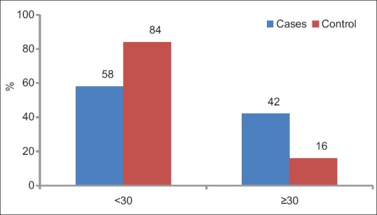
Age distribution of the cases and controls
Sex distribution
The sex distribution of the patients and controls is presented in Table 2 and Figure 2. More than half of the cases (84%) and controls (78%) were males. The percentage of male/female ratio was almost similar (P > 0.05) in both the groups; thus, both groups were comparable in terms of sex.
Table 2.
Sex distribution of the patients and controls

Figure 2.
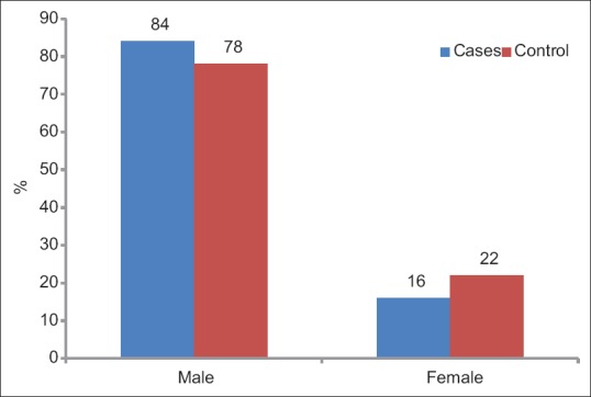
Sex distribution of the cases and controls
Comparison of trace elements
The level of serum zinc was significantly (P < 0.0001) lower among cases (73.48 ± 24.21) compared with controls (119.48 ± 52.78). However, the serum copper level was significantly (P < 0.0001) higher among the cases (155.50 ± 40.13) than controls (100.40 ± 24.52). The level of serum iron was observed to be lower among the cases (66.57 ± 27.76) as compared to controls (94.19 ± 35.70), and the difference was statistically significant [Table 3 and Figure 3].
Table 3.
Comparison of trace elements between cases and controls

Figure 3.
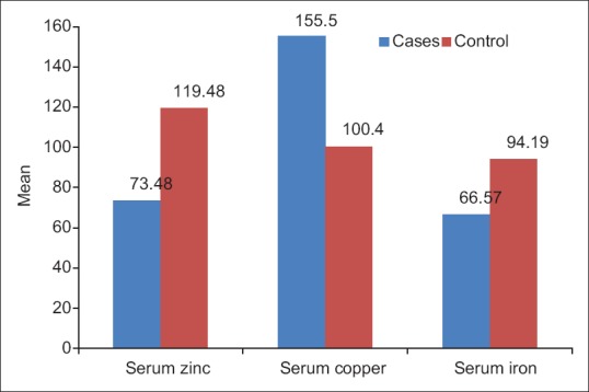
Comparison of trace elements between cases and controls
Comparison of trace elements according to grade
The comparison of trace elements according to grade among the cases is given in Table 4 and Figure 4. Analysis of variance revealed that there was a significant difference in all the trace elements among all the grades. Since there is only one case in the Grade IVa, the post hoc multiple comparison test could not be done to compare the grades.
Table 4.
Comparison of trace elements according to grade among the cases
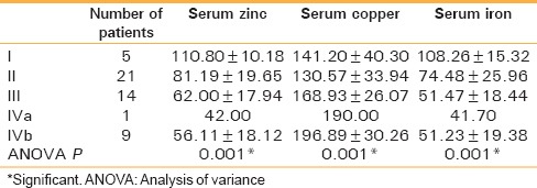
Figure 4.
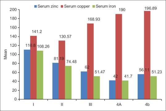
Comparison of trace elements according to grade among the cases
DISCUSSION
OSMF is a well-recognized, potentially malignant condition of the oral cavity. Controlling the devastating, widespread consequences of OSMF requires interventions in at-risk persons ideally before the disease becomes invasive. Detection of the premalignancies and preventing them from malignant transformation seem to be the best available tool in the fight against oral cancer. Very few studies have been conducted to find out the role of different trace elements in oral precancer and cancer. Hence, a comprehensive study has been carried out to estimate levels of serum zinc, copper, and iron in patients with OSMF in the population of Lucknow District.
Age
In the present study, fifty subjects with OSMF were in the age range of 17–55 years with a mean age of 28.64 years. This is comparable to mean age of 28 years observed by Kumar et al.,[3] 28.8 years by Hazarey et al.,[4] Maher et al.,[5] and Borle and Borle.[6]
The age range seen in our study can be attributed to changing lifestyle of youngsters and increased in the usage of Gutkha/pan masala [Table 1 and Figure 1].
Sex
Among the fifty OSMF subjects, 42 were male and 8 were female patients, thus showing an extreme male predominance over female with the ratio of 5.25:1. A similar male predominance was reported by Sinor et al.,[7] Pindborg et al.,[8] Ahmad et al.,[9] and Hazarey et al.[4] [Table 2 and Figure 2].
Male predominance can be related to easy accessibility for males to use these products more frequently than females in our society.
Comparison of trace elements between cases and controls
Serum copper levels in oral submucous fibrosis patients
In our study, the serum copper level was significantly (P < 0.0001) higher among the cases (155.50 ± 40.13) than controls (100.40 ± 24.52). It was similar to the study by Balpande et al.[10] and Shetty et al.[11] [Tables 3, 4 and Figures 3, 4]
Increased serum copper in OSMF can be correlated to copper present in areca nut increases the collagen production in oral fibroblasts by upregulating lysyl oxidase leading to crosslinking of collagen and elastin[11] Trivedy et al. has also reported on the copper-induced mutagenesis through the p53 aberrations in OSMF, which may be critical in the progression of the potentially malignant lesions to squamous cell carcinoma.[12]
Serum zinc levels in oral submucous fibrosis patients
The level of serum zinc was significantly (P < 0.0001) lower among cases (73.48 ± 24.21) compared with controls (119.48 ± 52.78). It was similar to the study done by Paul et al.,[13] Nayak et al.,[14] Varghese et al.,[15] Balpande et al.,[10] Kode and Karjodkar,[16] and Shettar.[17]
This could be because the malignant cells probably require more zinc which is taken up from the serum causing low levels of zinc in it. As there is negative interaction between copper and zinc, an increase in copper level may cause subsequent reduction in zinc level as well.[10]
Serum iron levels in oral submucous fibrosis patients
The serum iron level was observed to lower among the cases (66.57 ± 27.76) as compared to controls (94.19 ± 35.70), and the difference was statistically significant. It was similar to the done by Paul et al.,[13] Balpande et al.,[10] Karthik et al., and[18] Shetty et al.[11] Decreased iron levels in OSMF patients might be due to utilization of iron in collagen synthesis.[10] It has been stated that the decrease in iron content leads to decrease in epithelial vascularity which results in increased penetration of arecoline which leads to fibrosis.
Cytochrome oxidase is an iron-dependent enzyme which is required for the normal maturation of the epithelium. In iron deficiency state, the levels of cytochrome oxidase are low, consequently leading to epithelial atrophy. An atrophic epithelium makes the oral mucosa vulnerable to the soluble irritants.[3]
SUMMARY AND CONCLUSION
In the recent times, although there has been a consistent rise in the number of patients with OSMF, there is still no detailed understanding of the mechanism directly delineating the etiopathogenetic mechanism involved in the formation of the condition. It can be concluded from this study that serum zinc, copper, and iron levels could be used as a potential prognostic and diagnostic markers in OSMF patients. However, as there are controversial reports on the association of OSMF and these trace elements, future studies are anticipated on a larger heterogeneous population to confirm the hypothesis.
Financial support and sponsorship
Nil.
Conflicts of interest
There are no conflicts of interest.
REFERENCES
- 1.Prabhu SR, Wilson DF, Daftary DK, Johnson NW. Oral Diseases in the Tropics. 1st ed. New Delhi: Oxford; 1993. [Google Scholar]
- 2.Khanna JN, Andrade NN. Oral submucous fibrosis: A new concept in surgical management. Report of 100 cases. Int J Oral Maxillofac Surg. 1995;24:433–9. doi: 10.1016/s0901-5027(05)80473-4. [DOI] [PubMed] [Google Scholar]
- 3.Kumar A, Sharma SC, Sharma P, Chandra OM, Singhal KC, Nagar A. Beneficial effect of oral zinc in the treatment of oral submucous fibrosis. Indian J Pharmacol. 1991;23:236–41. [Google Scholar]
- 4.Hazarey VK, Erlewad DM, Mundhe KA, Ughade SN. Oral submucous fibrosis: Study of 1000 cases from central India. J Oral Pathol Med. 2007;36:12–7. doi: 10.1111/j.1600-0714.2006.00485.x. [DOI] [PubMed] [Google Scholar]
- 5.Maher R, Lee AJ, Warnakulasuriya KA, Lewis JA, Johnson NW. Role of areca nut in the causation of oral submucous fibrosis: A case-control study in Pakistan. J Oral Pathol Med. 1994;23:65–9. doi: 10.1111/j.1600-0714.1994.tb00258.x. [DOI] [PubMed] [Google Scholar]
- 6.Borle RM, Borle SR. Management of oral submucous fibrosis: A conservative approach. J Oral Maxillofac Surg. 1991;49:788–91. doi: 10.1016/0278-2391(91)90002-4. [DOI] [PubMed] [Google Scholar]
- 7.Sinor PN, Gupta PC, Murti PR, Bhonsle RB, Daftary DK, Mehta FS, et al. A case-control study of oral submucous fibrosis with special reference to the etiologic role of areca nut. J Oral Pathol Med. 1990;19:94–8. doi: 10.1111/j.1600-0714.1990.tb00804.x. [DOI] [PubMed] [Google Scholar]
- 8.Pindborg JJ, Murti PR, Bhonsle RB, Gupta PC, Daftary DK, Mehta FS. Oral submucous fibrosis as a precancerous condition. Scand J Dent Res. 1984;92:224–9. doi: 10.1111/j.1600-0722.1984.tb00883.x. [DOI] [PubMed] [Google Scholar]
- 9.Ahmad MS, Ali SA, Ali AS, Chaubey KK. Epidemiological and etiological study of oral submucous fibrosis among gutkha chewers of Patna, Bihar, India. J Indian Soc Pedod Prev Dent. 2006;24:84–9. doi: 10.4103/0970-4388.26022. [DOI] [PubMed] [Google Scholar]
- 10.Balpande AR, Sathawane RS. Estimation and comparative evaluation of serum iron, copper, zinc and copper/zinc ratio in oral leukoplakia, submucous fibrosis and squamous cell carcinoma. J Indian Acad Oral Med Radiol. 2010;22:73–6. [Google Scholar]
- 11.Shetty SR, Babu S, Kumari S, Shetty P, Hegde S, Karikal A. Role of serum trace elements in oral precancer and oral cancer – A biochemical study. J Cancer Res Treat. 2013;1:1–3. [Google Scholar]
- 12.Trivedy CR, Warnakulasuriya KA, Peters TJ, Senkus R, Hazarey VK, Johnson NW. Raised tissue copper levels in oral submucous fibrosis. J Oral Pathol Med. 2000;29:241–8. doi: 10.1034/j.1600-0714.2000.290601.x. [DOI] [PubMed] [Google Scholar]
- 13.Paul RR, Chatterjee J, Das AK, Dutta SK, Roy D. Zinc and iron as bioindicators of precancerous nature of oral submucous fibrosis. Biol Trace Elem Res. 1996;54:213–30. doi: 10.1007/BF02784433. [DOI] [PubMed] [Google Scholar]
- 14.Nayak AG, Chatra L, Shenai KP. Analysis of copper and zinc levels in the mucosal tissue and serum of oral submucous fibrosis patients. World J Dent. 2010;1:75–80. [Google Scholar]
- 15.Varghese I, Sugathan CK, Balasubramoniyan G, Vijayakumar T. Serum copper and zinc levels in premalignant and malignant lesions of the oral cavity. Oncology. 1987;44:224–7. doi: 10.1159/000226482. [DOI] [PubMed] [Google Scholar]
- 16.Kode MA, Karjodkar FR. Estimation of the serum and the salivary trace elements in OSMF patients. J Clin Diagn Res. 2013;7:1215–8. doi: 10.7860/JCDR/2013/5207.3023. [DOI] [PMC free article] [PubMed] [Google Scholar]
- 17.Shettar SS. Estimation of serum copper and zinc levels in patients with oral submucous fibrosis. J Indian Acad Oral Med Radiol. 2010;22:193. [Google Scholar]
- 18.Karthik H, Nair P, Gharote HP, Agarwal K, Ramamurthy Bhat G, Kalyanpur Rajaram D. Role of hemoglobin and serum iron in oral submucous fibrosis: A clinical study. ScientificWorldJournal 2012. 2012 doi: 10.1100/2012/254013. 254013. [DOI] [PMC free article] [PubMed] [Google Scholar]


