Abstract
This article addresses recent advances in the application of microscopy techniques to characterize crystallization processes as they relate to biomineralization and bio-inspired materials synthesis. In particular, we focus on studies aimed at revealing the role organic macromolecules and functionalized surfaces play in modulating the mechanisms of nucleation and growth. In nucleation studies, we explore the use of methods such as in situ transmission electron microscopy, atomic force microscopy, and cryogenic electron microscopy to delineate formation pathways, phase stabilization, and the competing effects of free energy and kinetic barriers. In growth studies, emphasis is placed on understanding the interactions of macromolecular constituents with growing crystals and characterization of the internal structures of the resulting composite crystals using techniques such as electron tomography, atom probe tomography, and vibrational spectromicroscopy. Examples are drawn from both biological and bio-inspired synthetic systems.
Keywords: Biomimetic (assembly), crystal, nucleation and growth, scanning probe microscopy, transmission electron microscopy
Introduction
One of the universal characteristics of biomineralizing systems is that macromolecular constituents, such as proteins and polysaccharides, are found in close association with the mineral constituents.1,2 Some of these organic components are water soluble, but others assemble to create an insoluble scaffold within which the minerals form. This intimate relationship between the matrix and mineral, along with the unusual morphologies of biominerals and the common occurrence of metastable mineral phases, suggests that the organic matrix actively controls mineral nucleation and growth. Moreover, significant quantities of the organic matrix are typically occluded within the resulting hierarchically-structured tissues (discussed further in this issue by Schenk and Kim).3 This composite architecture endows biominerals with remarkable mechanical properties and, as a result, during the past few decades, biomineral systems have inspired developments in materials synthesis of new composite materials.4,5 However, truly emulating biological processes for novel material creation has yet to be fully realized, because both the mechanisms by which the macromolecular matrix assembles and mineralizes and the principles underlying the structure-function relationships of mineralized tissues are poorly understood. Recent advances in the application of microscopy techniques to define both the processes of matrix assembly and mineralization and the hierarchical distribution of matrix and mineral are beginning to fill the gaps in our understanding of both formation and properties. Here we review some of these advances, using examples from biological as well as bio-inspired systems to illustrate the insights they have enabled. Table I provides a summary of the microscopy techniques discussed, including their benefits and limitations within the scope of analyzing biominerals and bio-inspired systems.
Table I.
Summary of the various techniques discussed and brief summary of advantages/disadvantages.
| Technique | Benefits | Limitations | References |
|---|---|---|---|
| AFM (ATOMIC FORCE MICROSCOPY) |
|
|
3–7, 9, 13, 14, 27, 71–74 |
| CRYOEM |
|
|
19–21, 23, 24, 75–78 |
| LIQUID CELL TEM |
|
|
25–28, 79, 80 |
| (S)TEM AND ELECTRON TOMOGRAPHY |
|
|
38, 39, 41, 42, 44–46, 81 |
| IMMUNOGOLD PROTEIN LABELING |
|
|
34–37 |
| APT (ATOM PROBE TOMOGRAPHY) |
|
|
47–52 |
| X-RAY SCATTERING BASED MICROSCOPY |
|
|
53–55, 57, 59, 31, 82–84 |
| VIBRATIONAL SPECTRO-MICROSCOPY |
|
|
62–66 |
Visualizing matrix assembly and mineral formation
What mechanisms and energetic factors control nucleation and growth?
In recent years, investigations of both matrix assembly and mineralization have been performed to better understand the in vivo mechanisms that control these processes. In situ methods have been particularly important, because dynamic behavior occurs in response to differences in energy states and the barriers that separate those states; thus, such studies provide an opportunity to probe the energy landscape across which matrix assembly and mineralization take place. For both processes, the important length scales are from the molecular scale to tens of nanometers. Consequently, atomic force microscopy (AFM) and transmission electron microscopy (TEM) have been the most significant techniques used, though the information gained is typically augmented with other types of data, both in situ and ex situ.
In situ AFM: Real-time imaging of assembly processes on surfaces
AFM is a type of scanning probe microscopy that uses a sharp tip on the end of a cantilever to sense changes in sample topography. When the tip is brought to within the range of the interatomic potential between the tip and surface, variations in the potential with position lead to vertical deflections of the cantilever. Thus the topography of the surface can be mapped by measuring the deflection of cantilever as the tip is raster-scanned over surface. Typically, this deflection is measured by reflecting a laser from the top of the cantilever onto a photodiode array, giving a vertical resolution of less than an Angstrom.
AFM can be applied to both organic matrices and mineral surfaces exposed to fluid, which is either static or flowing.6,7 Consequently, it enables direct observations of matrix assembly on substrates,8–10 mineral nucleation on organic matrices,11,12 and post-nucleation growth and interaction with organic constituents.7,13–15 These measurements can be complemented by a number of other techniques to obtain a comprehensive picture of chemical interactions, energetic drivers, and mechanisms of formation. The first, cryogenic electron microscopy (cryoEM), is a form of transmission electron microscopy (TEM) in which a sample of solution is frozen so rapidly in liquid N2 or liquid ethane that it forms a thin layer of vitrified water. Cryo-EM enables the observation of specimens in their native environment without any staining or fixation, thus providing high-resolution structural information. The second, in situ dynamic force spectroscopy (DFS) is a special application of AFM that records the force required to break the bond between a functionalized AFM tip and a surface. When this rupture force is measured as a function of the bond extension rate, the resulting relationship provides characteristic parameters of the intermolecular and mineral-matrix bonds such as the binding free energy per molecule. Vibrational and electronic spectroscopy can be used to the reveal the functional groups responsible for the observed changes in free energies and barriers, and molecular simulations can be employed to test proposed mechanisms and determine underlying structural reasons for the dominant interactions.
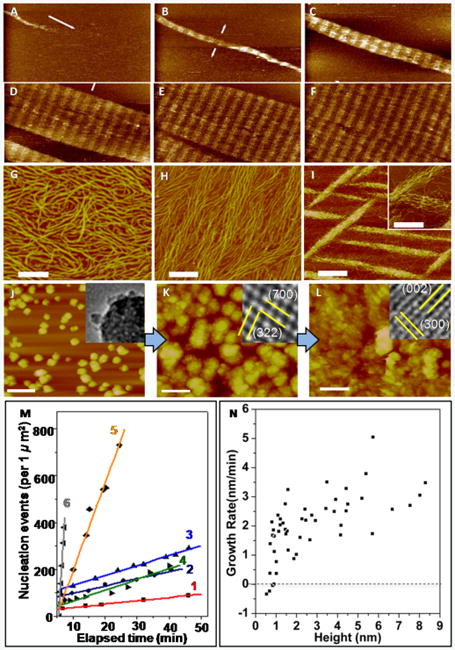
AFM has been used to investigate assembly of protein matrices by introducing aqueous solutions of protein into a sample chamber referred as a fluid cell, which contains substrates that promote assembly.8,9 In vivo, collagen molecules, which form the organic matrix of bone, align and intertwine to form microfibrils containing “hole zones,” where there are gaps between the N- and C-termini of successive collagen molecules.16 Observations of collagen assembly on mica revealed formation of ordered fibrils via two distinct steps that resulted in the same periodicity observed in collagen fibrils formed in vivo (Figure 1A–F).8 First, three single strand collagen molecules associate with one another to form 1.5 nm-high triple-helices called topocollagen molecules. These then assembled into ordered, 3 nm-high microfibrils, which formed the building blocks of the larger scale fibrils. Other studies recorded development of alternative architectures that depended strongly on the choice of pH and salt concentration (Figure 1G–I). For example, while the ordered structure seen in Figure 1A-F is obtained at pH 9.0 for K+ concentrations of 200 mm and above, at pH 4.0 the architecture evolves from a monolayer of randomly oriented molecules to a monolayer of co-aligned molecules to 3D bundles of co-aligned molecules as the K+ concentration is increased from 100 to 300 mM.10 Comparing the in situ results with coarse-grained molecular models of assembly provided insights into the energetic underpinnings of collagen architecture.10 Predictions based on variations of the relative strengths of the inter-collagen and collagen-substrate binding through a periodic set of strong and weak inter-collagen binding sites gave the observed evolution in collagen architecture.
Figure 1.

In situ AFM can be used to investigate matrix self-assembly and mineralization. (A)–(F) Self-assembly of Type I collagen on mica into an ordered array with periodicity of 67 nm found in natural collagen. Times (min) are: (A) 12.9, (B) 25.8, (C) 43.0, (D) 55.9, (E) 60.2, and (F) 77.4. (G)–(I) Architecture of collagen on mica at pH 4.0 for K+ concentrations (mMol) of (G) 100 (H) 200 (I) 300. (J)–(L) Nucleation of calcium phosphate on collagen at a solute concentration above the ACP solubility limit. (J) ACP at 24 min then transforms to octacalcium phosphate at 64 min (K) and then HA at 98 min (L). Time 0 corresponds to the moment solution was injected into the atomic force microscopy fluid cell. Insets show transmission electron microscopy images collected on samples taken from the three stages of development. (M) Dependence of nucleation rate on time at six different supersaturations σi. Details of the supersaturation values can be found in Reference 8. Analysis of the data gives interfacial energies αi of αACP = 40 mJ • m−2 and αHA = 90 mJ • m−2. (N) Relationship between growth rate and particle height on surface of collagen with σHA = 3.31, σOCP = 1.71, σACP = –0.02. A critical size of 0.7 nm is determined from the zero crossing of the average growth rate (dashed line) All scale bars are 200 nm except in (I) where it is 500 nm in the main image only. Adapted and reprinted from 8 with permission from Elsevier. Reprinted with permission from Macmillan Publishers Ltd 11
Mineral nucleation on organic matrices can also be visualized by AFM.11,12 Organic matrices are first assembled on flat substrates. Aqueous solutions of the mineral phase are then passed through the fluid cell and formation of mineral nuclei is observed (Figure 1J–L). If the nucleation process is arrested by switching from flow of reagents to a liquid in which the mineral has no solubility, such as alcohol, the sample can be collected and analyzed by TEM or Raman spectroscopy to determine the mineral phase at a single point in time.11 The use of DFS to measure the binding energy of the protein to single crystal substrates of the mineral allows one to correlate the interfacial energy with matrix-mineral binding free energy.12,17,18
In bone, nonstoichiometric hydroxyapatite (Ca10(PO4)6(OH)2; HA) crystals form within a highly organized matrix of collagen.19 There is an ongoing debate regarding the nucleating potential of collagen fibrils in the absence of any other proteins.20–22 Recently, AFM measurements of heterogeneous calcium phosphate nucleation rates on collagen showed that solutions stable for days in the absence of collagen produced nuclei in hours or less on collagen matrices.11 At supersaturations below the solubility limit of amorphous calcium phosphate (ACP), HA formed directly, but when the concentration was raised to slightly above the solubility limit of ACP, the amorphous phase formed first (Figure 1J) before transforming to octacalcium phosphate (Ca8H2(PO4)6 • 5H2O; OCP) (Figure 1K) and, finally, HA (Figure 1L). Analysis of nucleation rates (Figure 1M) gave interfacial energies two to three times less than those estimated for homogeneous nucleation and barriers that were smaller by more than an order of magnitude. Analysis of the size distribution of particles that, on average, grew versus those that dissolved (Figure 1N), provided an estimate of the critical nucleus size. However, neither the change from HA nucleation to ACP nucleation nor the small values of critical size could be understood within classical nucleation theory. Comparison to cryoEM and titration analyses revealed a formation pathway involving assembly of multi-ion complexes, which offered a rationale for the unexpected formation of ACP precursor particles and the low value of the critical size.
Cryogenic Electron Microscopy (cryoEM) and tomography: High-resolution, three-dimensional (3D) imaging of hydrated samples
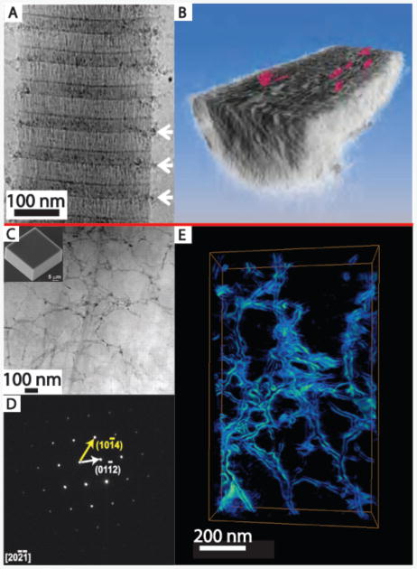
CryoEM techniques23 can provide insights into tissue structures at close to native state.24 They can also be used to provide quasi-time resolved studies of nucleation and growth processes.25–27 These studies can complement in situ AFM studies, and provide a more detailed picture of mineralization occurring within an organic matrix. For example, cryoEM studies of synthetic intrafibrillar HA formation in collagen fibrils revealed the early stages of infiltration of ACP particles at the hole zones in the fibrils (Figure 2A,B).28 CryoEM tomography of the fully mineralized fibrils revealed embedded nm-thick plate-shaped HA crystals with their c-axis parallel to the fibril axis. Similar cryoEM approaches have been taken to study other composite systems such as calcium carbonate nucleation under Langmuir–Blodgett monolayers.25,29 In these studies, amorphous calcium carbonate (ACC) forms and then transforms to either calcite (in the absence of the monolayer) or vaterite and then calcite (with the monolayer present) by dissolution/reprecipitation.25
Figure 2.

(A, B) Cryogenic electron microscopy yields insights into the mineralization of hydrated samples. (A) CryoTEM of a collagen fibril (from horse tendon) with amorphous calcium phosphate (ACP) particles collecting near the hole regions of the collagen (white arrows) after mineralization for 24 hrs in buffered CaCl2, KPO4, and 10 μg/mL of pAsp. (B) Tomographic reconstruction of the fibril after the apatite has been fully crystallized (72 hrs in buffered solution) reveals platelets (colored red) of mineral within the fibril itself. (C)–(E) Electron microscopy and tomography reveal details of incorporated macromolecular additives. (C) ADF-scanning transmission electron microscopy (STEM) of a focused ion beam section of a calcite crystal (scanning electron microscopy, inset) grown in a 1 w/v% agarose gel. Despite well-developed rhombohedral crystal, agarose gel fibers are clearly visible (darker contrast) randomly distributed within the thin section. (D) Selected-area electron diffraction of the area imaged in (C) demonstrating that the gel-grown crystal diffracts electrons as a single crystal. (E) ADF-STEM tomographic reconstruction of an area similar to (C) which emphasizes the network of agarose fibers incorporated within the crystal. Adapted and reprinted by permission from Macmillan Publishers Ltd: 28. Also from 55 with permission from AAAS
In situ TEM: Direct observation of nucleation in bulk solutions and within organic matrices
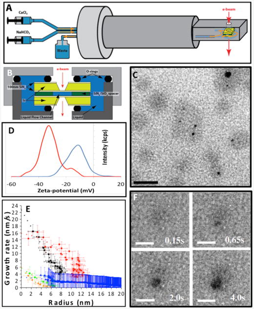
When nucleation occurs in bulk solution or within a biomimetic matrix, AFM cannot be used to observe the nucleation process. In situ liquid phase TEM provides a novel technique in these instances.30–33 A sealed liquid cell on a specialized TEM holder (Figure 3A,B) allows for mixing reagents at the entrance to the cell and flow of the mixture through the cell.34 Alternatively, the cell can be filled with one reagent through one of the two flow lines, and the second reagent can be introduced by gaseous diffusion through the second line.35 To observe nucleation within a macromolecular matrix, the macromolecules are added to one of the two reagents and transferred into the cell prior to introduction of the second reagent. The appearance of mineral nuclei and the spatial relationship to an organic matrix can then be observed by TEM imaging. Electron diffraction is used to determine the phase of the mineral and its evolution. Low-dose methods, beam shuttering, and examination of regions not previously exposed to the beam are required to separate electron beam effects from intrinsic phenomena.36,37 Liquid phase TEM was used to observe calcium carbonate (CaCO3) nucleation both in organic-free solutions34 and in the presence of an organic matrix.35 Results on pure solutions produced by mixing of two aqueous reagents demonstrated that multiple nucleation pathways can be simultaneously operative, including formation both directly from solution and indirectly through transformation of initially formed amorphous or crystalline precursors.34 When crystalline phases formed as secondary phases through transformation of primary ACC, the secondary nuclei appeared at the surface of the ACC particles and remained in direct contact until the ACC was consumed. When ACC dissolution was induced, the observed behavior suggested ACC comprises a spectrum of structures ranging from dense liquids to solids.
Figure 3.

In situ liquid phase transmission electron microscopy (TEM) enables observation of nucleation and phase evolution for both mineral and organic components. Schematic of (A) TEM liquid cell holder and (B) liquid cell. (C) Globules (gray) formed through Ca2+ counter-ion condensation on polystyrene sulfonate (PSS), some containing newly formed amorphous calcium carbonate (ACC) particles (black). (D) Zeta potential showing shift to more positive values due to Ca2+ binding to PSS to form globules (red—pure CaCl2 solution, blue— CaCl2 solution containing PSS). (E) Growth rates of vaterite particles (orange, green, blue) formed in the absence of PSS and ACC particles (black and red) within Ca-PSS globules. (F) Time series showing ACC nucleation and growth within a globule. In absence of PSS, vaterite forms randomly (not shown). Scale bars: (C) 50 nm, (F) 20 nm. Adapted and reprinted by permission from Macmillan Publishers Ltd: 35. Also adapted and reprinted from 34 with permission from AAAS.
To investigate the role of macromolecular matrices in directing mineralization, liquid phase TEM was also used to observe the nucleation and growth of CaCO3 in the presence of poly(styrene sulfonate) (PSS).35 This acidic macromolecule was chosen to mimic the polysaccharides believed important in the formation of certain marine biominerals.38–40 In the PSS-free system, the configuration used for these experiments led to random nucleation with vaterite, a metastable CaCO3 polymorph, being the dominant phase. When PSS was first introduced in the CaCl2 reagent, calcium binding to the polymer resulted in formation of Ca-PSS globules with an average diameter of approximately 10 nm (Figure 3C). These observations were complemented by titration calorimetry, which showed that more than 50% of the Ca2+ ions were complexed by PSS, zeta potential measurements (Figure 3D), and in situ Fourier transform infrared spectroscopy (FTIR), which demonstrated that the Ca2+ ions bound to the sulfate groups. Upon introduction of carbonate, ACC particles nucleated first but only formed within the globules (Figure 3C–F). Vaterite appeared at much later times but only formed outside of the globules. Both phases grew at diffusion-limited rates (Figure 3E), from which the supersaturation at the time of nucleation could be determined. The findings demonstrated that ion binding can play a significant role in directing nucleation, independent of any control over interfacial free-energy, and that the acidic polymers redirect the nucleation pathway to ACC formation.
Macromolecular inclusions in biominerals and bioinspired materials
How are large additives incorporated into crystalline lattices?
Understanding the internal structure of biominerals, and the interactions between biomacromolecules and growing inorganic crystals, is of great interest for developing bio-inspired synthetic materials with improved property profiles, as discussed in more detail in the article by Schenck and Kim in this issue. One outstanding challenge is the compositional and structural characterization of hierarchically-structured, hydrated, biomineralized tissues, which typically have significant quantities of nonperiodically arranged organics occluded within crystalline matrices. While in situ techniques provide insights into the very early stages of crystal nucleation and growth, due to sample size limitations of AFM fluid cells and in situ TEM, the final product cannot be structurally characterized. Additionally, for biological tissues, we often only have the final mineral product since we currently cannot image the formation processes in vivo.
Fortunately, in recent years, advanced characterization techniques have provided unprecedented resolution of the internal structure of biominerals and unique insights into the mechanisms of formation of these structures. The most useful techniques enable simultaneous imaging of the organic and inorganic components, with nanometer-scale resolution, and when possible, chemically rich information.
Electron Tomography: 3D structural information
Electron microscopy, in particular scanning TEM (STEM), is particularly well-suited to simultaneously imaging organic and inorganic components with nanometer-scale resolution.41 In addition, local crystallographic information can be obtained by selected-area electron diffraction (SAED), and where possible, high-resolution lattice imaging can provide additional insight into crystal structure.42 Immunogold labeling can be used in conjuation with TEM to visualize the locations of specific proteins within a tissue sample.43–46 Briefly, small (<10 nm) gold nanoparticles are functionalized with antibodies and allowed to bind to the target proteins within tissue thin sections. The high electron density of the gold nanoparticles facilitates imaging by TEM.
More recently, electron tomography, in which a series of images of the sample are taken at varying tilt angles with respect to the electron beam and reconstructed into a 3D image of the sample,47,48 has emerged as a technique for obtaining additional spatial information about the distribution of organic inclusions within biominerals. Energy dispersive spectroscopy (EDS) and electron energy-loss spectroscopy (EELS) can provide additional chemical information about the samples, but both techniques require high accelerating voltages. One of the main challenges in performing these types of experiments on biomineralized tissues is that samples must be electron transparent, which often requires thinning of samples via focused ion beam (FIB) milling. As alternatives to FIB, other techniques can be used to obtain ultrathin sections such as cutting with a (cryo)ultramicrotome or wedge polishing hard tissue samples.17,49 Additionally, beam damage can be a problem for samples with high organic content as it can introduce imaging artifacts such as amorphization of crystalline regions, crystallization of amorphous regions, and destruction of organic regions.
Mollusk shells are a model biomineral that have been extensively studied due to their dramatically higher hardness and fracture toughness as compared to geologic minerals.50 Electron microscopy and tomography have revealed details regarding the incorporation of biomacromolecules within the CaCO3 single crystals that make up both the prismatic (calcitic) and nacreous (aragonitic) layers.51–53 In all of these studies, a common picture emerges of single-crystal components (from SAED) with nanoscale inclusions, which are often arranged along preferred crystallographic orientations (from electron tomography). In another study, a combination of annular dark field STEM, electron tomography, SAED, and EELS were used to analyze the remarkably structured calcite building blocks of coccolithophores (single-celled algae).54
Inspired by biominerals such as mollusk shells, there have been several recent studies on the incorporation of nanoscale aggregates of organic macromolecules into synthetic single crystals of calcite (see Schenck and Kim article for additional discussion).55,56 The internal structure of calcite crystals grown within an agarose gel was revealed by electron tomography (Figure 2c–e). Similar to biogenic crystals, the presence of the nanoscale gel fibers within the crystal did not appear to disrupt the local crystalline structure. Even more interestingly, the cavities in which the fibers were encased appeared to be faceted and contained high-energy homocharged {012} facets as well as the expected {104} cleavage planes.
These studies reveal the power of electron tomography for revealing the internal structure of biominerals and bio-inspired crystals. What these static techniques cannot capture, however, are the mechanisms by which the organic macromolecules become trapped within the crystals without significantly disrupting the lattice. Future studies will aim to apply AFM, cryoEM and/or liquid cell TEM to visualize the growth of crystals in the presence of these nanoscale additives.
Atom Probe Tomography (APT): Spatially-resolved chemical information at the nanoscale
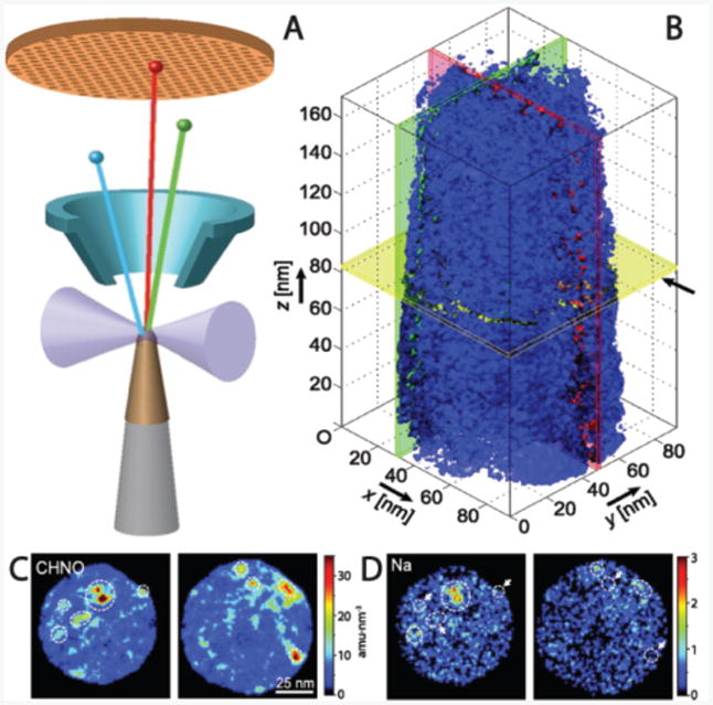
Atom probe tomography offers both 3D imaging and chemical composition measurements at the atomic scale (around 0.1-0.3 nm resolution in depth and 0.3-0.5 nm laterally).57,58 This technique is currently the only way to chemically map inclusions in biominerals, in detail, at the nanoscale. Using FIB and nanomanipulators, needle-shaped samples are milled and mounted to a Si post array. Then a pulsed ultraviolet laser sequentially triggers field evaporation of single atoms or small clusters from the surface of the tip. The resulting ions are projected onto a position sensitive detector (PSD) and their time-of-flight, and thus chemical identity, is determined. The detector simultaneously measures 1) the time between the laser flash and the arrival on the PSD to determine the mass over charge ratio of the ions and 2) the X-Y position and the order of arrival of the ions on the PSD to reconstruct the original position of the atoms in the sample. By repeating this operation, the atoms are progressively removed from the sample, and a 3D image of the material can be reconstructed at the atomic scale (Figure 4A). The small size of the sample, however, limits the overall image size and since the ejected organic fragments tend to be larger than single ions, the resolution of their location is slightly reduced. Though this technique was originally developed for studies of metals and semiconductors, recent advancements have made studies of composite and biological materials possible58–60 and, despite challenges, the results reported so far are groundbreaking.
Figure 4.

(A) Schematic of an atom probe tomography experiment in which the sample is ablated by a laser (forming ions) onto a mass spectrometer to determine the type of atoms within the removed section. (B) Tomographic reconstruction of an elephant dentin sample shows the isosurface for the organic fragment number density (blue surface is the threshold for dense organic) revealing fibers aligned along the z axis. (C) and (D) Selected sections from the reconstruction in (B) with mass density for organic fragments (C) and Na+ (D) locations shown. The highest density regions for each are often, but not exclusively, the same, which implies that something in the organic macromolecules may bind the ions. Scale for (C) and (D) are the same as (B) x–y plane. Adapted and reprinted with permission from 62.
The first pioneering application of APT to the study of biomineral composites was to the tooth of a chiton, a small marine mollusk whose teeth are designed to scrape algae off of rocks.61 Their teeth have a hard magnetite exterior and several organic-inorganic interfaces that are of interest. The APT revealed a 2–4 nm boundary-like region where the organic and inorganic materials are interspersed. The authors speculate that this interphase likely improves adhesion between the chemically disparate regions. A similar approach has been applied to dentin from elephant teeth.62 APT revealed long organic fibers running parallel to the tooth direction with cross-sectional dimensions on the order of the width of a collagen microfibril (Figure 4b). They also found that the fibers have locally increased concentrations of Mg2+ and Na+ ions (Figure 4c–d) and theorize that the exterior of the fibers may be coated with noncollagenous proteins, which selectively bind ions and may aid in mineralization. In other work, details of the chemical composition and spatial heterogeneity of rodent enamel have been elucidated using APT.63 The unique chemical insights from APT allow for conclusions to be drawn about mechanisms of biomineral formation and function based on spatially resolved chemical information.
X-ray spectroscopy: Mapping amorphous to crystalline transformations
X-ray spectroscopy techniques, including x-ray absorption fine structure (EXAFS) and x-ray absorption near-edge spectroscopy (XANES), coupled with x-ray photoelectron emission microscopy (X-PEEM), have yielded many insights into the structures of biominerals.64–68 Sample preparation is relatively minimal compared to other techniques (typically embedding in resin and polishing), but some of the techniques are surface-sensitive (depending on the wavelength of x-rays used), requiring additional attention to avoid artifacts from surface-layers left by polishing.
EXAFS spectra contain information about local structure and therefore can distinguish between amorphous and crystalline phases. X-PEEM studies have provided a detailed picture of the amorphous to crystalline transformation in a variety of biomineralized tissues. The use of amorphous precursors in biomineralization has attracted much attention as a synthetic route to morphologically complex single crystals.69 For example, a recent study of larval sea urchin spicules, mineralized skeletal components, revealed that ACC nanoparticles unexpectedly persisted despite crystallization of the surrounding material.70 To-date, most studies have focused on characterizing the inorganic component of biominerals, however, the organic component can also be probed by looking at the carbon K-edge in the EXAFS spectra, as shown in several recent studies.71–73
Vibrational spectromicroscopy: Chemically rich 3D maps of tissue composition
Vibrational (Raman and infrared) spectromicroscopy can non-destructively provide spatially-resolved (at the micrometer scale) compositional information about the organic and inorganic components of biominerals.74,75 These techniques couple a Raman (or IR) spectrometer to a standard optical microscope, allowing imaging of a sample and Raman (or IR) analysis with a microscopic laser spot. Two-dimensional maps of tissue samples are typically generated by raster-scanning the laser over a selected area to generate a 2D area map with a spectrum at each pixel. Both organic matrix and inorganic mineral components have distinct spectral signatures. Confocal Raman microscopy refers to the ability to spatially filter the analysis volume of the sample, in the XY (lateral) and Z (depth) axes. The limits of spatial resolution are defined principally by the laser quality and wavelength and the type of microscope objective. Typical spatial resolution is on the order of 0.5-1 μm.
Stomatopod raptorial appendages, which are used by the organism to “club” or “spear” prey, (depending on the species) at very high velocities,76 have recently been analyzed by Raman microscopy.77 In this study, confocal Raman microscopy was used to map the compositional gradients present in these unique appendages, These maps revealed a transition from ACC to amorphous calcium phosphate at the bulk-impact interface, and finally, to highly crystalline sulfate-containing fluorapatite (FAP) along the actual impact surface. Importantly, the sensitivity of the Raman technique allowed discernment of FAP from apatite, which could not be done on the basis of standard x-ray diffraction alone.
Confocal Raman spectromicroscopy can also be used to characterize compositional changes within 3D structures.78–81 In addition to 2D and 3D compositional maps, polarized Raman spectroscopy can provided quantification of molecular and crystallographic orientation. For example, Masic et al. were able to observe multiscale orientational changes in rat tail tendon (Type I) collagen under stress using polarized Raman spectroscopy.82 Finally, one of the most promising applications of Raman is in vivo imaging, and the capability to couple Raman microscopes with other techniques, such as fluorescence microscopy. For example, bone formation in zebrafish larvae was recently observed in vivo.83,84 Anesthetized fish were analyzed under a water-immersion objective, using a fluorescence-enabled modified Raman setup. Spectra were acquired along growing bones in the fin rays and showed a clear transition from little or no calcium phosphate to a predominance of calcium phosphate, correlated with calcium fluorescence (a calcium-binding marker). Furthermore, the peaks suggested that octacalcium phosphate (OCP), or an OCP-like intermediate phase, may be present, a finding previously undocumented in vivo.
Conclusion
The development and application of advanced microscopy and spectroscopy techniques, some of which can be performed in situ, have been essential for understanding the structure and formation of biominerals and bio-inspired materials. The information provided by these techniques about the chemistry, crystallography, phase distribution, structure, energetics, and kinetics of these materials forms the basis for an increasingly diverse understanding of the fundamental mechanisms by which natural systems can create structures. Yet we are only beginning to effectively mimic these processes synthetically. Fully understanding incorporated organic materials and the role they play in composite crystals will lead to further developments at the leading edge of characterization and synthesis.
Numerous recent advances in AFM imaging promise to provide new insights into biomineralization processes. High speed imaging85–87 with image collection times below 100 ms should enable researchers to probe the conformational fluctuations and transformations that control the emergence of order during the assembly of an organic matrix. Frequency Modulated AFM (FM-AFM)88–90 is capable of both imaging the structure of the near-surface water layers at solid interfaces in solutions and measuring the small attractive forces barriers between the tip and sample during the approach to the surface. These capabilities will lead to new insights into the role of solvation forces and solvent-mediated matrix-mineral interactions in directing biomineral formation.
Several of the techniques described in this review, such as liquid phase TEM, are still in their infancy. Development of liquid cells for TEM instrumented for pH measurement, heating and cooling, and quantitative mixing will enable a wider range of experiments with well-defined solution saturation states. Solution supersaturation is a critical parameter to quantify in order for the resulting data regarding nucleation and growth modes to be interpreted in terms of theoretical predictions or compared to computer simulations.
New techniques, beyond those described here, are also emerging that have the potential to provide even more detailed spatial and temporal resolution of biomineralization processes in hydrated (fluid) environments. For example, the recent implementation of scanning transmission x-ray microscopy end stations on synchrotron sources with resolution down to 10–20 nm91 has enabled elemental mapping of hybrid organic/inorganic structures in fluid.92,93 Another development is a new type of scanning electron microscope (e.g., airSEM™) in which fully-hydrated and uncoated samples can be imaged in air, allowing in situ observation of processes, such as calcium transport, in living organisms.84,94 The airSEM enables correlative imaging of tissues under ambient conditions by light and fluorescence microscopy and electron microscopy with elemental analysis by EDS.83,95 Finally, new sample preparation techniques such as cryo-FIB, in which cryogenically frozen samples are thinned by a focused ion beam will enhance our ability to prepare electron-transparent samples of biological samples under near-native conditions.96
The future holds the promise of integrating several of these techniques to allow for correlative imaging and, for example, link structural and compositional variations to changes in properties, such as elastic moduli or hardness. Ultimately, the knowledge gained through such studies will enable materials scientists to reproduce the remarkable properties of biominerals through bio-inspired approaches to materials synthesis.
Acknowledgments
The authors acknowledge support from the National Science Foundation (DMR 1210304) and the National Institutes of Health (CA173083). Additional support for this work was provided by the US Department of Energy, Office of Basic Energy Sciences, Materials Science and Engineering Division, as well as the Laboratory Directed Research and Development Initiative on Materials Synthesis and Simulation across Scales, at the Pacific Northwest National Laboratory (PNNL). PNNL is operated by Battelle for the US Department of Energy under Contract DE-AC05–76RL01830.
Footnotes
References
- 1.Lowenstam H, Weiner S. On Biomineralization. Oxford University Press Inc; 1989. [Google Scholar]
- 2.Dove PM, De Yoreo JJ, Weiner S, editors. Mineral Society of America. Washington, DC: 2003. p. 54. [Google Scholar]
- 3.Asenath-Smith E, Li H, Keene EC, Seh ZW, Estroff La. Adv Funct Mater. 2012;22(14):2891–2914. [Google Scholar]
- 4.Nudelman F, Sommerdijk NaJ. M Angew Chemie - Int Ed. 2012;51:6582–6596. doi: 10.1002/anie.201106715. [DOI] [PubMed] [Google Scholar]
- 5.Arakaki A, Shimizu K, Oda M, Sakamoto T, Nishimura T, Kato T. Org Biomol Chem. 2014;13(4):974–989. doi: 10.1039/c4ob01796j. [DOI] [PubMed] [Google Scholar]
- 6.Franz CM, Muller DJ. Methods Mol Biol. 2011;736:97–107. doi: 10.1007/978-1-61779-105-5_7. [DOI] [PubMed] [Google Scholar]
- 7.De Yoreo JJ. AIP Conference Proceedings. Vol. 916. AIP; 2007. pp. 416–438. [Google Scholar]
- 8.Cisneros DA, Hung C, Franz CM, Muller DJ. J Struct Biol. 2006;154(3):232–245. doi: 10.1016/j.jsb.2006.02.006. [DOI] [PubMed] [Google Scholar]
- 9.Chung S, Shin SH, Bertozzi CR, De Yoreo JJ. Proc Natl Acad Sci U S A. 2010;107(38):16536–16541. doi: 10.1073/pnas.1008280107. [DOI] [PMC free article] [PubMed] [Google Scholar]
- 10.Narayanan B, Gilmer GH, Tao J, De Yoreo JJ, Ciobanu CV. Langmuir. 2014;30(5):1343–1350. doi: 10.1021/la4043364. [DOI] [PubMed] [Google Scholar]
- 11.Habraken WJEM, Tao J, Brylka LJ, Friedrich H, Bertinetti L, Schenk AS, Verch A, Dmitrovic V, Bomans PHH, Frederik PM, Laven J, van der Schoot P, Aichmayer B, de With G, DeYoreo JJ, Sommerdijk NAJM. Nat Commun. 2013;4:1507. doi: 10.1038/ncomms2490. [DOI] [PubMed] [Google Scholar]
- 12.Hamm LM, Giuffre AJ, Han N, Tao J, Wang D, De Yoreo JJ, Dove PM. Proc Natl Acad Sci U S A. 2014;111(4):1304–1309. doi: 10.1073/pnas.1312369111. [DOI] [PMC free article] [PubMed] [Google Scholar]
- 13.Rimer JD, An Z, Zhu Z, Lee MH, Goldfarb DS, Wesson JA, Ward MD. Science. 2010;330(6002):337–341. doi: 10.1126/science.1191968. [DOI] [PMC free article] [PubMed] [Google Scholar]
- 14.Orme Ca, Noy a, Wierzbicki a, Mc Bride MT, Grantham M, Teng HH, Dove PM, De Yoreo JJ. Nature. 2001;411(6839):775–779. doi: 10.1038/35081034. [DOI] [PubMed] [Google Scholar]
- 15.Olafson KN, Ketchum MA, Rimer JD, Vekilov PG. Proc Natl Acad Sci U S A. 2015:1501023112. doi: 10.1073/pnas.1501023112. [DOI] [PMC free article] [PubMed] [Google Scholar]
- 16.Orgel JPRO, Irving TC, Miller A, Wess TJ. Proc Natl Acad Sci U S A. 2006;103(24):9001–9005. doi: 10.1073/pnas.0502718103. [DOI] [PMC free article] [PubMed] [Google Scholar]
- 17.Tao J, Battle KC, Pan H, Salter EA, Chien YC, Wierzbicki A, De Yoreo JJ. Proc Natl Acad Sci U S A. 2014;112(2):326–331. doi: 10.1073/pnas.1404481112. [DOI] [PMC free article] [PubMed] [Google Scholar]
- 18.De Yoreo JJ, Chung S, Friddle RW. Adv Funct Mater. 2013;23(20):2525–2538. [Google Scholar]
- 19.Weiner S, Wagner HD. Annu Rev Mater Sci. 1998;28(1):271–298. [Google Scholar]
- 20.Landis WJ, Silver FH. Cells Tissues Organs. 2009;189(1-4):20–24. doi: 10.1159/000151454. [DOI] [PMC free article] [PubMed] [Google Scholar]
- 21.Wang Y, Azaïs T, Robin M, Vallée A, Catania C, Legriel P, Pehau-Arnaudet G, Babonneau F, Giraud-Guille MM, Nassif N. Nat Mater. 2012;11(8):724–733. doi: 10.1038/nmat3362. [DOI] [PubMed] [Google Scholar]
- 22.Olszta MJ, Cheng X, Jee SS, Kumar R, Kim YY, Kaufman MJ, Douglas EP, Gower LB. Mater Sci Eng R Reports. 2007;58(3-5):77–116. [Google Scholar]
- 23.Nudelman F, de With G, Sommerdijk NAJM. Soft Matter. 2011;7(1):17. [Google Scholar]
- 24.Quan BD, Sone ED. Methods Enzymol. 2013;532:189–205. doi: 10.1016/B978-0-12-416617-2.00009-6. [DOI] [PubMed] [Google Scholar]
- 25.Pichon BP, Bomans PHH, Frederik PM, Sommerdijk NAJM. J Am Chem Soc. 2008;130(12):4034–4040. doi: 10.1021/ja710416h. [DOI] [PubMed] [Google Scholar]
- 26.Carcouët CCMC, van de Put MWP, Mezari B, Magusin PCMM, Laven J, Bomans PHH, Friedrich H, Esteves ACC, Sommerdijk NAJM, van Benthem RATM, de With G. Nano Lett. 2014;14(3):1433–1438. doi: 10.1021/nl404550d. [DOI] [PubMed] [Google Scholar]
- 27.Frandsen C, Legg BA, Comolli LR, Zhang H, Gilbert B, Johnson E, Banfield JF. CrystEngComm. 2014;16(8):1451–1458. [Google Scholar]
- 28.Nudelman F, Pieterse K, George A, Bomans PHH, Friedrich H, Brylka LJ, Hilbers PAJ, de With G, Sommerdijk NAJM. Nat Mater. 2010;9(12):1004–1009. doi: 10.1038/nmat2875. [DOI] [PMC free article] [PubMed] [Google Scholar]
- 29.Pouget EM, Bomans PHH, Goos JACM, Frederik PM, de With G, Sommerdijk NAJM. Science. 2009;323(5920):1455–1458. doi: 10.1126/science.1169434. [DOI] [PubMed] [Google Scholar]
- 30.Williamson MJ, Tromp RM, Vereecken PM, Hull R, Ross FM. Nat Mater. 2003;2(8):532–536. doi: 10.1038/nmat944. [DOI] [PubMed] [Google Scholar]
- 31.Wang CM, Liao HG, Ross FM. MRS Bull. 2015;40(01):46–52. [Google Scholar]
- 32.Zheng H, Meng YS, Zhu Y. MRS Bull. 2015;40(01):12–18. [Google Scholar]
- 33.Sutter EA, Sutter PW. J Am Chem Soc. 2014;136(48):16865–16870. doi: 10.1021/ja508279v. [DOI] [PubMed] [Google Scholar]
- 34.Nielsen MH, Aloni S, De Yoreo JJ. Science (80-) 2014;345(6201):1158–1162. doi: 10.1126/science.1254051. [DOI] [PubMed] [Google Scholar]
- 35.Smeets PJM, Cho KR, Kempen RGE, Sommerdijk NAJM, De Yoreo JJ. Nat Mater. 2015 doi: 10.1038/nmat4193. advance on. [DOI] [PubMed] [Google Scholar]
- 36.Woehl TJ, Evans JE, Arslan I, Ristenpart WD, Browning ND. ACS Nano. 2012;6(10):8599–8610. doi: 10.1021/nn303371y. [DOI] [PMC free article] [PubMed] [Google Scholar]
- 37.Zhang H, De Yoreo JJ, Banfield JF. ACS Nano. 2014;8(7):6526–6530. doi: 10.1021/nn503145w. [DOI] [PubMed] [Google Scholar]
- 38.Addadi L, Moradian J, Shay E, Maroudas NG, Weiner S. Proc Natl Acad Sci. 1987;84(9):2732–2736. doi: 10.1073/pnas.84.9.2732. [DOI] [PMC free article] [PubMed] [Google Scholar]
- 39.Marsh ME. Protoplasma. 1994;177(3-4):108–122. [Google Scholar]
- 40.Schenk AS, Zlotnikov I, Pokroy B, Gierlinger N, Masic A, Zaslansky P, Fitch AN, Paris O, Metzger TH, Cölfen H, Fratzl P, Aichmayer B. Adv Funct Mater. 2012;22(22):4668–4676. [Google Scholar]
- 41.Sousa Aa, Leapman RD. Ultramicroscopy. 2012;123:38–49. doi: 10.1016/j.ultramic.2012.04.005. [DOI] [PMC free article] [PubMed] [Google Scholar]
- 42.Kabalah-Amitai L, Mayzel B, Kauffmann Y, Fitch AN, Bloch L, Gilbert PUPA, Pokroy B. Science. 2013;340(6131):454–457. doi: 10.1126/science.1232139. [DOI] [PubMed] [Google Scholar]
- 43.Osuna-Mascaró A, Cruz-Bustos T, Benhamada S, Guichard N, Marie B, Plasseraud L, Corneillat M, Alcaraz G, Checa A, Marin F. Comp Biochem Physiol B Biochem Mol Biol. 2014;168:76–85. doi: 10.1016/j.cbpb.2013.11.009. [DOI] [PubMed] [Google Scholar]
- 44.Mass T, Drake JL, Peters EC, Jiang W, Falkowski PG. Proc Natl Acad Sci U S A. 2014;111(35):12728–12733. doi: 10.1073/pnas.1408621111. [DOI] [PMC free article] [PubMed] [Google Scholar]
- 45.Ameye L, Hermann R, Killian C, Wilt F, Dubois P. J Histochem Cytochem. 1999;47(9):1189–1200. doi: 10.1177/002215549904700911. [DOI] [PubMed] [Google Scholar]
- 46.Seto J, Zhang Y, Hamilton P, Wilt F. J Struct Biol. 2004;148:123–130. doi: 10.1016/j.jsb.2004.04.001. [DOI] [PubMed] [Google Scholar]
- 47.Midgley PA, Weyland M. Ultramicroscopy. 2003;96(3-4):413–431. doi: 10.1016/S0304-3991(03)00105-0. [DOI] [PubMed] [Google Scholar]
- 48.Leary R, Midgley P, Thomas J. Accounts Chem …. 2012;45(10) doi: 10.1021/ar3001102. [DOI] [PubMed] [Google Scholar]
- 49.Voyles PM, Grazul JL, Muller DA. Ultramicroscopy. 2003;96(3-4):251–273. doi: 10.1016/S0304-3991(03)00092-5. [DOI] [PubMed] [Google Scholar]
- 50.Kunitake ME, Mangano LM, Peloquin JM, Baker SP, Estroff LA. Acta Biomater. 2013;9(2):5353–5359. doi: 10.1016/j.actbio.2012.09.030. [DOI] [PubMed] [Google Scholar]
- 51.Li H, Xin HL, Kunitake ME, Keene EC, Muller Da, Estroff La. Adv Funct Mater. 2011;21(11):2028–2034. [Google Scholar]
- 52.Gries K, Kröger R, Kübel C, Fritz M, Rosenauer A. Acta Biomater. 2009;5(8):3038–3044. doi: 10.1016/j.actbio.2009.04.017. [DOI] [PubMed] [Google Scholar]
- 53.Okumura T, Suzuki M, Nagasawa H, Kogure T. J Cryst Growth. 2013;381:114–120. [Google Scholar]
- 54.van de Locht R, Slater TJAT, Verch A, Young JR, Haigh SJ, Kröger R. Cryst Growth Des. 2014:140320115609006. [Google Scholar]
- 55.Li H, Xin HL, Muller Da, Estroff La. Science. 2009;326(5957):1244–1247. doi: 10.1126/science.1178583. [DOI] [PubMed] [Google Scholar]
- 56.Kim YY, Ganesan K, Yang P, Kulak AN, Borukhin S, Pechook S, Ribeiro L, Kröger R, Eichhorn SJ, Armes SP, Pokroy B, Meldrum FC. Nat Mater. 2011;10(11):890–896. doi: 10.1038/nmat3103. [DOI] [PubMed] [Google Scholar]
- 57.Seidman DN. Annu Rev Mater Res. 2007;37(1):127–158. [Google Scholar]
- 58.Miller MK, Forbes RG. Mater Charact. 2009;60(6):461–469. [Google Scholar]
- 59.Kelly TF, Nishikawa O, Panitz Ja, Prosa TJ. MRS Bull. 2009 Oct;34:744–750. [Google Scholar]
- 60.Marquis EA, Bachhav M, Chen Y, Dong Y, Gordon LM, McFarland A. Curr Opin Solid State Mater Sci. 2013;17(5):217–223. [Google Scholar]
- 61.Gordon LM, Joester D. Nature. 2011;469(7329):194–197. doi: 10.1038/nature09686. [DOI] [PubMed] [Google Scholar]
- 62.Gordon L, Tran L, Joester D. ACS Nano. 2012;No. 12:10667–10675. doi: 10.1021/nn3049957. [DOI] [PubMed] [Google Scholar]
- 63.Gordon LM, Cohen MJ, MacRenaris KW, Pasteris JD, Seda T, Joester D. Science. 2015;347(6223):746–750. doi: 10.1126/science.1258950. [DOI] [PubMed] [Google Scholar]
- 64.Tamura N, Gilbert PUPA. Methods Enzymol. 2013;532:501–531. doi: 10.1016/B978-0-12-416617-2.00021-7. [DOI] [PubMed] [Google Scholar]
- 65.Gilow C, Zolotoyabko E, Paris O, Fratzl P, Aichmayer B. Cryst Growth Des. 2011;11(6):2054–2058. [Google Scholar]
- 66.Beniash E, Metzler RA, Lam RSK, Gilbert PUPA. J Struct Biol. 2009;166(2):133–143. doi: 10.1016/j.jsb.2009.02.001. [DOI] [PMC free article] [PubMed] [Google Scholar]
- 67.Weiner S, Mahamid J, Politi Y, Ma Y, Addadi L. Front Mater Sci China. 2009;3(2):104–108. [Google Scholar]
- 68.Levi-Kalisman Y, Raz S, Weiner S, Addadi L, Sagi I. J Chem Soc Dalt Trans. 2000;No. 21:3977–3982. [Google Scholar]
- 69.Weiner S, Addadi L. Annu Rev Mater Res. 2011;41(1):21–40. [Google Scholar]
- 70.Gong YUT, Killian CE, Olson IC, Appathurai NP, Amasino AL, Martin MC, Holt LJ, Wilt FH, Gilbert PUPA. Proc Natl Acad Sci U S A. 2012;109(16):6088–6093. doi: 10.1073/pnas.1118085109. [DOI] [PMC free article] [PubMed] [Google Scholar]
- 71.Metzler RA, Tribello GA, Parrinello M, Gilbert PUPA. J Am Chem Soc. 2010;132(33):11585–11591. doi: 10.1021/ja103089r. [DOI] [PubMed] [Google Scholar]
- 72.Metzler RA, Evans JS, Killian CE, Zhou D, Churchill TH, Appathurai NP, Coppersmith SN, Gilbert PUPA. J Am Chem Soc. 2010;132(18):6329–6334. doi: 10.1021/ja909735y. [DOI] [PubMed] [Google Scholar]
- 73.Lam RSK, Metzler Ra, Gilbert PUPa, Beniash E. ACS Chem Biol. 2012;7:476–480. doi: 10.1021/cb200260d. [DOI] [PMC free article] [PubMed] [Google Scholar]
- 74.Gamsjaeger S, Mendelsohn R, Boskey AL, Gourion-Arsiquaud S, Klaushofer K, Paschalis EP. Curr Osteoporos Rep. 2014;12(4):454–464. doi: 10.1007/s11914-014-0238-8. [DOI] [PMC free article] [PubMed] [Google Scholar]
- 75.Talari ACS, Movasaghi Z, Rehman S, Rehman I ur. Appl Spectrosc Rev. 2014;50(1):46–111. [Google Scholar]
- 76.Weaver JC, Milliron GW, Miserez A, Evans-Lutterodt K, Herrera S, Gallana I, Mershon WJ, Swanson B, Zavattieri P, DiMasi E, Kisailus D. Science. 2012;336(6086):1275–1280. doi: 10.1126/science.1218764. [DOI] [PubMed] [Google Scholar]
- 77.Amini S, Masic A, Bertinetti L, Teguh JS, Herrin JS, Zhu X, Su H, Miserez A. Nat Commun. 2014 May;5:3187. doi: 10.1038/ncomms4187. 2013. [DOI] [PubMed] [Google Scholar]
- 78.Lee K, Wagermaier W, Masic A, Kommareddy KP, Bennet M, Manjubala I, Lee SW, Park SB, Cölfen H, Fratzl P. Nat Commun. 2012;3:725. doi: 10.1038/ncomms1720. [DOI] [PMC free article] [PubMed] [Google Scholar]
- 79.Eder SHK, Gigler AM, Hanzlik M, Winklhofer M. PLoS One. 2014;9(9):e107356. doi: 10.1371/journal.pone.0107356. [DOI] [PMC free article] [PubMed] [Google Scholar]
- 80.Omelon S, Georgiou J, Variola F, Dean MN. Acta Biomater. 2014;10(9):3899–3910. doi: 10.1016/j.actbio.2014.06.008. [DOI] [PubMed] [Google Scholar]
- 81.Lloyd AA, Wang ZX, Donnelly E. J Biomech Eng. 2015;137(1):010801. doi: 10.1115/1.4029046. [DOI] [PMC free article] [PubMed] [Google Scholar]
- 82.Masic A, Bertinetti L, Schuetz R, Galvis L, Timofeeva N, Dunlop JWC, Seto J, Hartmann Ma, Fratzl P. Biomacromolecules. 2011;12:3989–3996. doi: 10.1021/bm201008b. [DOI] [PubMed] [Google Scholar]
- 83.Bennet M, Akiva a, Faivre D, Malkinson G, Yaniv K, Abdelilah-Seyfried S, Fratzl P, Masic a. Biophys J. 2014;106(4):L17–L19. doi: 10.1016/j.bpj.2014.01.002. [DOI] [PMC free article] [PubMed] [Google Scholar]
- 84.Akiva A, Malkinson G, Masic A, Kerschnitzki M, Bennet M, Fratzl P, Addadi L, Weiner S, Yaniv K. Bone. 2015;75:192–200. doi: 10.1016/j.bone.2015.02.020. [DOI] [PubMed] [Google Scholar]
- 85.Ando T, Kodera N, Naito Y, Kinoshita T, Furuta K, Toyoshima YY. ChemPhysChem. 2003;4(11):1196–1202. doi: 10.1002/cphc.200300795. [DOI] [PubMed] [Google Scholar]
- 86.Shibata M, Yamashita H, Uchihashi T, Kandori H, Ando T. Nat Nanotechnol. 2010;5(3):208–212. doi: 10.1038/nnano.2010.7. [DOI] [PubMed] [Google Scholar]
- 87.Kodera N, Yamamoto D, Ishikawa R, Ando T. Nature. 2010;468(7320):72–76. doi: 10.1038/nature09450. [DOI] [PubMed] [Google Scholar]
- 88.Fukuma T, Jarvis SP. Rev Sci Instrum. 2006;77(4):043701. [Google Scholar]
- 89.Kilpatrick JI, Loh SH, Jarvis SP. J Am Chem Soc. 2013;135(7):2628–2634. doi: 10.1021/ja310255s. [DOI] [PubMed] [Google Scholar]
- 90.Fukuma T, Higgins MJ, Jarvis SP. Biophys J. 2007;92(10):3603–3609. doi: 10.1529/biophysj.106.100651. [DOI] [PMC free article] [PubMed] [Google Scholar]
- 91.Obst M, Schmid G. In: Electron Microscopy: Methods and Protocols, Methods in Molecular Biology. Kuo J, editor. SPRINGER; 233 SPRING ST, NEW YORK, NY 10013 USA: New York: 2014. pp. 757–781. [DOI] [PubMed] [Google Scholar]
- 92.Cosmidis J, Benzerara K, Nassif N, Tyliszczak T, Cosmidis J, Benzerara K, Nassif N, Tyliszczak T, Char FB. Acta Biomater. 2015;12:260–269. doi: 10.1016/j.actbio.2014.10.003. [DOI] [PubMed] [Google Scholar]
- 93.Li Y, El Gabaly F, Ferguson TR, Smith RB, Bartelt NC, Sugar JD, Fenton KR, Cogswell Da, Kilcoyne aLD, Tyliszczak T, Bazant MZ, Chueh WC. Nat Mater. 2014 Sep 13;:1149–1156. doi: 10.1038/nmat4084. [DOI] [PubMed] [Google Scholar]
- 94.Vidavsky N, Addadi S, Mahamid J, Shimoni E, Ben-Ezra D, Shpigel M, Weiner S, Addadi L. Proc Natl Acad Sci U S A. 2014;111(1):39–44. doi: 10.1073/pnas.1312833110. [DOI] [PMC free article] [PubMed] [Google Scholar]
- 95.Solomonov I, Talmi-Frank D, Milstein Y, Addadi S, Aloshin A, Sagi I. Sci Rep. 2014;4:5987. doi: 10.1038/srep05987. [DOI] [PMC free article] [PubMed] [Google Scholar]
- 96.Rigort A, Bäuerlein FJB, Villa E, Eibauer M, Laugks T, Baumeister W, Plitzko JM. Proc Natl Acad Sci U S A. 2012;109(12):4449–4454. doi: 10.1073/pnas.1201333109. [DOI] [PMC free article] [PubMed] [Google Scholar]
- 97.Rashkovich LN, Yoreo JJ, Orme CA, Chernov AA. Crystallogr Reports. 2006;51(6):1063–1074. [Google Scholar]
- 98.De Yoreo JJ, Zepeda-Ruiz LA, Friddle RW, Qiu SR, Wasylenki LE, Chernov AA, Gilmer GH, Dove PM. Cryst Growth Des. 2009;9(12):5135–5144. [Google Scholar]
- 99.Friddle RW, Weaver ML, Qiu SR, Wierzbicki a, Casey WH, De Yoreo JJ. Proc Natl Acad Sci U S A. 2010;107(1):11–15. doi: 10.1073/pnas.0908205107. [DOI] [PMC free article] [PubMed] [Google Scholar]
- 100.Wallace AF, Hedges LO, Fernandez-Martinez A, Raiteri P, Gale JD, Waychunas Ga, Whitelam S, Banfield JF, De Yoreo JJ. Science (80-) 2013;341(6148):885–889. doi: 10.1126/science.1230915. [DOI] [PubMed] [Google Scholar]
- 101.Sone ED, Weiner S, Addadi L. J Struct Biol. 2007;158(3):428–444. doi: 10.1016/j.jsb.2007.01.001. [DOI] [PubMed] [Google Scholar]
- 102.Fang PA, Margolis HC, Conway JF, Simmer JP, Beniash E. J Struct Biol. 2013;183(2):250–257. doi: 10.1016/j.jsb.2013.05.011. [DOI] [PMC free article] [PubMed] [Google Scholar]
- 103.Fang PA, Conway JF, Margolis HC, Simmer JP, Beniash E. Proc Natl Acad Sci U S A. 2011;108(34):14097–14102. doi: 10.1073/pnas.1106228108. [DOI] [PMC free article] [PubMed] [Google Scholar]
- 104.Lucić V, Förster F, Baumeister W. Annu Rev Biochem. 2005;74:833–865. doi: 10.1146/annurev.biochem.73.011303.074112. [DOI] [PubMed] [Google Scholar]
- 105.Li D, Nielsen MH, De Yoreo JJ. Methods Enzymol. 2013;532:147–164. doi: 10.1016/B978-0-12-416617-2.00007-2. [DOI] [PubMed] [Google Scholar]
- 106.Nielsen MH, Li D, Zhang H, Aloni S, Han TYJ, Frandsen C, Seto J, Banfield JF, Cölfen H, De Yoreo JJ. Microsc Microanal. 2014;20(2):1–12. doi: 10.1017/S1431927614000294. [DOI] [PubMed] [Google Scholar]
- 107.Okuda M, Ogawa N, Takeguchi M, Hashimoto A, Tagaya M, Chen S, Hanagata N, Ikoma T. Microsc Microanal. 2011;17:788–798. doi: 10.1017/S1431927611011949. [DOI] [PubMed] [Google Scholar]
- 108.Mahamid J, Aichmayer B, Shimoni E, Ziblat R, Li C, Siegel S, Paris O, Fratzl P, Weiner S, Addadi L. Proc Natl Acad Sci U S A. 2010;107(14):6316–6321. doi: 10.1073/pnas.0914218107. [DOI] [PMC free article] [PubMed] [Google Scholar]
- 109.Gilbert PUPA. J Electron Spectros Relat Phenomena. 2012;185(10):395–405. [Google Scholar]
- 110.Ma Y, Aichmayer B, Paris O, Fratzl P, Meibom A, Metzler RA, Politi Y, Addadi L, Gilbert PUPA, Weiner S. Proc Natl Acad Sci U S A. 2009;106(15):6048–6053. doi: 10.1073/pnas.0810300106. [DOI] [PMC free article] [PubMed] [Google Scholar]


