

Introduction
Left Bochdalek hernia is a life threatening condition with a high mortality if presented in the neonatal period and not managed appropriately. Rarely the presentation may be delayed, in which case the symptomatology is different and with timely surgical intervention, prognosis is excellent [1]. We present a case of congenital left Bochdalek hernia with delayed presentation in view of its rarity and pitfalls in diagnosis due to misleading symptomatology.
Case Report
A 4 year old male child presented with complaints of vague pain chest and dry, non-productive cough along with moderate grade fever of 5 months duration. A medical practitioner diagnosed him as a case of left sided pleural effusion on clinical and radiological grounds & started him on anti tubercular treatment (ATT) with SHRZ. He became asymptomatic after about 10 days. Repeat X-ray chest done after 3 weeks however showed non-resolution of the opacity and later clarithromycin was added. He was brought to our hospital when child did not show any radiological resolution even after 2 months of ATT.
Examination revealed a child weighing 14kg with normal vital parameters. Examination of the respiratory system revealed bilaterally symmetrical chest with decreased movements on left side. Breath sounds and vocal fremitus were reduced in left infrascapular, inter scapular and infra axillary regions. Gurgling sounds were heard over the left lower chest. Other systems were essentially normal.
Investigations revealed normal Hb, blood counts and ESR. Mantoux and sputum for AFB were negative. Chest X-ray and barium meal contrast study showed herniated intestinal loops in the left hemithorax.
He was disgnosed as a case of left Bochdalek hernia on clinical & radiological findings. Corrective surgery was carried out; child had an uneventful postoperative period and has been found to be thriving well on follow up visits.
Discussion
Bochdalek hernia is a congenital diaphragmatic prolapse and usually, when left sided, presents with a clinical picture that demands urgent medical attention. Embryologically, the diaphragm develops by fusion of various components during ninth week of intrauterine life. The right hemi-diaphragm completes its closure before the left. Last area to close on either side is the postero-lateral portion, known as foramen of Bochdalek [2].
The occurrence rate is 1 in 7000 live births, of which the presentation is delayed in 5-10% of patients. The defect commonly occurs on the left side and there is male preponderance with male to female ratio of 2:1 [2]. When the abdominal organs herniate in to the pleural cavity, the development of the lung is disturbed and the degree of pulmonary hypoplasia depends upon the stage of pulmonary development at the time of visceral herniation. Early visceral herniation into the pleural cavity worsens pulmonary hypoplasia and prognosis [3]. Typically, in the neonatal period, congenital diaphragmatic hernia produces respiratory distress, cyanosis and acidosis as herniated bowel distends with swallowed air and compromises diaphragmatic breathing producing atelectasis and respiratory acidosis, making this presentation a true emergency with high mortality [4]. The serious respiratory distress is related to pulmonary hypoplasia, often bilateral, combined with persistent fetal circulation and mechanical respiratory disorders [5].
If the newborn does not become symptomatic within 8 hours of birth, symptoms that appear later are probably due to simple lung compression [6, 7]. Late presentation of congenital diaphragmatic hernia poses diagnostic difficulty due to its rarity and misleading clinical features. These children remain asymptomatic for months to years and are often picked up incidentally when X-ray chest is done for some other purpose. They usually present with gastrointestinal as well as respiratory symptoms [4]. Respiratory symptoms are chronic or recurrent and less severe than in a newborn. The decreased severity of respiratory symptoms may be due to a good respiratory compensatory mechanism. This difference in symptoms may delay the correct diagnosis [8]. Chest X-ray in such cases is diagnostic and at times upper gastro-intestinal contrast studies may be resorted to confirm the diagnosis [9].
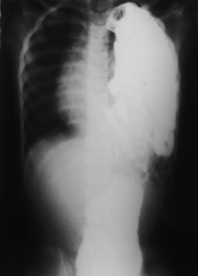
Fig. 1.

X-ray chest showing partially opaque left hemithorax with multiple ring opacities suggestive of bowel loops and mediastinal shift to right
The definitive treatment is surgery and needs thoracotomy / laparotomy and repair of the defect in diaphragm. The herniated loops may be incarcerated or strangulated if early surgical intervention is not resorted to [10]. The post-operative recovery and long-term outcome depends upon the developmental status of the lung. In delayed presentation, the lungs are usually mature and long-term outcome is excellent. Chest physiotherapy enhances the speed of recovery.
Out patient had a congenital defect of left diaphragm, which was detected when chest skiagram was taken because of respiratory symptoms. It is also possible that the congenital defect may have been present between the rims of diaphragm or may have been obscured by spleen or left lobe of liver. At the time of acute respiratory infection, increased intra abdominal pressure due to cough might have led to herniation of the abdominal contents through the defect leading to typical X-ray picture.
Diagnosis of this condition may not have been entertained earlier because of its rarity and misleading clinical features. Possibility of Bochdalek hernia with delayed presentation should always be kept in mind in the differential diagnosis of a child presenting with respiratory distress.
Fig. 2.
Barium meal study showing contrast filled herniated intestinal loop in the left hemithorax
References
- 1.Vukic Zoran. Left Bochdalek hernia with delayed presentation: Report of two cases. CMJ. 2001;42(5):569–571. [PubMed] [Google Scholar]
- 2.Graivier L, Dorman GW, Votteler TP. Congenital diaphragmatic hernia in Children. Surg Gynecol Obstet. 1971;132:408–410. [PubMed] [Google Scholar]
- 3.Thibeault DW, Haney B. Lung volume, pulmonary vasculature and affecting survival in congenital diaphragmatic hernia. Pediatrics. 1998;101:289–295. doi: 10.1542/peds.101.2.289. [DOI] [PubMed] [Google Scholar]
- 4.Osebold WR, Soper RT. Congenital diaphragmatic hernia past infancy. Am J Surg. 1976;131:748–754. doi: 10.1016/0002-9610(76)90194-x. [DOI] [PubMed] [Google Scholar]
- 5.Levin DL. Morphologic analysis of pulmonary vascular bed in congenital left sided diaphragmatic hernia. J Pediatr. 1978;92:805–809. doi: 10.1016/s0022-3476(78)80162-0. [DOI] [PubMed] [Google Scholar]
- 6.Alagappan A, Malloy MH. Outcome of congenital diaphragmatic hernia: ten years experience after ECMO. Pediatrics. 1997;100(Suppl 2):517–518. [Google Scholar]
- 7.Manthei U, Vaucher Y, Crowe CP. Congenital diaphragmatic hernia. Immediate pre-operative and post-operative oxygen gradients identify patients requiring prolonged respiratory support. Surgery. 1983;93:83–87. [PubMed] [Google Scholar]
- 8.Kirkland JA. Congenital postero-lateral diaphragmatic hernia in the adult. Br J Surg. 1959;47:16. doi: 10.1002/bjs.18004720103. [DOI] [PubMed] [Google Scholar]
- 9.Ruff SJ, Campbell JR, Harrison MW, Campbell TJ. Pediatric diaphragmatic hernia: an 11-year experience. Am J Surg. 1980;139:641–645. doi: 10.1016/0002-9610(80)90353-0. [DOI] [PubMed] [Google Scholar]
- 10.Ban JL, Moore TC. Intra-thoracic tension incarceration of stomach and liver through right-sided congenital postero-lateral hernia. J Thorac Cardiovasc Surg. 1973;66:969–973. [PubMed] [Google Scholar]