

Introduction
The Ehlers-Danlos Syndromes (EDS) are a genetically, biochemically and clinically diverse group of heritable connective tissue disorders having joint laxity and dermal features in common. Prior classifications of EDS have included upto eleven disorders. Table 1 provides the currently accepted classification. Many patients with features of EDS do not fit readily into one of the well-defined subtypes. Some cases have been reported from India [1, 2, 3]. EDS Type V described here is a rare variety. There is only one single large, well-documented family with EDS type V [4]. The diagnosis is clinical.
Table 1.
Classification of Ehlers Danlos Syndrome
| Type | Clinical features | Inheritance | Biochemical defects |
|---|---|---|---|
| I (Gravis) | Soft, hyperextensible skin; easy bruising; thin, atrophic scars; hypermobile joints; varicose veins; premature birth | AD | Mutations in pro X1 (V) or pro X2 (V) chains of type V collagen |
| II (Mitis) | Similar to EDS type I but less severe | AD | Same as EDS I |
| III (Familial hypermobility) | Soft skin; large and small joint hypermobility | AD | Not known |
| IV (Arterial (ecchymotic) | Thin, transluscent skin with visible veins, easy bruising; absence of skin and joint extensibility; arterial, bowel, and uterine rupture Similar to EDS type II | AD | Mutations in COL3 AI; abnormal type III collagen synthesis, secretion or structure |
| V (X-linked) | XRL | Not known | |
| VI (Ocular) | Soft skin muscle hypotonia; scoliosis; joint laxity; hyper-extensible skin | AR | Lysyl hydroxylase deficiency; mutations in PLOD gene |
| VII (Arthrochalasis multiplex congenita) | Soft skin with normal scarring, congenital hip dislocation, severe joint hyper-mobility | AD/AR |
|
| VIII (Periodontal) | Hyperelastic, fragile, skin, moderate joint hyper mobility, severe periodontitis | AD | Reduced type III collagen |
| IX | Hernias, bladder diverticulae, mild skin laxity, skeletal defects | ? | Increased copper incorporation and lysyl-oxidase deficiency |
| X | Similar to EDS type II, with abnormal clotting studies | AR | Proposed defect in fibronectin |
Case Report
On 25 July 2001 a 4 year old son of a serving Army soldier was brought to the skin OPD by his mother with complaints of easy bruisability, delayed wound healing, excessive scar formation at injury sites since 1 year of age. This second born male child was delivered vaginally at home after 8.5 months gestation. Gestation period and delivery were normal. There was no history of any drug intake by mother during pregnancy. Child was apparently asymptomatic till about 1 year of age when he started having presenting complaints. There was no history of seizures/mental retardation/delayed milestones/prolonged bleeding from wounds or body orifices. There was no history of passing high colour or orangish red urine. There was no history of repeated bony fractures. Personal/family history revealed that the child was born out of non-consanguineous marriage. No family history of similar complaints was present in parents, grandparents, other relatives, siblings (2 sisters - 6 years and 2 years). No history of maternal abortions was elicited. General physical and systemic examination revealed no abnormality. The child was normal in growth and intelligence. All relevant investigations including blood coagulation profile were normal. Skin biopsy revealed wavy collagen fibrils in dermis. Dermatological examination revealed velvety, hyperelastic skin with normal recovery time, easy bruisability, multiple hypertrophic scars/cigarette paper scars at previous wound sites over forehead and limbs, molluscoid pseudotumours over both shins. Widely spaced eyes, broad nasal bridge, hyperextensible joints and muscle hypotonia were present (Figs 1 & 2). Hair/mucosa/nails/eyes/oral cavity/dentition - was normal. Limb span : body segment ratio was normal. A clinical diagnosis of Ehlers-Danlos Syndrome Type V, X-linked recessive (XLR) was made based upon the following findings - parents and relatives not affected, 2 sisters not affected, broad nasal bridge, hyperelastic velvety skin with normal recovery time, easy bruisability, multiple hypertrophic cigarette paper scars at previous wound sites, hyperextensible joints, muscle hypotonia and molluscoid pseudotumours over shins.
Fig. 1.

Excessive scarring over forhead from previous wounds, widely spaced eyes, broad nasal bridge and hyperelastic skin is clearly seen
Fig. 2.
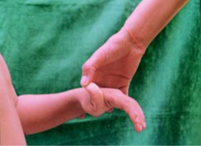
Hyperextensible thumb
Parents were advised to protect the child from trauma/physical abuse and tablet Vit C- 500 mg twice daily for 6 weeks was given.
Discussion
Virtually all forms of EDS consisting of a group of inherited disorders of collagen tissue are the result of mutations that code for fibrillar collagen genes or for enzymes that catalyze the intracellular or extracellular post translational modifications [5]. The basic abnormality is deficient collagen biosynthesis, apparently of type 1 and type III. Most of the clinical manifestations are secondary to involvement of skin, joints, cardiovascular system, gastrointestinal tract and eyes. Its severity ranges from mild to lethal. EDS may be inherited as an autosomal dominant (AD), autosomal recessive (AR) or XLR trait. Biochemical defects have been identified in some. Histological examination reveals scanty, whorled and disorderly dermal collagen. Elastic tissue is usually but not invariably increased. Pseudotumours consisting of fat and mucoid material cuffed by fibrous capsule are present, these may be calcified. Similar changes are observed in bone collagen. An abnormal bleeding tendency in EDS is attributed to defects in platelets. However, no such platelet abnormality was detected in this case.
In EDS type V, the exact biochemical defect is not known. EDS type V is a rare X-linked disorder characterised by mild skin hyperelasticity, abnormal scarring, and joint hyperextensibility. Female carriers are asymptomatic. It clinically resembles EDS type II except that the latter disorder has AD inheritance. Features observed in this case included characteristic velvety hyperextensible skin which when released returned quickly to its normal position, easy bruisability, delayed wound healing leaving atrophic scars and molluscoid pseudotumours, widely spaced eyes and broad nasal bridge, hypermobile joints and muscle hypotonia.
EDS has to be differentiated from cutis laxa, where the skin is lax and hangs in folds. Cutis laxa, pseudoxanthoma elasticum, osteogenesis imperfecta, renaltubular acidosis, medullary sponge kidney, osteolysis of terminal phalynx and Marfan's syndrome have been associated with EDS type 1 [6]. However no such association was seen in this case. EDS-II, V & VI may respond to oral ascorbic acid (1 gm/day) [7]. Plastic surgery may be performed for cosmetic benefit. Physical and mental development and life expectancy is normal in EDS type V as opposed to some of the other types of EDS [6]. Marriage counselling may help in reducing the incidence.
References
- 1.Chakraborty AN, Bannerjee AK, Ghosh S. Ehlers-Danlos Syndrome. J Indian Med Assoc. 1954;23:344–345. [PubMed] [Google Scholar]
- 2.Sepna GG, Jain SR, Manjrekar A. Ehlers-Danlos Syndrome. J Indian Med Assoc. 1966;47:355–356. [PubMed] [Google Scholar]
- 3.Singh SD, Munjal M, Manshalmni RK. Ehlers-Danlos Syndrome. Indian J Paed. 1964;31:1–2. doi: 10.1007/BF02751671. [DOI] [PubMed] [Google Scholar]
- 4.Wenstrup Richard J. Ehlers-Danlos Syndrome. In: Freedberg Irwin M., editor. 5th ed. Vol II. McGraw-Hill; 1999. p. 1840. (Dermatology in General Medicine). [Google Scholar]
- 5.Byers PH. Disorders of collagen biosynthesis and structure. In: Scriver CR, editor. The Metabolic and Molecular Bases of Inherited Diseases. 7th- ed. McGraw-Hill; New York: 1995. p. 4029. chapter 134. [Google Scholar]
- 6.Shah BH, Talati NK. Disorders of connective tissue. In: Valia RG, Valia AR, editors. Textbook and Atlas of Dermatology. 1st ed. Bhalani Publishing House; Mumbai: 1994. pp. 800–824. [Google Scholar]
- 7.Basak P, Kanwar AJ, Kaur S, Nanda A. Ehlers-Danlos Syndrome. Ind J Dermatol Venereol Leprol. 1989;55:324–326. [PubMed] [Google Scholar]