Abstract
Background
Hepatitis C virus is a worldwide problem. Noninvasive methods for liver fibrosis assessment as ultrasound-based approaches have emerged to replace liver biopsy. The aim of this study was to evaluate the diagnostic accuracy of real-time elastography (RTE) in the assessment of liver fibrosis in patients with chronic hepatitis C (CHC), compared with transient elastography and liver biopsy.
Methods
RTE, FibroScan and liver biopsy were performed in 50 CHC patients. In addition, aspartate aminotransferase to platelet ratio index (APRI) and routine laboratory values were included in the analysis.
Results
RTE was able to diagnose significant hepatic fibrosis (F ≥2) according to METAVIR scoring system at cut-off value of 2.49 with sensitivity 100%, specificity 66%, and area under the receiver-operating characteristics (AUROC) 0.8. FibroScan was able to predict significant fibrosis at cut-off value 7.5 KPa with sensitivity 88%, specificity 100%, and AUROC 0.94.APRI was able to predict significant hepatic fibrosis (F ≥2) with sensitivity 54%, specificity 80%, and AUROC 0.69. There was a significant positive correlation between the FibroScan score and RTE score (r=0.6, P=0.001).
Conclusions
Although FibroScan is superior in determining significant hepatic fibrosis, our data suggest that RTE may be a useful and promising noninvasive method for liver fibrosis assessment in CHC patients especially in cases with technical limitations for FibroScan.
Keywords: Hepatitis C virus, liver fibrosis, real-time elastography, transient elastography, liver biopsy, aspartate aminotransferase to platelet ratio index
Introduction
Hepatitis C virus (HCV) is a global disease with serious effects [1]. The highest prevalence of HCV infection (14.7%) is reported in Egypt [2], mostly by genotype 4 (90%) [3]. Liver fibrosis is part of the structural and functional alterations in HCV-related chronic liver diseases (CLD). Moreover, in chronic hepatitis C (CHC), prognosis and management is influenced mainly by the extent of fibrosis [4].
Although liver biopsy remains the gold standard for hepatic fibrosis assessment, it is painful and invasive, with rare but potentially life-threatening complications in addition to some limitations of this technique including interobserver variation and sampling variability which may lead to understaging of cirrhosis [5-7]. Furthermore, liver biopsy cannot be used for mass screening in a country with a very high prevalence of HCV like Egypt.
Limitations of the liver biopsy have motivated research for noninvasive methods for measuring hepatic fibrosis that are less invasive and of equal accuracy [8]. Transient elastography (TE) has emerged as the noninvasive method of reference. It is the most widely used and validated technique that measures liver stiffness based on using elastic shear waves emitted from the vibrator attached to the ultrasound transducer probe. Pulse-echo ultrasound acquisitions follow the shear waves, and the velocity of such waves, directly related to tissue stiffness, is measured. The harder the tissue, the faster the shear wave propagates [8,9]. However, it cannot be applied in obese and patients with ascites [10]. Failure to obtain any measurement had been reported in 3.1% of cases and unreliable results (not meeting the manufacturer’s recommendations) were reported in 15.8% [11]. Failure to obtain any measurement and unreliable results rates were 2.7% and 11.6% respectively as reported by another study [12].
Real-time elastography (RTE) is technically different from FibroScan. It captures 2-dimensional (2D) strain images induced by internal heartbeats, and the strain images show progressively increasing patchiness with increasing severity of hepatic fibrosis [13,14]. Therefore, it can be used in obese patients and those with ascites [15].
In the current study, we aimed to evaluate the value of RTE for the assessment of liver fibrosis in Egyptian patients with HCV-related CLD. RTE results were compared with fibrosis stage obtained by assessing liver biopsy by METAVIR scoring system, TE and aspartate aminotransferase (AST) to platelet ratio index (APRI).
Patients and methods
This prospective study was conducted in 50 CHC patients, recruited from the outpatient clinics of the National Hepatology and Tropical Medicine Research Institute, Egypt. All patients were positive for HCV antibodies and HCV RNA by polymerase chain reaction (PCR). All patients with hepatitis B virus (HBV) co-infection, decompensated liver disease, hepatocellular carcinoma, history of previous antiviral therapy, body mass index (BMI) >30 and presence of absolute contraindication for liver biopsy were excluded from this study. An informed written consent was obtained from all patients according to the 1975 Helsinki Declaration.
All patients were subjected to detailed history, thorough clinical examination, and basic laboratory tests including: complete blood count, AST, alanine aminotransferase, alkaline phosphatase, serum albumin, total bilirubin, INR, α-fetoprotein, hepatitis seromarkers for HCV (anti-HCV) and HBV (HBsAg, anti HBc, and anti-HBs) using ELISA technique. HCV RNA was tested by quantitative PCR. The APRI index was calculated as follows: AST (/upper limit of normal range) × 100/platelet count (109/L) [16].
All patients were then subjected to abdominal ultrasonography and liver stiffness measurments using TE (FibroScan, Echosens, France) and RTE (Hitachi, Hi vision Avius, Linear probe (EUP - L 52).
TE
TE (FibroScan, Echosens, Paris, France) was used following the technical background and examination procedure as described previously [17-19]. Interquartile range was lower than 30%. Results were expressed in kilopascals (kPa). The median value of 10 successful measurements was considered as the representative of the liver stiffness of the median value. All examinations were performed by a single experienced operator.
RTE
RTE (Hitachi, Hi vision Avius, Linear probe EUP - L 52) was used. The tissue elasticity was calculated by the strain and stress of the examined tissue. In first step, the amount of displacement of the reflected ultrasound echoes before and under compression were measured (stress field). In the second step, a strain field was reconstructed from the measured displacements (strain image). High elasticity areas (i.e. soft tissue) showed as places of high strain and low elasticity areas (i.e. hard tissue) showed as places of low strain. Ten valid measurements were performed in each subject and recorded as color-coded images.
Ultrasound-guided liver biopsies were performed within a week of liver stiffness measurements. All samples were examined by a single pathologist, blind to the results of the elastography-based techniques. The grade of activity was evaluated using a modified hepatic activity index: mild (0-6), moderate (7-12) and severe (13-18). Fibrosis was staged according to the METAVIR scoring system from F0 to F4 [20]. Based on the results obtained from histopathological assessment of their liver biopsies, patients were divided into two groups: the significant fibrosis group (F ≥2) (n=26), and the nonsignificant fibrosis group (F <2) (n=24).
Statistical analysis
Continuous data were presented as mean ± standard deviation while categorical data were presented as number (percent). A P value less than 0.05 was considered statistically significant. All statistical calculations were done using computer programs SPSS (Statistical Package for the Social Science) for Microsoft Windows. The diagnostic performance of TE and RTE were assessed by comparison with liver histology and by measuring the area under the receiver-operating characteristics (AUROC). Diagnostic accuracy was also evaluated by comparing the sensitivity, specificity, positive and negative predictive values (PPV and NPV respectively).
Results
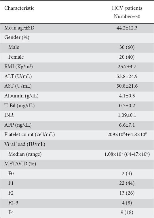
The present study was conducted on 50 HCV patients. Their age ranged from 27-65 years. The mean age was 44.2±12. Demographic features, laboratory data and histopathological features of the included patients are shown in Table 1.
Table 1.
Demographic features, laboratory data and histopathological features of the included patients
The analysis of the accuracy of RTE, FibroScan and APRI in predicting liver fibrosis in 50 HCV patients is shown in Table 2. At cut-off value 7.5 KPa, FibroScan could diagnose significant fibrosis (F ≥2) with sensitivity 88%, specificity 100%, PPV 100%, NPV 89.3%, and AUROC 0.94 (Table 2, Fig. 1). RTE could diagnose significant fibrosis (F ≥2) at cut-off value 2.49 with sensitivity 100%, specificity 66%, PPV 74.6%, NPV 100%, and AUROC 0.8 (Table 2, Fig. 2). APRI, at cut off value 0.65, could predict significant fibrosis (F ≥2) with sensitivity 54%, specificity 80%, PPV 72.9%, NPV 63.5%, and AUROC 0.69 (Table 2, Fig. 3). There was a significant positive correlation between the FibroScan score and RTE score (r=0.6, P=0.001) (Fig. 4).
Table 2.
Diagnostic accuracy of FibroScan, aspartate aminotransferase to platelet ratio index (APRI), real-time elastography (RTE) compared to histopathology in 50 HCV patients in prediction of significant fibrosis (≥F2)
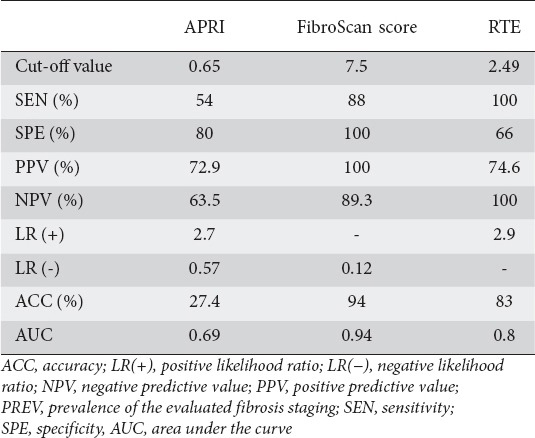
Figure 1.
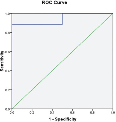
ROC curve for FibroScan
Figure 2.
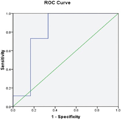
ROC curve for real-time elastography
Figure 3.
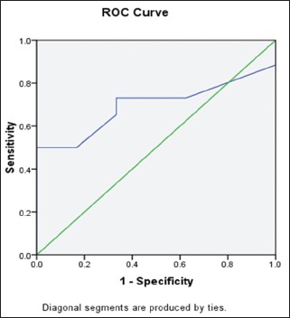
ROC curve for aspartate aminotransferase to platelet ratio index
Figure 4.

Correlation between real-time elastography and FibroScan score
Discussion
Ultrasonography-based noninvasive approaches are increasingly considered to assess parenchymal stiffness and progression of CLD [21]. FibroScan measures the propagation speed of shear waves [22-24]. RTE captures 2D strain images induced by internal heart beats, and the strain images show progressively increasing patchiness with increasing severity of fibrosis [13,14]. Therefore, it is possible to perform in obese patients or with ascites. In the current study, the diagnostic accuracy of RTE and TE in assessing significant liver fibrosis was compared against histopathology in CHC patients with BMI ≤30.
In CHC patients, prognosis and management strongly depend on the degree of liver fibrosis [4]. Treatment should be initiated promptly in those with severe fibrosis (F3-F4) and should be strongly considered in CHC patients with significant fibrosis (F >2). Our study showed that TE was able to diagnose the presence of significant fibrosis at a cut off value of 7.5 kPa with a sensitivity of 88%, specificity of 100%, PPV 100%, and NPV 89.3%. The overall accuracy was found to be 94% with no failure of TE was recorded. This high diagnostic performance of TE was probably explained by exclusion of patients with BMI >30 from this study. Likewise, previous studies showed that the AUROCs ranged from 0.79 to 0.83 for the prediction of significant hepatic fibrosis and were over 0.95 for the identification of cirrhosis [25,26].
Other studies reported that TE showed a good diagnostic performance in predicting significant fibrosis, and disease progression to advanced fibrosis and cirrhosis when compared to other noninvasive tests [25,27-30]. Friedrich-Rust et al [29] assessed the overall performance of TE for the diagnosis of liver fibrosis; the AUROCs were 0.84 for significant fibrosis (F ≥2), 0.89 for severe fibrosis (F ≥3), and 0.94 for the diagnosis of cirrhosis (F=4).
RTE had been reported to be useful for assessment of hepatic fibrosis in patients with CHC while is not in patients with nonalcoholic fatty liver disease [15]. In the work of Tatsumi et al (2008), RTE correlated well with liver stiffness measured by FibroScan [31]. In our study, RTE was able to diagnose significant fibrosis (≥F2) with a sensitivity of 100%, specificity of 66%, and an overall accuracy of 83%. Although inferior to TE in determining significant hepatic fibrosis, RTE still showed a highly significant positive correlation with TE (P=0.001). Thus, RTE may be useful for assessing hepatic fibrosis in patients for whom the application of FibroScan may be limited.
FibroScan had some limitations in special patients [32]. Furthermore, examination with FibroScan often requires the use of ultrasonography to find the good window because there is no B-mode and around 20% of the cases, reliable measurements cannot be obtained by TE using the standard M-probe [11]. On the contrary, RTE displays in real time the relative strain of the tissue by measuring its displacement and it can easily find the most appropriate region and capture the value. Better results may be achieved by a combination of FibroScan (a technology based on shear wave propagation) and RTE (a technology based on tissue distortion).
In the current study, APRI at cutoff value 0.65 could predict significant fibrosis (F ≥2) with sensitivity 54%, specificity 80% and AUROC 0.69; at cut-off 0.5 the sensitivity was 65.4% and specificity was 66.7%, and at cutoff 1.5 the sensitivity was 30.8% and specificity of 100%. The diagnostic accuracy of APRI in the current study was lower than previous studies comparing APRI with FibroScan or FibroTest [16,25,33].
Ferraioli et al (2012) suggested that real-time strain elastography can be used in the same way as TE is being used for the assessment of severe fibrosis and cirrhosis, with the benefit of improved assessment of significant fibrosis as no significant difference was observed between AUROCs of TE and real-time SWE for severe fibrosis (0.96 and 0.98, respectively) [34].
In conclusion, although FibroScan is superior in determining significant hepatic fibrosis, our data suggest that RTE may be a useful and promising noninvasive method for liver fibrosis assessment in CHC patients, especially in cases with technical limitations for FibroScan.
Summary Box.
What is already known:
Noninvasive methods for liver fibrosis assessment as ultrasound-based approaches have emerged to replace liver biopsy
FibroScan is the most widely used and proved technique that measures liver stiffness based on the propagation speed of shear waves
Real-time elastography (RTE) is technically different (a technology based on tissue distortion)
What the new findings are:
The overall diagnostic accuracy of FibroScan was found to be 94%, while in RTE it was 83%
Although RTE was less than FibroScan in determining significant liver fibrosis, our data suggest that RTE may be a useful for liver fibrosis assessment in chronic hepatitis C patients especially in cases with technical limitations for FibroScan
There was a significant positive correlation between the FibroScan score and RTE score (r=0.6, P=0.001)
Acknowledgment
All authors would like to thank Dr Mohamed Hassany for his cooperation and paying much effort to regulate the work
Biography
National Hepatology and Tropical Medicine Research Institute; Cairo University, Cairo, Egypt
Footnotes
Conflict of Interest: None
References
- 1.WHO |Hepatitis C. World Health Organization. 2012 Fact sheet no.164. [Google Scholar]
- 2.Mohamoud YA, Mumtaz GR, Suzanne R, Miller D. The epidemiology of hepatitis C virus in Egypt:a systematic review and data synthesis. BMC Infect Dis. 2013;13:288. doi: 10.1186/1471-2334-13-288. [DOI] [PMC free article] [PubMed] [Google Scholar]
- 3.Nguyen MH, Keeffe EB. Prevalence and treatment of hepatitis C virus genotypes 4, 5, and 6. Clin Gastroenterol Hepatol. 2005;3(Suppl 2):S97–S101. doi: 10.1016/s1542-3565(05)00711-1. [DOI] [PubMed] [Google Scholar]
- 4.European Association for the Study of the Liver. EASL Clinical Practice Guidelines:management of hepatitis C virus infection. J Hepatol. 2011;55:245–264. doi: 10.1016/j.jhep.2011.02.023. [DOI] [PubMed] [Google Scholar]
- 5.Friedman SL, Bansal MB. Reversal of hepatic fibrosis-fact or fantasy? Hepatology. 2006;43:82–88. doi: 10.1002/hep.20974. [DOI] [PubMed] [Google Scholar]
- 6.Bravo AA, Sheth SG, Chopra S. Liver biopsy. N Engl J Med. 2001;344:495–500. doi: 10.1056/NEJM200102153440706. [DOI] [PubMed] [Google Scholar]
- 7.Colloredo G, Guido M, Sonzogni A. Impact of liver biopsy size on histological evaluation of chronic viral hepatitis:the smaller the sample, the milder the disease. J Hepatol. 2003;39:239–244. doi: 10.1016/s0168-8278(03)00191-0. [DOI] [PubMed] [Google Scholar]
- 8.Foucher J, Chanteloup E, Vergniol J. (FibroScan):Diagnosis of cirrhosis by transient elastography (FibroScan):a prospective study. Gut. 2006;55:403–408. doi: 10.1136/gut.2005.069153. [DOI] [PMC free article] [PubMed] [Google Scholar]
- 9.Takeda T, Yasuda T, Nakayama Y. Usefulness of noninvasive transient elastography for assessment of liver fibrosis stage in chronic hepatitis C. World J Gastroenterol. 2006;12:7768–7773. doi: 10.3748/wjg.v12.i48.7768. [DOI] [PMC free article] [PubMed] [Google Scholar]
- 10.European Association for the Study of the Liver. J Hepatology. 2015;63:237–264. doi: 10.1016/j.jhep.2022.10.006. [DOI] [PubMed] [Google Scholar]
- 11.Castera L, Foucher J, Bernard PH, et al. Pitfalls of liver stiffness measurement:a 5-year prospective study of 13,369 examinations. Hepatology. 2010;51:828–835. doi: 10.1002/hep.23425. [DOI] [PubMed] [Google Scholar]
- 12.Wong GL, Wong VW, Chim AM, et al. Factors associated with unreliable liver stiffness measurement and its failure with transientelastography in the Chinese population. J Gastroenterol Hepatol. 2011;26:300–305. doi: 10.1111/j.1440-1746.2010.06510.x. [DOI] [PubMed] [Google Scholar]
- 13.Friedrich-Rust M, Ong M, Herrmann E, et al. Real-time elastography for noninvasive assessment of liver fibrosis in chronic viral hepatitis. AJR. 2007;188:758–764. doi: 10.2214/AJR.06.0322. [DOI] [PubMed] [Google Scholar]
- 14.Morikawa H, Fukuda K, Kobayashi S, et al. Real-time tissue elastography as a tool for the noninvasive assessment of liver stiffness in patients with chronic hepatitis C. Gastroenterology. 2011;46:350–358. doi: 10.1007/s00535-010-0301-x. [DOI] [PubMed] [Google Scholar]
- 15.Tomeno W, Yoneda M, Imajo K, et al. Evaluation of the liver fibrosis index calculated by using real-time tissue elastography for the non-invasive assessment of liver fibrosis in chronic liver diseases. Hepatol Res. 2013;43:735–742. doi: 10.1111/hepr.12023. [DOI] [PubMed] [Google Scholar]
- 16.Wai CT, Greenson JK, Fontana RJ, et al. A simple noninvasive index can predict both significant fibrosis and cirrhosis in patients with chronic hepatitis C. Hepatology. 2003;38:518–526. doi: 10.1053/jhep.2003.50346. [DOI] [PubMed] [Google Scholar]
- 17.Ziol M, Handra-Luca A, Kettaneh A, et al. Noninvasive assessment of liver fibrosis by measurement of stiffness in patients with chronic hepatitis C. Hepatology. 2005;41:48–54. doi: 10.1002/hep.20506. [DOI] [PubMed] [Google Scholar]
- 18.Castera L, Forns X, Alberti A. Non-invasive evaluation of liver fibrosis using transient elastography. J Hepatol. 2008;48:835–847. doi: 10.1016/j.jhep.2008.02.008. [DOI] [PubMed] [Google Scholar]
- 19.Kettaneh A, Marcellin P, Douvin C, et al. Features associated with success rate and performance of fibroscan measurements for the diagnosis of cirrhosis in HCV patients:a prospective study of 935 patients. J Hepatol. 2007;46:628–634. doi: 10.1016/j.jhep.2006.11.010. [DOI] [PubMed] [Google Scholar]
- 20.Bedossa P, Poynard T. The METAVIR cooperative study group. An algorithm for the grading of activity in chronic hepatitis C. Hepatology. 1996;24:289–293. doi: 10.1002/hep.510240201. [DOI] [PubMed] [Google Scholar]
- 21.Paparo F, Corradi F, Cevasco L, et al. Real-time elastography in the assessment of liver fibrosis:a review of qualitative and semi-quantitative methods for elastogram analysis. Ultrasound Med Biol. 2014;40:1923–1933. doi: 10.1016/j.ultrasmedbio.2014.03.021. [DOI] [PubMed] [Google Scholar]
- 22.Ferraioli G, Tinelli C, Dal Bello B, Zicchetti M, Filice G, Filice C. Accuracy of real-time shear wave elastography for assessing liver fibrosis in chronic hepatitis C:a pilot study. Hepatology. 2012;56:2125–2133. doi: 10.1002/hep.25936. [DOI] [PubMed] [Google Scholar]
- 23.Friedrich-Rust M, Wunder K, Kriener S, et al. Liver fibrosis in viral hepatitis:noninvasive assessment with acoustic radiation force impulse imaging versus transient elastography. Radiology. 2009;252:595–604. doi: 10.1148/radiol.2523081928. [DOI] [PubMed] [Google Scholar]
- 24.Sandrin L, Fourquet B, Hasquenoph J, et al. Transient elastography:a new noninvasive method for assessment of hepatic fibrosis. Ultrasound Med Biol. 2003;29:1705–1713. doi: 10.1016/j.ultrasmedbio.2003.07.001. [DOI] [PubMed] [Google Scholar]
- 25.Castera L, Vergniol J, Foucher J, et al. Prospective comparison of transient elastography, Fibrotest, APRI, and liver biopsy for the assessment of fibrosis in chronic hepatitis C. Gastroenterology. 2005;128:343–350. doi: 10.1053/j.gastro.2004.11.018. [DOI] [PubMed] [Google Scholar]
- 26.Ziol M, Handra-Luca A, Kettaneh A, et al. Noninvasive assessment of liver fibrosis by measurement of stiffness in patients with chronic hepatitis C. Hepatology. 2005;41:48–54. doi: 10.1002/hep.20506. [DOI] [PubMed] [Google Scholar]
- 27.Shaheen AA, Myers RP. Diagnostic accuracy of the aspartate aminotransferase-to-platelet ratio index for the prediction of hepatitis C-related fibrosis:a systematic review. Hepatology. 2007;46:912–921. doi: 10.1002/hep.21835. [DOI] [PubMed] [Google Scholar]
- 28.Poynard T, Morra R, Halfon P, et al. Meta-analyses of FibroTest diagnostic value in chronic liver disease. BMC Gastroenterol. 2007;7:40. doi: 10.1186/1471-230X-7-40. [DOI] [PMC free article] [PubMed] [Google Scholar]
- 29.Friedrich-Rust M, Ong MF, Martens S, et al. Performance of transient elastography for the staging of liver fibrosis:a meta-analysis. Gastroenterology. 2008;134:960–974. doi: 10.1053/j.gastro.2008.01.034. [DOI] [PubMed] [Google Scholar]
- 30.Castèra L, Le Bail B, Roudot-Thoraval F, et al. Early detection in routine clinical practice of cirrhosis and oesophageal varices in chronic hepatitis C:comparison of transient elastography (FibroScan) with standard laboratory tests and non-invasive scores. J Hepatol. 2009;50:59–68. doi: 10.1016/j.jhep.2008.08.018. [DOI] [PubMed] [Google Scholar]
- 31.Tatsumi R, Kudo M, Ueshima K, et al. Noninvasive evaluation of liver fibrosis Using serum fibrotic markers, transient elastography (FibroScan) and real-time tissue elastography. Intervirology. 2008;51(Suppl 1):27–33. doi: 10.1159/000122602. [DOI] [PubMed] [Google Scholar]
- 32.Fraquelli M, Rigamonti C, Casazza G, et al. Reproducibility of transient elastography in the evaluation of liver fibrosis in patients with chronic liver disease. Gut. 2007;56:968–973. doi: 10.1136/gut.2006.111302. [DOI] [PMC free article] [PubMed] [Google Scholar]
- 33.Le Calvez S, Thabut D, Messous D, et al. The predictive value of Fibrotest vs. APRI for the diagnosis of fibrosis in chronic hepatitis C. Hepatology. 2004;39:862–863. doi: 10.1002/hep.20099. [DOI] [PubMed] [Google Scholar]
- 34.Ferraioli G, Tinelli C, Malfitano A, Dal Bello B, Filice G, Filice C. Liver Fibrosis Study Group. Above E, Barbarini G, Brunetti E, et al. Performance of real-time strain elastography, transient elastography, and aspartate to- platelet ratio index in the assessment of fibrosis in chronic hepatitis C. AJR Am J Roentgenol. 2012;199:19–25. doi: 10.2214/AJR.11.7517. [DOI] [PubMed] [Google Scholar]


