Fig. 3.

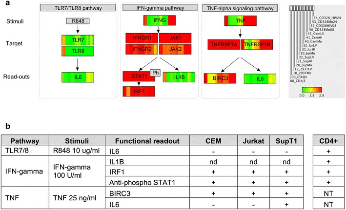
Defects in 3 selected innate immunity pathways in cell lines. a The figure represents a simplified view of the TLR7/TLR8, IFN-gamma and TNF-alpha signaling pathways. Boxes representing genes display the transcriptional levels detected in RNA-seq libraries of resting CD4+ T cells, the four human laboratory cell lines HEK293T, Jurkat, SupT1 and CEM -mock (MO), heat-inactivated (HI) and HIV-infected (HIV)- and 4 samples corresponding to Activated CD4+ T cells at 8 and 24 h after TCR activation. Inset describes the order of the libraries as well as the color-code scale of expression levels (log10 transformation of the number of library size-normalized reads per kilobase of exonic sequence) ranging from 0 (green) to ≥2.8 (red; lower limit of the 9th-decile of expression values). The expression levels indicated for IFNG and TNF convey the basal expression level before adding the stimuli. b Experimental validation of the functional integrity of the selected innate immunity pathways. The table reports the stimuli applied and the functional read-out measured 24 h after stimulation. A positive sign indicates positive detection of functional read-outs (transcript levels by RT-qPCR or phosphorylation of STAT1 by Western blot analysis). NT not tested, nd not detected
