Erratum
Following the publication of our article in Stem Cell Research & Therapy [1], we have become aware that errors were introduced inadvertently in Fig. 2.
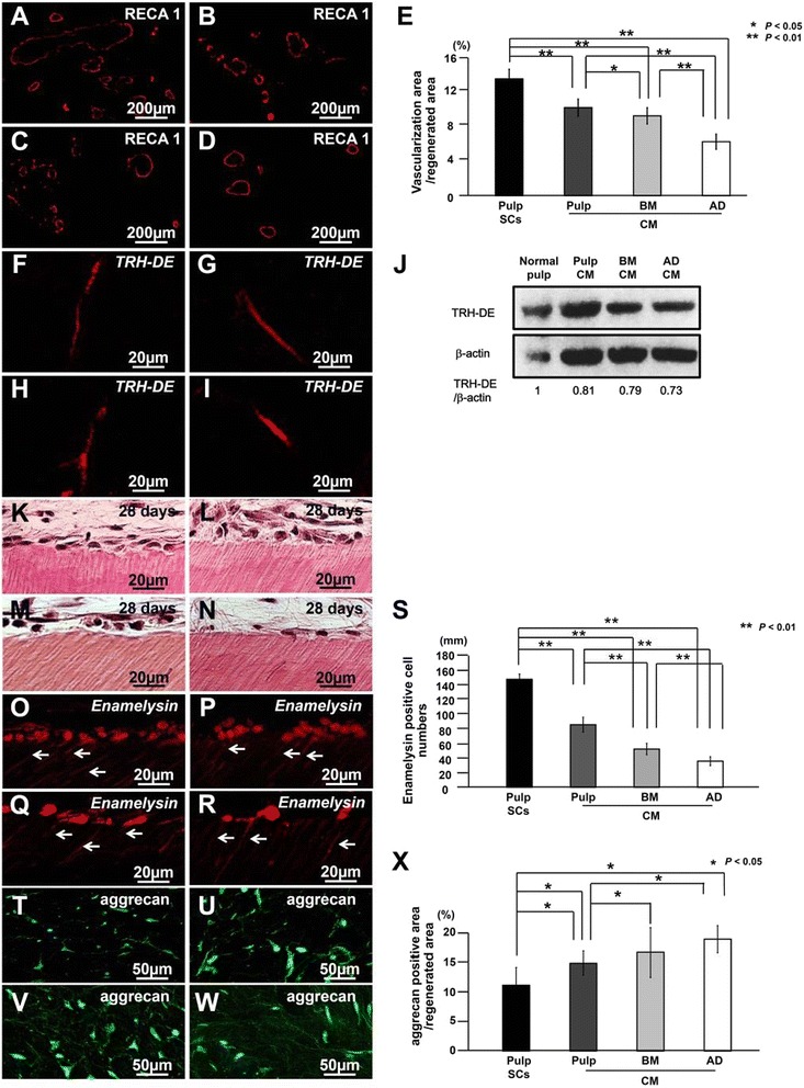
Fig. 2.

Characterization of regenerated tissue on day 28 in an ectopic tooth root transplantation model. a, f, k, o, t Transplant of pulp CD31− side population (SP) cells (Pulp SCs). b, g, l, p, u Transplant of conditioned medium (CM) from pulp CD31− SP cells (Pulp CM). c, h, m, q, v Transplant of CM from bone marrow CD31− SP cells (BM CM). d, i,n, r, w Transplant of CM from adipose CD31− SP cells (AD CM). a-d Immunostaining with rat endothelial cell antigen 1 (RECA1). e Ratio of vascularization area to the total regenerated area. (f-i) In situ hybridization analysis of expression of thyrotropin-releasing hormone-degrading enzyme (TRH-DE) as a pulp marker using an anti-sense probe reactive to both porcine and mouse genes. j Protein expression of TRH-DE in regenerated pulp after transplantation of CM from pulp, bone marrow (BM), and adipose (AD) CD31− SP cells. k-s Odontoblastic differentiation potential in the regenerated pulp. k-n Odontoblastic cells along with the dentinal wall. o-r In situ hybridization analysis of enamelysin. Odontoblastic process extending into the tubular dentin (arrows). s Comparison of the numbers of enamelysin-positive cells along the dentinal wall. t-w Immunostaining with aggrecan (green) merged with Hoechst 33342 (Blue). x Ratio of aggrecan-positive area to the total regenerated area. Data are expressed as mean ± standard deviation of four determinations. *P < 0.05, **P < 0.01
During the preparation of panel J, the bands of β-actin were cut out and attached in the middle to remove non-specific bands. The bands of all samples were located on the same membrane. Unfortunately this panel was included in our article by mistake.
We are now providing a new version of Fig. 2 below which presents in panel J experimental data which were obtained at the same time.
The protein band intensity was re-quantified by densitometry (CS Analyzer) using the correct TRH-DE and β-actin band. Relative protein expression level was evaluated on the basis of band intensity of TRH-DE/β-actin. Each expression level of normal pulp was defined as 1.0.
We apologize for this error and confirm that the conclusions of the article are not affected.
Footnotes
The online version of the original article can be found under doi:10.1186/s13287-015-0088-z.
References
- 1.Hayashi Y, Murakami M, Kawamura R, Ishizaka R, Fukuta O, Nakashima M. CXCL14 and MCP1 are potent trophic factors associated with cell migration and angiogenesis leading to higher regenerative potential of dental pulp side population cells. Stem Cell Res Ther. 2015;6:111. doi: 10.1186/s13287-015-0088-z. [DOI] [PMC free article] [PubMed] [Google Scholar]


