Abstract
Background
Gastric antral vascular ectasia (GAVE) is characterized by mucosal and submucosal vascular ectasia causing recurrent hemorrhage and thus, chronic anemia, in patients with cirrhosis. Treatment with argon plasma coagulation (APC) is an effective and safe method, but requires multiple sessions of endoscopic therapy. Endoscopic band ligation (EBL) was found to be a good alternative for APC as a treatment for GAVE, especially in refractory cases. The aim of this prospective randomized controlled study was to evaluate the safety and efficacy of EBL, as compared to APC, in the treatment of GAVE and gastric fundal vascular ectasia (GFVE).
Patients and methods
A total of 88 cirrhotic patients with GAVE were prospectively randomized to endoscopic treatment with either EBL or APC, every 2 weeks until complete obliteration was accomplished; then they were followed up endoscopically after 6 months, plus they had monthly measurement of hemoglobin levels during that period.
Results
We describe the presence of mucosal and submucosal lesions in the gastric fundal area that were similar to those found in GAVE in 13 patients (29.5%) of the EBL group and 9 patients (20.5%) of the APC group; we named this GFVE. In these cases, we treated the fundal lesions with the same techniques we had used for treating GAVE, according to the randomization. We found that EBL significantly decreased the number of sessions required for complete obliteration of the lesions (2.98 sessions compared to 3.48 sessions in the APC group (p < 0.05)). Hemoglobin levels increased significantly after obliteration of the lesions in both groups, compared to pretreatment values (p < 0.05), but with no significant difference between the two groups (p > 0.05); however, the EBL group of patients required a significantly smaller number of units of blood transfusion than the APC group of patients (p < 0.05). There were no significant differences in adverse events nor complications between the two groups (p > 0.05).
Conclusions
This study described and histologically proved the presence of GFVE occurring comcomitantly with GAVE in cirrhotic patients. We showed that GFVE can be successfully managed by EBL or APC. Our study revealed that EBL is more effective and is comparable in safety to APC, in the treatment of GAVE and GFVE in cirrhotic patients.
Keywords: Anemia, argon plasma coagulation, cirrhosis, comparative study, endoscopic band ligation, gastric antral vascular ectasia, gastric fundal vascular ectasia, transfusion, treatment techniques
Introduction
Gastric antral vascular ectasia (GAVE), or watermelon stomach, is an uncommon cause of non-variceal gastrointestinal hemorrhage.1,2 GAVE is characterized by mucosal and submucosal vascular ectasia causing recurrent gastrointestinal hemorrhage; and consequently, chronic anemia, in patients with cirrhosis.3
The majority of patients present with iron-deficiency anemia, secondary to occult blood loss. Indeed, 60–70% of patients are transfusion-dependent due to recurrent anemia, despite iron supplementation. Patients also present with positive fecal occult blood on routine check-up. In addition, some patients present with overt gastrointestinal bleeding, in the form of intermittent melena and, occasionally, hematemesis.4 The classic features of GAVE include red, often hemorrhagic lesions, aggregated in linear strips or diffusely, predominantly located in the gastric antrum; which can result in significant blood loss.3 Histologically, dilated mucosal capillaries with fibrin thrombi and fibromuscular hyperplasia of the lamina propria are seen, without inflammation.5
GAVE has been associated with liver cirrhosis, renal failure, bone marrow transplantation, scleroderma, systemic lupus erythematosus (SLE), ischemic heart disease, valvular heart disease, hypertension, familial Mediterranean fever, hypothyroidism, diabetes, hypergastrinemia and acute myeloid leukemia.6–8
Considering the treatment options for GAVE, the non-endoscopic treatments that aimed at reducing the bleeding without ablative therapy, such as beta-blockers, octreotide, thalidomide or tranexamic acid have provided little benefit.9 Treatment with argon plasma coagulation (APC) is a safe, effective method to decrease the blood loss in patients with GAVE.10 On medium and long-term follow-up after treatment, it has been found that APC has a high recurrence rate. Therefore, endoscopic band ligation (EBL) was thought to be effective for refractory GAVE, as it may lead to obliteration of the submucosal vascular plexus.11 EBL was recently found to be useful as a treatment for GAVE12 and a good alternative for APC,10 especially in refractory cases11 and in cases complicated with polyp formation.12
This prospective, randomized controlled study aimed at comparing the safety and efficacy of EBL and APC in the treatment of GAVE, and of gastric fundal vascular ectasia (GFVE).
Patients and methods
Study design and end points
This is a prospective, open-label, randomized controlled trial. The included patients were randomized into either the EBL or APC group, using a computerized random number generator to select randomly permuted blocks with a block size of six and an equal allocation ratio. Sequentially numbered, opaque, sealed envelopes were used to ensure concealment. Study collaborators who were not involved in the upper endoscopy procedures recruited, enrolled and assigned the participants to a computer-generated randomization sequence, held by an independent observer. The intensity of ablation, number of bands and efficacy of obliteration of lesions were judged by two observing endoscopists, in communication with the operating endoscopist, every individual session. The primary endpoint was the number of treatment sessions needed for obliteration of GAVE.
Patients
We conducted this study on 88 cirrhotic patients, whom were admitted to the departments of Tropical Medicine and Internal Medicine, and whom were found to have gastric antral and fundal (around the cardia) vascular ectasia (Figure 1), from December 2012 to December 2013. Endoscopic biopsies were taken from the affected pericardial fundal lesions and the antrum, before commencing a treatment, in order to compare the pathologic findings.
Figure 1.
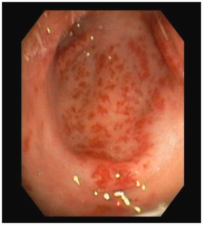
GAVE before treatment.
GAVE: gastric antral vascular ectasia.
Methods
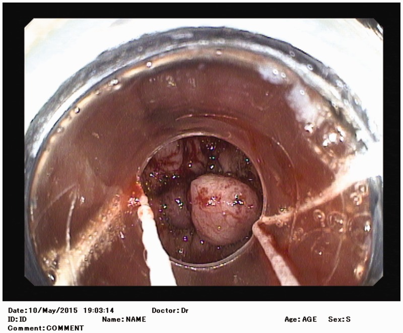
Patients were randomized into two groups: Group I: patients were treated with EBL. Averages of 8 bands (range: 6–12) were applied every 2 weeks until complete obliteration of lesions. (Figure 2) Group II: patients were treated with APC. Forced APC (60 W with argon gas flow of 2 L/min) was applied dynamically to the antral lesions beginning near the pylorus and proceeding proximally for antral lesions. The same settings were applied to lesions in the cardiac fundus starting from just below the Z-line in the retroflex position. The procedure was repeated every two weeks until complete endoscopic obliteration was accomplished. Then, all patients had a monthly follow up of their clinical and laboratory parameters for 6 months.
Figure 2.
Endoscopic band ligation for GAVE.
GAVE: Gastric antral vascular ectasia.
Post-treatment follow up included recording cessation of bleeding, hemoglobin measurement, transfusion requirements, number of treatment sessions and endoscopic recurrence.
Institution ethical committee approval and an informed consent from every patient were obtained.
Power estimates and statistical methods
A sample size of 88 patients (44 patients in each treatment group) was estimated, based on a previous study,13 so that the treatment of GAVE associated with liver diseases with EBL had a mean of 3 (SD 0.9) sessions, compared to 2.3 (SD 0.9) with APC; with a power of 90% and a significance level of 5% (2-sided).
Our statistical data were reported as the mean ± SD; with frequencies (n) and percentages (%), when appropriate. A comparison of the numerical variables, including the primary end point between the study groups, was performed using the Student’s t-test to compare independent samples from the two groups, when the samples were normally distributed. The Mann-Whitney U test was used to compare independent samples, when these samples were not normally distributed. To compare categorical data, the chi square test was performed. P-values of < 0.05 (2-tailed) were considered statistically significant. All our statistical calculations were performed using the computer program Statistical Package for the Social Science (SPSS, Chicago, IL, USA) version15 for Microsoft Windows.
Results
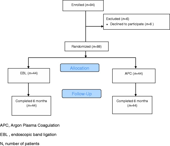
A total number of 94 patients with GAVE associated with liver disease were enrolled in our study. Of these, 88 patients were randomized to be treated with either APC or EBL (Figure 3).
Figure 3.
Study flow chart.
APC: argon plasma coagulation; EBL: endoscopic band ligation; n: number of patients
The demographic and endoscopic profiles of the studied groups are summarized in Table 1. There was no statistically significant difference between the groups regarding age, gender, the presence of diabetes or hypertension (p > 0.05).
Table 1.
Demographic and endoscopic profile of the studied groups
| EBL (n = 44) n (%) | APC (n = 44) n (%) | P-value | |
|---|---|---|---|
| Age(mean ± SD) | 51.41 ± 7.54 | 53.09 ± 7.16 | 0.233 |
| Gender(M/F) | 19/25 | 15/29 | 0.943 |
| HTN | 4/44 (9.1%) | 8/44 (18.2%) | 0.819 |
| DM | 11/44 (25%) | 14/44 (31.8%) | 0.973 |
| Child-Pugh classification | |||
| A | 2 (4.6%) | 0 (0%) | |
| B | 22 (50%) | 23 (52.3%) | 0.915 |
| C | 20 (45.4%) | 21 (47.7%) | |
| Mean score | 9.46 ± 1.72 | 9.52 ± 1.69 | 0.910 |
| Endoscopic findings of EV | |||
| Grade I | 3 (6.8%) | 2 (4.6%) | |
| Grade II | 7 (15.9%) | 7 (15.9%) | |
| Grade III | 3 (6.8%) | 6 (13.6%) | 0.999 |
| Grade IV | 3 (6.8%) | 3 (6.8 %) | |
| No varices | 28 (63.6%) | 26 (59.1%) | |
| Previous treatment for EV | 6/44 (13.6%) | 14/44 (31.8%) | 0.387 |
| Fundal ectasia | 13/44 (29.5%) | 9/44 (20.5%) | 0.914 |
| Previous treatment for GAVE | |||
| APC | 9/44 (20.5%) | 3/44 (6.8%) | 0.482 |
| Blood transfusion | 2/44 (4.5%) | 5/44 (11.4%) | 0.845 |
| Blood transfusion units(mean ± SD) | 2.5 ± 0.707 | 4.6 ± 0.894 | 0.033* |
| Treatment sessions(mean ± SD) | 2.93 ± 0.846 | 3.48 ± 0.902 | 0.007* |
| Complications | 6/44 (13.6%) | 9/44 (20.5%) | 0.948 |
HTN, hypertension;DM,diabetes mellitus; APC, argon plasma coagulation; EBL, endoscopic band ligation;EV, esophageal varices;GAVE, gastric antral vascular ectasia. P < 0.05 *significant.
The mean Child Turgotte Pugh (CTP) score was comparable in both the EBL (9.46 ± 1.72) and the APC groups (9.52 ± 1.69), with p > 0.05 (Table 1).
As regards the presence of esophageal varices (EV) and the history of previous endoscopic treatment for esophageal varices, there was no significant difference between either of the groups (p > 0.05) (Table 1).
GFVE was found in 13 out of 44 patients (29.5%) in the EBL group and in 9 out of 44 patients (20.5%) in the APC group, with no statistically significant difference (p > 0.05) (Table 1).
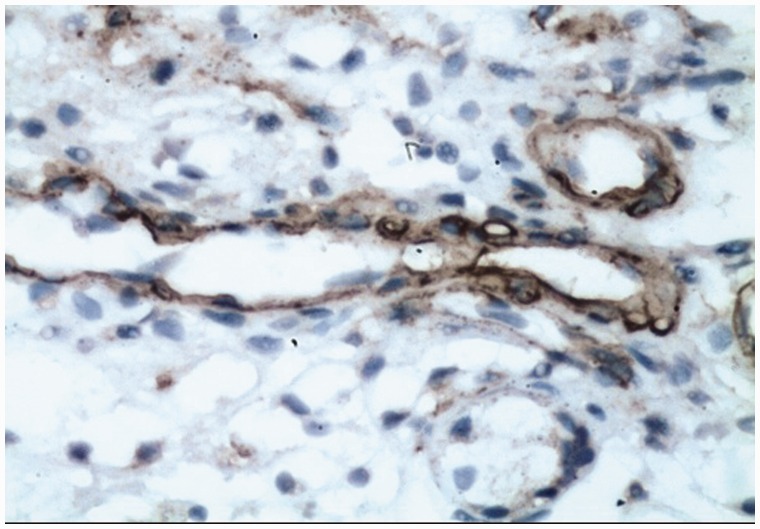
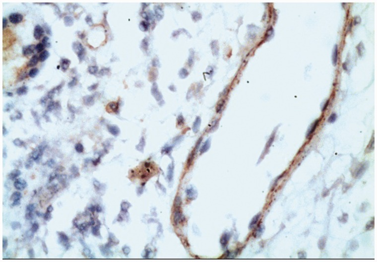
Histopathologic examination revealed similar pathologic findings of dilated capillaries in the submucosa of the pericardial fundal lesions (GFVE) (Figure 4) and the GAVE lesions (Figure 5).
Figure 4.
Dilated capillaries within the mucosa of GFVE. SMA immunostain shows the wall muscle layer (streptavidin biotin X250).
GFVE: Gastric fundal vascular ectasia; SMA: smooth muscle actin.
Figure 5.
Dilated capillaries within the mucosa of GAVE. SMA immunostain shows a continuous muscle layer (streptavidin biotin X250).
GAVE: Gastric antral vascular ectasia; SMA: smooth muscle actin.
The number of treatment sessions ranged from 2–5 sessions in the EBL group, with a mean of 2.93 ± 0.846; while in the APC group, the number of treatment sessions had a mean of 3.48 ± 0.902. The EBL group showed a statistically significant lower number of treatment sessions, when compared to the APC group (p = 0.007) (Table 1).
In the EBL group, the average hemoglobin levels increased from 6.73 ± 0.991 (range: 5–9 gm/dL) before treatment, to 10.31 ± 1.01 (range: 8.5–12 gm/dL) after treatment; and the difference was statistically significant (p < 0.001). In the APC group, hemoglobin levels increased from 6.72 ± 0.905 (range 5–9 gm/dL) before treatment, to 9.85 ± 0.906 (range 8–11 gm/dL) after treatment; and the difference was statistically significant (p < 0.001). Comparison of hemoglobin levels between the two groups, both before and after treatment, did not show any significant difference (p > 0.05) (Table 2).
Table 2.
Comparison between hemoglobin before and after treatment in the studied groups
| Hemoglobin g/dl | EBL (n = 44) | APC (n = 44) | P-value |
|---|---|---|---|
| Before (mean ± SD) | 6.73 ± 0.991 | 6.73 ± 0.905 | 0.851 |
| After (mean ± SD) | 10.31 ± 1.01 | 9.85 ± 0.906 | 0.051 |
| P-value | <0.001* | <0.001* |
APC, argon plasma coagulation; EBL, endoscopic band ligation. P < 0.05 *significant.
The patients of the EBL group required a significantly lower number of blood units transfused than in the APC group, with a mean of 2.5 units versus 4.6 units, respectively (p = 0.033) (Table 1).
Mild adverse events were recorded in 6 out of 44 patients (13.6%) in the EBL group, in the form of fever in two patients, mild bleeding from a post-band ulcer in one and epigastric pain in three patients. In the APC group, 9 out of 44 patients (20.5%) had adverse events, in the form of fever in two patients, abdominal distension in four patients and epigastric pain in three patients. No statistically significant difference was found (p > 0.05) (Table 1).
Discussion
Treatment of GAVE with APC requires multiple sessions for the management of vascular ectasia and control of bleeding. EBL is proposed as an alternative for APC in the treatment of GAVE that is causing recurrent gastrointestinal (GI) bleeding10 or is complicated with post-APC polyp formation.12 EBL was first reported in 2006 as a treatment for refractory GAVE in patients whom failed other treatment modalities, such as APC or hormonal therapy, by Sinha et al.14
Wells et al.10 performed an observational comparative study in 2008 that included nine patients treated with EBL and 13 patients treated with endoscopic thermal therapy (ETT). Their initial experience shows the superiority of EBL over ETT, as regards to reduction of treatment sessions, control of bleeding, period of hospitalization, need for transfusion and increase in hemoglobin values. This was in agreement with the results of the present study. Our study showed that EBL treatment significantly reduced the number of treatment sessions, compared to APC treatment sessions. Also, our study showed that EBL significantly reduced the need for transfusion, compared to APC.
Both Sato et al.13 in 2012 and Prachayakul et al.15 in 2013 conclude that EBL may be useful in the treatment of GAVE, to avoid the high recurrence rate after APC. Our study found that EBL had comparable safety and efficacy than APC. In agreement with these results, although they had fewer patients, Keohane et al.16 conclude in their retrospective study in 2013 that EBL is a safe and effective treatment of GAVE.
Recently in 2015, Zeped-Gomez et al.17 published a prospective study on a small number of patients using EBL in the treatment of GAVE: They found a significant increase in hemoglobin levels and a significant reduction in the need for transfusion.
In our work, we have described and histologically proven the presence of GFVE in 20.5–29.5% of our patients with GAVE. It was successfully treated with the same treatment modalities, according to randomization. This lesion should be sought for carefully, as we believe it may contribute to more bleeding and anemia. We do not know if GFVE can be present without GAVE, so future studies are needed to study a larger number of these cases, to discover if GFVE can be present alone and if GFVE can be present in non-cirrhotic patients.
The limitations of our study were that we did not widen the parameters of comparison, such as with the procedure time, length of hospital stay, patient satisfaction, how quality of life was affected, cost effectiveness, nor have a longer follow-up period.
In conclusion, our study described and histologically proved the presence of GFVE occurring concomitantly with GAVE in cirrhotic patients, and that GFVE can be successfully managed by EBL or APC. Our study revealed that EBL was more effective and that it is comparable in safety to APC, in the treatment of GAVE and GFVE, in cirrhotic patients.
References
- 1.Selinger CP, Ang YS. Gastric antral vascular ectasia (GAVE): An update on clinical presentation, pathophysiology and treatment. Digestion 2008; 77: 131–137. [DOI] [PubMed] [Google Scholar]
- 2.Rider JA, Klotz AP, Kirsner JB. Gastritis with veno-capillary ectasia as a source of massive gastric hemorrhage. Gastroenterology 1953; 24: 118–123. [PubMed] [Google Scholar]
- 3.Burak KW, Lee SS. Portal hypertensive gastropathy and gastric antral vascular ectasia (GAVE) syndrome. Gut 2001; 49: 866–872. [DOI] [PMC free article] [PubMed] [Google Scholar]
- 4.Sebastian S, O’Morain CA, Buckley MJ. Review article: Current therapeutic options for gastric antral vascular ectasia. Aliment Pharmacol Ther 2003; 18: 157–165. [DOI] [PubMed] [Google Scholar]
- 5.Suit PF, Petras RE, Bauer TW, et al. Gastric antral vascular ectasia. A histologic and morphometric study of watermelon stomach. Am J Surg Pathol 1987; 11: 750–757. [PubMed] [Google Scholar]
- 6.Ward EM, Raimondo M, Rosser BG, et al. Prevalence and natural history of gastric antral vascular ectasia (GAVE) in patients undergoing orthotopic liver transplantation. J Clin Gastroenterol 2004; 38: 898–900. [DOI] [PubMed] [Google Scholar]
- 7.Tobin RW, Hackman RC, Kimmey MB, et al. Bleeding from gastric antral vascular ectasia in marrow transplant patients. Gastrointest Endosc 1996; 44: 223–229. [DOI] [PubMed] [Google Scholar]
- 8.Takahashi T, Miya T, Oki M, et al. Severe hemorrhage from gastric antral vascular ectasia developed in a patient with AML. Int J Hematol 2006; 83: 467–468. [DOI] [PubMed] [Google Scholar]
- 9.Cho S, Zanati S, Yong E, et al. Endoscopic cryotherapy for the management of gastric antral vascular ectasia. Gastrointest Endosc 2008; 68: 895–902. [DOI] [PubMed] [Google Scholar]
- 10.Wells CD, Harrison ME, Gurudu SR, et al. Treatment of gastric antral vascular ectasia (watermelon stomach) with endoscopic band ligation. Gastrointest Endosc 2008; 68: 231–236. [DOI] [PubMed] [Google Scholar]
- 11.Sato T, Yamazaki K, Akaike J, et al. Endoscopic band ligation for refractory gastric antral vascular ectasia associated with liver cirrhosis. Clin J Gastroenterol 2011; 4: 108–111. [DOI] [PubMed] [Google Scholar]
- 12.Shah N, Cavanagh Y, Kaswala DH, et al. Development of hyperplastic polyps following argon plasma coagulation of gastric antral vascular ectasia. J Nat Sci Biol Med 2015; 6: 479–482. [DOI] [PMC free article] [PubMed] [Google Scholar]
- 13.Sato T, Yamazaki K, Akaike J. Endoscopic band ligation versus argon plasma coagulation for gastric antral vascular ectasia associated with liver diseases. Dig Endosc 2012; 24: 237–242. [DOI] [PubMed] [Google Scholar]
- 14.Sinha SK, Udawat HP, Varma S, et al. Watermelon stomach treated with endoscopic band ligation. Gastrointest Endosc 2006; 64: 1028–1031. [DOI] [PubMed] [Google Scholar]
- 15.Prachayakul V, Aswakul P, Leelakusolvong S. Massive gastric antral vascular ectasia successfully treated by endoscopic band ligation as the initial therapy. World J Gastrointest Endosc 2013; 5: 135–137. [DOI] [PMC free article] [PubMed] [Google Scholar]
- 16.Keohane J, Berro W, Harewood GC, et al. Band ligation of gastric antral vascular ectasia is a safe and effective endoscopic treatment. Dig Endoscopy 2013; 25: 392–396. [DOI] [PubMed] [Google Scholar]
- 17.Zepeda-Gomez S, Sultanian R, Teshima C, et al. Gastric antral vascular ectasia: A prospective study of treatment with endoscopic band ligation. Endoscopy 2015; 47: 538–540. [DOI] [PubMed] [Google Scholar]


