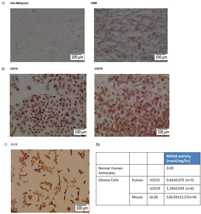
Figure 1. MAO A expression and activity are increased in mouse and human glioma cell lines and glioma tissues.

A. Non-malignant brain and glioma (GBM) tissue specimens, B. U251S (TMZ-sensitive) and U251R (TMZ-resistant) human glioma cells, and C. GL26 mouse glioma cells were stained for MAO A. The red precipitate denotes positive staining. Scale bars represent 100 μm. D. MAO A catalytic activity in U251S, U251R, GL26, and normal astrocytes was determined.
