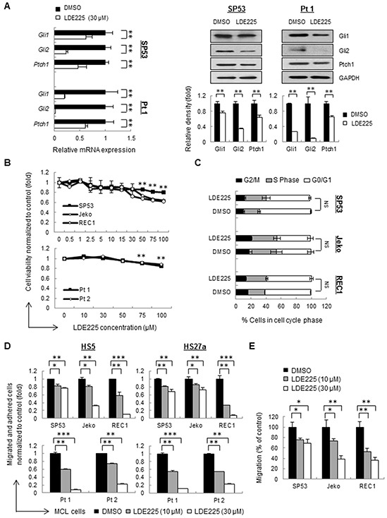
Figure 1. Hedgehog inhibitor LDE225 inhibits cell adhesion and migration without affecting cell viability in MCL cells.

A. The mRNA levels (Left) and protein levels (Right) of components in the Hh signaling pathway were measured by qRT-PCR and immunoblots. MCL cells (SP53) and primary patient cells were treated with LDE225 (30 μM) or DMSO. Each value in qRT-PCR was normalized to GAPDH and represents the mean ± S.D. from three independent experiments. GAPDH was used as a loading control, and the protein levels were quantified using a Gel-Pro Analysis software from three independent immunoblots. B. Dose-dependent LDE225-induced cytotoxicity (0-100 μM) at 48 h (primary cells) or 72 h (cell lines) was determined by MTT (3-(4,5-dimethythiazol-2-yl)-2,5-diphenyltetrazolium bromide) assays. Peripheral blood mononuclear cells (PBMC) were isolated from MCL patient aphaeresis blood by standard Ficoll gradient methods and CD19+ cells were purified using CD19-MicroBeads (Miltenyi). Data represent the mean ± S.D. from three independent experiments.**p < 0.01 (average viability of LDE225-treated cells vs. average viability of DMSO-treated cells; Student's t-test). C. Propidium iodide (PI) staining of MCL cells exhibited no obvious changes in the percentages of cells in the S-phase and G0/G1 of the cell cycle after treatment with LDE225 (30 μM). NS, not significant (vs. cells treated with DMSO; Student's t-test). D. MCL cell adhesion before and after LDE225 treatment (10 μM or 30 μM) was measured using cell lines and patients, which were stained with PKH26 prior to drug treatment. After 72 h of treatment, the cells were seeded onto a pre-established monolayer of HS5 or HS27a BMSCs. PKH26 dye intensity was analyzed and shown as the mean ± S.D. from three independent experiments. E. MCL cell migration is significantly reduced after LDE225 treatment. PKH26+ MCL cells were treated with LDE225 (10 μM or 30 μM) or DMSO for 72 h and then transferred to the transwell inserts. PKH26 dye intensity of migrated cells in the lower chamber was measured after incubation for 3 hours at 37°C in 5% CO2. The percentage of migrated cells relative to the cells treated with DMSO is indicated as the mean ± S.D. *p < 0.05, **p < 0.01, ***p < 1E-05 (vs. cells treated with DMSO; Student's t-test).
