Abstract
Fractures of hyoid bone resulting from trauma other than strangulation are very rare; hyoid bone fracture associated with panfacial trauma are even rarer. They occur more frequently in young individuals, and in men more than in women [1]. We report a comprehensive review of a case of hyoid bone fracture associated with head and neck trauma, induced by a direct blunt trauma onto the anterior neck.
Keywords: Hyoid bone, Head and neck trauma, Diagnosis, Treatment
Introduction
Injuries to the hyoid bone are rare. The most commonly reported injury is fracture, yet this is often a post-mortem finding, with an incidence of between 17–76 %, in victims of strangulation and hanging. In survivors it is more often associated with a trauma other than manual strangulation. The fracture of this bone is very rare accounting for only 0.002 % of all fractures. The hyoid bone is a U-shaped mobile bone situated in the anterior portion of the neck at the level of the C3 vertebra, in the angle between the mandible and the thyroid cartilage. It’s name originated from the Greek word hyoeides, which means “shaped like the letter upsilon” which represents the 20th letter in the Greek alphabets. It is composed of a body, two greater and two lesser horns and is a unique bone which is articulated to other bones by muscles or ligaments. The greater and lesser cornua fuse to body of hyoid bone between 40 and 60 years of age although non-fusion has been found even after the age of 60 years [1–4].
The primary function of hyoid bone is to provide attachment to the tongue, the larynx and the pharynx. Because of its morphology and its position amongst the other anatomical structures of the neck, it is associated not only with the sound production and the fully articulate human speech, but also with the smooth function of the airway and possibly with the normal swallowing. The following paper presents a thorough review of case of hyoid bone fracture associated with panfacial trauma in a 24 year old male.
Case Report
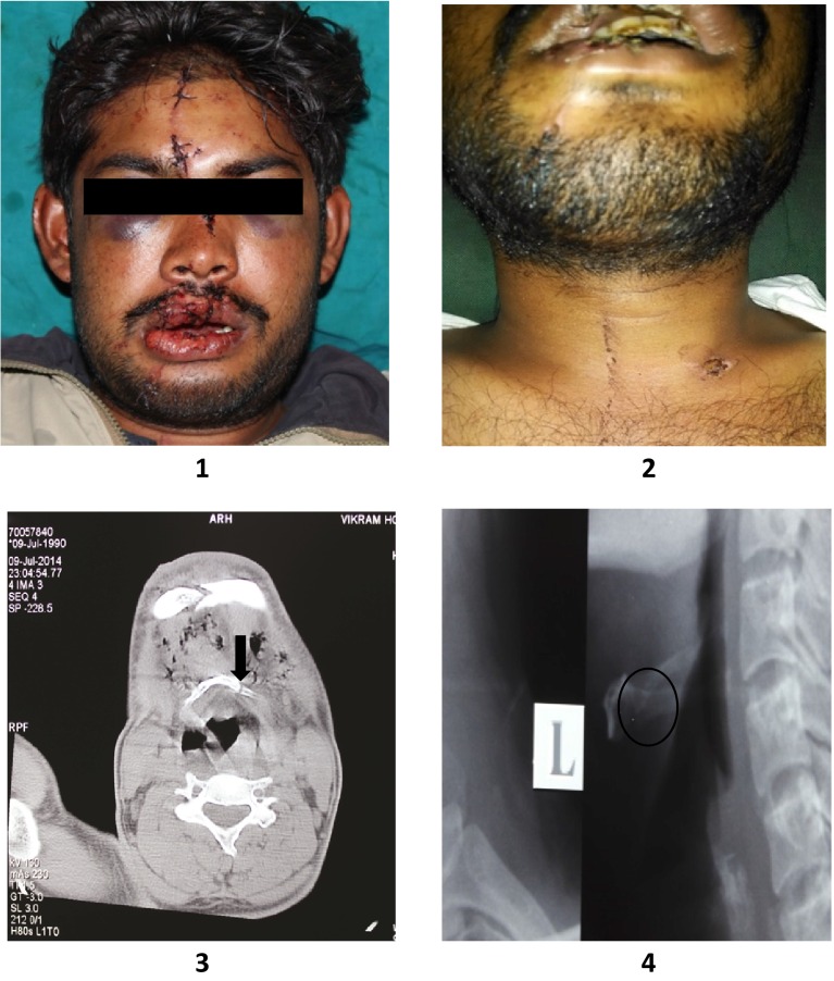
A 24 year old man presented to the casualty and emergency care with a complaint of pain over the face and neck region following a road traffic accident. His symptoms on presentation were a constant and severe neck pain, localized on the left side, with marked odynophagia when opening his mouth, swallowing or speaking. The patient had no dyspnoea or signs of airway compromise. Physical examination revealed tenderness over right chin and malar region, entire right side of maxilla was mobile with a midline split of maxilla, avulsion of mandibular incisors, tenderness on palpation of left side of hyoid bone with no external wounds noticed on neck except for some blunt traumatic injuries. Occlusion was deranged with maximal mouth opening of 30 mm; opening the mouth over this limit was painful.
Flexible nasal endoscopy was done in ENT outpatient department revealed a patent airway and a edematous vallecula on left side. The mechanism of injury and examination findings raised a clinical suspicion of a severe traumatic insult, and he was admitted to the oral and maxillofacial surgery ward to undergo an urgent CT scan of the head and neck. Diagnosis was achieved by a CT scan, orthopantomographic radiograph, lateral and antero-posterior view of cervical radiographs, which showed a fracture of the hyoid bone with panfacial fractures. Open reduction and internal fixation with mini plates was done for panfacial fractures, and the hyoid bone fracture was treated conservatively under general anaesthesia without any complications while intubation (Fig. 1–4).
Figs. 1–4.
1 PRE-OP frontal view, 2 PRE-OP frontal neck view, 3 CT scan axial view at C-3 and 4 Left lateral C-spine
Discussion
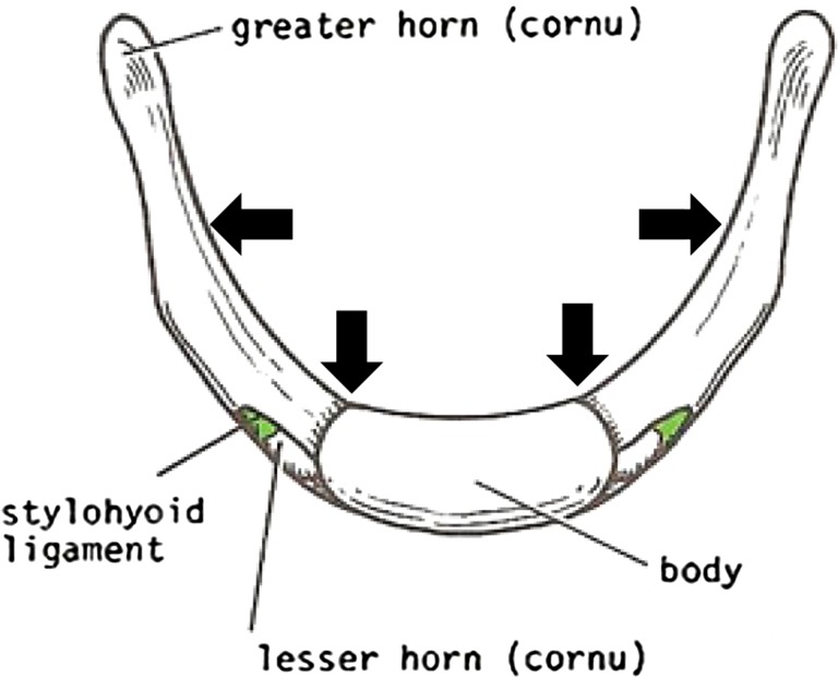
A fracture of hyoid bone secondary to trauma other than strangulation is a rare incident with only 31 cases being reported so far in the world literature [5]. This fracture accounts for 0.002 % of all fractures, but a higher incidence of 1.15 % was found by Stiebler and Maxeine [6]. Suspended from the styloid process, the hyoid bone is not susceptible to fractures owing to it’s inherent protection which is secured by its safe position and reinforced by its mobility. The hyoid bone is encircled, in normal anatomic position, by bones: anteriorly and laterally by the mandible and posteriorly by the cervical spine. Therefore, it is a well protected bone in the neck. However, when the neck is hyperextended, the protection is diminished, which allows the trauma to cause an isolated fracture of the hyoid. Fractures are seen frequently in fusion of greater cornua and body and mid-portion of greater cornua (Fig. 5). Patient’s age ranges between 15 and 55 years [7]. Most patients are under 35 years old, which may refer to the elastic cartilage-structure of the hyoid bone. They affect more frequently young individuals with a greater prevalence in the male gender.
Fig. 5.
Schematic diagram depicting hyoid bone composition and weak areas of hyoid bone leading to fracture (arrow marks)
Causes and Mechanism
In general hyoid bone fractures are reported to occur in 50 % of cases of manual strangulation or of ligature strangulation and in 27 % of hanging [8]. It can be classified into [8]:
Inward compression fractures with outside periosteal tears.
Antero-posterior compression fractures with inside periosteal tears.
Avulsion fractures.
Most cases of hyoid bone fractures are presented due to road traffic accidents, gunshot and knife wounds, others have happened during basketball, karate, martial arts or ice hockey as a result of falling assault, iatrogenically created following cervical spine surgery, or during attempts of resuscitation. Vomiting was also reported to be a causative factor [1, 2, 9].
Diagnosis
Diagnosis is difficult and is usually made with a strong degree of suspicion. Patients may suffer from severe pain in the area of throat, which is exaggerated by swallowing, nose blowing, or coughing. The most frequently presenting signs include anterior neck tenderness, swelling, ecchymosis, subcutaneous emphysema and crepitus, on palpation. Related symptoms such as dysphagia, severe pain especially by swallowing, or even dyspnoea may occur. Patient may suffer suffocation by tongue protrusion. Profuse bleeding may result from penetration of the bone fragments into the soft tissue and the pharynx.
Physical examination may reveal swelling in the area of trauma, crepitus in the neck, tenderness on palpation, and pain on moving the head. If a hyoid bone fracture is clinically strongly suspected in a patient with neck injury, diagnosis is made by radiographs, computed tomography, laryngoscopy, and surgical inspection [10] (penetrating trauma). Certain criteria are made for radiographic diagnosis including: radiolucent line, interruption of the cortical continuity, or displacement of the fragments. Laryngoscopy and CT scan are recommended for dyspnoea, haemoptysis, or bleeding from laryngeal lacerations, in order to assess the extent of injury.
Complications
Complications of hyoid bone fracture are divided into early and late. Early complications include subcutaneous emphysema, dyspnoea, pharyngeal tears and thyroid cartilage injury. Late complications are dysphagia, stridor, pseudoaneurysm of the external carotid artery, and crepitus by neck flexion [1]. Dysphagia, dysphonia, and dyspnoea with stridor, may develop quickly. Therefore, close observation of the patient for a minimal of 48–72 h is strongly recommended [11].
Treatment
Treatment depends on the severity of affliction, symptoms and signs. However, it has been suggested to achieve the laryngoscopic examination in all suspected cases of hyoid bone fracture [12], unless cervical spine injury is present.
Hyoid bone fracture treatment can be divided clinically into [1]:
Asymptomatic: usually needs no treatment.
- Symptomatic: treatment is organized according to the presence of:
- Mild or severe pain on turning head and swallowing: analgesics for few days, limitation of the movement of head and a soft diet may be useful.
- Pharyngeal laceration: Suturing the deep wounds, removing the fragments of hyoid bone fracture if present, and smoothing the ends are suggested. Fixation of fractured fragments by means of wiring is recommended especially when the fracture line runs through the curved greater cornua, at its junction with the body. Patients with dysphagia may need gastric tube feeding.
- External laceration of the neck: primary wound care and excision of the fragments of hyoid bone fracture if present have been suggested.
- Respiratory distress: may develop rapidly and without warning and can end with a cardiovascular collapse. For airway obstruction, endotracheal intubation or tracheostomy must be applied. However, in massive subcutaneous emphysema, surgical exposure and drainage of retro-pharyngeal space is mandatory. The associated injuries must be taken to consideration in the treatment plan.
Prognosis
Even in cases of non-union, the prognosis for hyoid bone fractures is good. Prognosis in cases involving laryngeal laceration may be less favourable due to underlying complications such as increased edema, haemoptysis, dyspnoea and risk of infection. While osseous healing in any fracture typically occurs in 6 weeks, symptom resolution for a hyoid fracture has been reported at 2–8 weeks [1–3]. The patient in the present case had full resolution of symptoms by 4 weeks.
Conclusion
Traumatic fractures of the hyoid bone are a rare entity, with potentially life threatening complications. Airway preservation is of the highest importance in these patients with a 48–72 h observational period recommended. Patients should be evaluated for adequate nutritional intake, a soft/liquid diet should be started if tolerated by the patient and Naso-Gastric Tube placement should be entertained if dysphagia is severe. Based on the available data, we recommend conservative management as the preferred treatment approach of hyoid bone fracture. Surgical intervention should be reserved for worsening symptoms.
Conflict of interest
The authors do not have any conflict of interest.
References
- 1.Dalati T. Isolated hyoid bone fracture. Review of an unusual entity. Int J Oral Maxillofac Surg. 2005;34:449–452. doi: 10.1016/j.ijom.2004.09.004. [DOI] [PubMed] [Google Scholar]
- 2.Iacovou E, Nayar M, Fleming JC, Lew-Gor S. A pain in the neck: a rare case of isolated hyoid bone trauma. JSCR. 2011;7:3. doi: 10.1093/jscr/2011.7.3. [DOI] [PMC free article] [PubMed] [Google Scholar]
- 3.Angoules AG, Boutsikari EC. Traumatic hyoid bone fractures: rare but potentially life threatening injuries. Emerg Med. 2013;3:e128. doi: 10.4172/2165-7548.1000e128. [DOI] [Google Scholar]
- 4.Porr J, Laframboise M, Kazemi M. Traumatic hyoid bone fracture–a case report and review of the literature. JCCA. 2012;56(4):269–274. [PMC free article] [PubMed] [Google Scholar]
- 5.Ml Bangnoli, Leban SG, Williams FA. Isolated fracture of the hyoid bone: report of a case. J Oral Maxillofac Surg. 1998;46:326–328. doi: 10.1016/0278-2391(88)90020-1. [DOI] [PubMed] [Google Scholar]
- 6.Stiebler A, Maxeiner H. Nicht strangulations bedingte Kehlkopf- und Zungenbeinverletzungen. Beitr Gerichtl Med. 1990;48:309. [PubMed] [Google Scholar]
- 7.Porrath S. Roentgenologic considerations of the hyoid apparatus. Am J Radiol. 1969;105:63–73. doi: 10.2214/ajr.105.1.63. [DOI] [PubMed] [Google Scholar]
- 8.Weintraub CM. Fractures of the hyoid bone. Med Leg J. 1961;29:209–216. doi: 10.1177/002581726102900405. [DOI] [PubMed] [Google Scholar]
- 9.Gupta R, Clarke DE, Wyer P. Stress fracture of the hyoid bone caused by induced vomiting. Ann Emerg Med. 1995;26:518–521. doi: 10.1016/S0196-0644(95)70123-0. [DOI] [PubMed] [Google Scholar]
- 10.Carroll B, Boulanger B, Gens D. Hyoid bone fracture secondary to gunshot. Am J Emerg Med. 1992;10:177–179. doi: 10.1016/0735-6757(92)90062-3. [DOI] [PubMed] [Google Scholar]
- 11.Padgham ND. Hyperextension fracture of the hyoid bone. J Laryngol Otol. 1988;102:1062–1063. doi: 10.1017/S0022215100107285. [DOI] [PubMed] [Google Scholar]
- 12.Eliachar I, Goldsher M, Gotz A, Joachimas HZ. Hyoid bone fracture with pharyngeal lacerations. J Laryngol Otol. 1980;94:331–335. doi: 10.1017/S0022215100088861. [DOI] [PubMed] [Google Scholar]


