

Abstract
Background
To evaluate the usefulness and limitations of graded compression ultrasonography in the diagnosis of clinically equivocal cases of suspected acute appendicitis at the setting of mid zonal military hospital of India.
Methods
A prospective study, graded compression ultrasonography with self localization was carried out with 3.5 MHz convex, 5 MHz convex and 7.5 MHz linear transducers (Wipro GE) in 69 clinically equivocal suspected cases of acute appendicitis. With maximal compression the anteroposterior diameter of appendix was measured from outer to outer wall. The main criterion for diagnosing appendicitis was demonstration of a non compressible appendix with anteroposterior dimension of 7mm or more.
Result
Sonologically 36 (52%) cases were diagnosed as appendicitis. Anteroposterior outer diameter of inflamed appendices ranged from 7mm to 21mm (mean 10.5mm). 30 (83%) of 36 patients could accurately self localize the point of maximum tenderness. There were 01 false positive and 04 false negative cases. Sensitivity and specificity were 89.7% and 96.6% respectively. Positive and negative predictive values were 97.2% and 87.8% respectively. Alternative diagnoses were offered in 33 (47.8%) cases. Amongst these 33 cases, 14(42.4%) had abdominal pain of unknown origin. Gynaecologic, urologic and gastrointestinal aetiologies were established in 10(30.3%), 07(21.2%) and 02(6%) cases respectively.
Conclusion
Graded compression ultrasonography superadded with self localization is an accurate means of diagnosing/excluding appendicitis in clinically equivocal cases of acute appendicitis and it is of great value in establishing alternative diagnoses.
Key Words: Ultrasonography, Acute appendicitis
Introduction
Appendicitis is one of the most common causes of acute abdomen requiring emergency surgical intervention. The preoperative clinical diagnosis is straightforward in 70-80% cases with an overall negative appendectomy rate of 20-25% [1, 2, 3, 4]. Accurate and prompt diagnosis followed by early surgery are essential to minimize morbidity. Because of wide spectrum of clinical presentation and a constant effort to reduce negative appendectomy rate, delay in diagnosis is not uncommon leading to unacceptable rise in perforation rates and significantly increased morbidity and mortality. Females of child bearing age have the highest negative appendectomy rate of 35-45% [4, 5] because of gynaecological conditions simulating appendicitis. High resolution ultrasonography (US) with graded compression is an accurate, non invasive, easily available imaging modality for the diagnosis of acute appendicitis [1, 6, 7]. In clinically suspected appendicitis, US can confirm or exclude appendicitis and can identify complications of acute appendicitis. In case there is no evidence of appendicitis sonologically, alternative diagnosis can be offered [8]. Thus, negative appendectomy rate can be significantly reduced by use of US along with clinical evaluation [7, 9]. We describe our experience of sonography in clinically equivocal suspected patients of acute appendicitis managed in a service hospital over a period of four years.
Material and Methods
From June 1999 to June 2003, sixty nine consecutive patients with right lower quadrant pain and clinically equivocal diagnosis of acute appendicitis were referred for ultrasonographic examination. A brief history was taken and focused clinical examination was carried out in each case prior to the sonographic examination. US was performed using 3.5 MHz convex, 5MHz convex and 7.5 MHz linear transducers (Wipro GE Logic α 100 V4, Wipro GE RT 3200 Advantage II).
Scanning was done in the transverse plane starting from the tip of the liver and proceeding caudally up to the right iliac fossa. Ascending colon was identified by its gas filled aperistaltic structure along with haustral pattern. Ascending colon was scanned caudally till the caecum. Visualisation of caecal tip, posas muscle, and iliac vessels were considered landmarks for identifying the appendix. Each patient was asked to localize the point of maximum tenderness and graded compression (slow and uniform compression) was applied to compress the bowel loops and to expel all gas and fluid contents. Parasagittal and oblique images were obtained until the entire region of interest was scanned. The appendix was identified as a blind ending non peristaltic tubular structure originating from the tip of caecum having sonographic bowel signature [5]. The appendix was examined in both long and short axis. The appendix was scanned in its entire length as far as possible. With maximal compression, the anteroposterior dimension of the appendix was measured from outer wall to outer wall. Integrity of the submucosa was also noted. Presence of faecolith, echogenic periappendiceal fat, or periappendiceal fluid collection were also noted. Pelvis was scanned for collection of fluid. When appendicitis was excluded sonologically, pelvis and upper adomen were scanned for alternative diagnosis. Whenever indicated, endovaginal probe (6.5 MHz, Wipro GE) was used to further evaluate uterus and adnexa.
Primary criterion for diagnosing appendicitis was demonstration of a noncompressible appendix with anteroposterior dimension of 7mm or more [5, 10]. Other supporting criteria were echogenic periappendiceal mesenteric/omental fat, localized periappendiceal fluid collection and mesenteric adenopathy. Sonographic findings of each patient was recorded. Peroperative and histopathological findings of all subjects who underwent appendectomy were also recorded separately. The investigators analyzing the data were unaware of the final diagnosis and out come in each case.
Result
A total of 69 clinically equivocal cases of suspected acute appendicitis were examined sonologically. There were 48 (69%) male and 21 (31%) female patients with mean age of 35 years (range 04 years to 88 years). Based on sonographic findings 36 (52%) patients were diagnosed to have appendicitis. Out of 36 patients, 30 (83%) could self localize the point of maximum tenderness with the tip of one finger. The sonographic findings (Fig. 1, Fig. 2, Fig. 3) in surgically confirmed cases of appendicitis are elaborated in Table-1. The appendicular anteroposterior outer diameter ranged from 07mm to 21mm with a mean of 10.5mm. Gangrenous appendicitis was suspected (based on loss of integrity of submucosa) in 07 cases and was confirmed surgically in 05 cases. Purulent fluid in the appendicular lumen was suspected in 05 cases and confirmed surgically in all cases. In three cases omental adhesions were found during surgery which could not be detected sonologically.
Fig. 1.

Inflamed appendix (classical US finding) in longitudinal (arrow) and cross section (curved arrow)
Fig. 2.
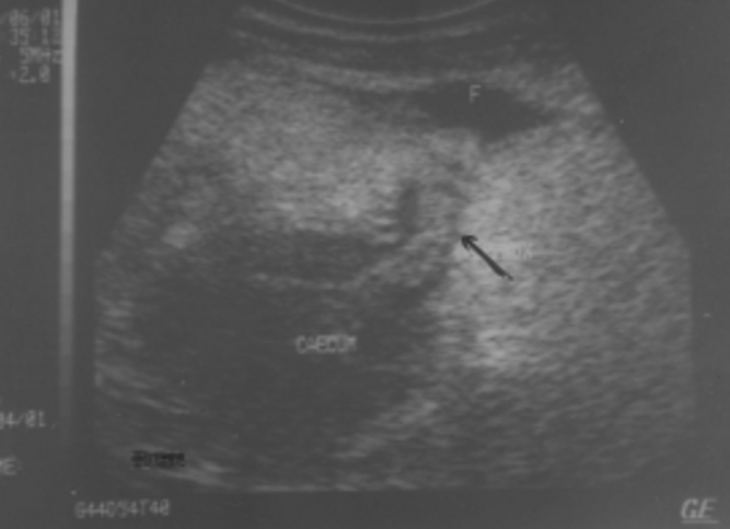
Inflamed appendix (arrow) with fluid collection (F) around tip of appendix. Note hypoechoic appearance of caecum due to inflammation
Fig. 3.

Gangrenous appendicitis. Note periappendiceal fluid collection (arrow) and loss in continuity of wall of appendix (curved arrow)
Table 1.
Sonographic findings in appendicitis
| Sonographic findings | No. of cases (Total no = 36) | Percentage |
|---|---|---|
| Classical inflamed appendix (uncomplicated) (Fig. 1) | 21 | 58.3% |
| Inflamed appendix along with inflamed caecum (Fig. 2) | 02 | 5.5% |
| Inflamed appendix with 06 periappendiceal fluid collection (Fig. 3) | ||
| Appendicular abscess | 03 | 8.3% |
| Appendicular lump | 04 | 11.1% |
| Dilated fluid filled aperistaltic terminal ileum | 03 | 8.3% |
| Appendicolith | 03 | 8.3% |
| Pelvic abscess | 01 | 2.7% |
Alternative diagnoses were made in 33 (47.8%) cases (Table-2). Surgery was performed in 33 cases (emergency appendectomy in 30 cases, interval appendectomy in 03 cases). Two cases resolved spontaneously on conservative treatment, one of these patients had recurrent appendicitis at the end of 09 months. One case scheduled for interval appendectomy was lost to follow up. There was one false positive case; a 04 year old girl diagnosed as appendicular abscess turned out to be a case of psoas abscess. There were 04 false negative cases: 01 was an obese individual with retrocaecal inflamed appendix, 02 had inflammation localized to the tip of appendix and one patient had a perforated appendix where the appendiceal diameter was <06mm. All these 4 cases were missed on ultrasound and proved at surgery. Overall sensitivity and specificity in our study were 89.7% and 96.6% respectively. The positive and negative predictive values were 97.2% and 87.8% respectively.
Table 2.
Alternative diagnosis in cases of suspected appendicitis
| Alternative diagnoses | No. of cases (Total no = 33) | Percentage |
|---|---|---|
| Abdominal pain of unknown origin | 14 | (42.4%) |
| Gynae: | 10 | (30.3%) |
| Pelvic inflammatory disease | 03 | |
| Endometriosis | 02 | |
| Ruptured follicle | 02 | |
| Ectopic pregnancy | 01 | |
| Ovarian cysts | 02 | |
| Urologic: | 07 | (21.2%) |
| Ureteric calculus | 02 | |
| Vesico ureteric junction calculus | 04 | |
| Cystitis | 01 | |
| GIT: | 02 | (6%) |
| Perforated duodenal ulcer | 01 | |
| Cholecystitis | 01 |
Discussion
Acute appendicitis remains the most common surgical emergency with a life-time occurrence of 07% [11]. The key to successful management of acute appendicitis depends on prompt diagnosis and early surgical intervention. The clinical diagnosis of acute appendicitis is often not straightforward and a negative laparotomy rate of 20-25% is not uncommon. There is an inverse relationship between negative appendectomy rate and perforation rate. A surgical specialist who manages appendicitis based solely on clinical criteria is at risk to face either increased rates of negative appendectomy (in case he/she is aggressive) or increased perforation and sequelae (in case he/she prefers conservative management). So the challenge for a surgical specialist is how to balance between an effort to reduce negative appendectomy rate without increasing the perforation rate. Imaging can play a great role in making an early diagnosis of appendicitis and also suggest alternative diagnosis thereby reducing both negative appendectomy rate as well as perforation rate. Graded compression sonography as popularized by Puylaert [1] superadded with self localization [2] is a readily available, non-invasive, highly accurate means of diagnosing appendicitis and a variety of relevant diaseases[8]. Prospective studies have shown that the overall accuracy of US in diagnosing acute appendicitis ranges between 87-96% (sensitivity 75-90%, specificity 86-100%) with positive and negative predictive values of 91-94% and 89-97% respectively [3, 4, 5, 12]. In experienced hands US significantly improves diagnostic accuracy in suspected appendicitis while reducing the negative laparotomy rate to 8-15% [13].
Graded compression US with gentle, slow and maintained pressure allows for a lengthy and successful evaluation of the area of interest even in the most uncomfortable and reluctant patients. Patients are also able to identify the point of maximum tenderness which often helps in focusing the examination in the correct area. Even in classical cases of appendicitis in young males where clinically there is no doubt about the diagnosis, US can provide valuable information regarding sonomorphology of inflamed appendix as well as surrounding area. This can help the surgeon in pre operative planning as well as prime the surgeon as to what he/she can expect during surgery. For example, information like location of the appendix, state of the appendix, contents of the appendicular lumen, periappendiceal collection, presence of abscess, appendicular lump, pelvic/generalized peritonitis may help the surgical specialist in overall management of the case.
We feel that even today there is a reservation amongst physicians/surgeons regarding routine use of ultrasonography in cases of suspected appendicitis. The value of US in the diagnosis and management of appendicitis is well established world wide. In fact computed tomography is also being widely and increasingly used in the diagnosis and management of appendicitis [14]. However, keeping in view of ready availability, high accuracy in diagnosing appendicitis and much less expenses involved, US becomes the first modality of imaging investigation of choice in our country. By routinely using US in cases of suspected appendicitis we would be able to reduce morbidity significantly.
Conclusion
US is an accurate means of diagnosing/excluding appendicitis or suggesting an alternate diagnosis. Routine and liberal use of US should be made in cases of suspected appendicitis to reduce both negative appendectomy rates as well as perforation rates. Our experience in this context is gratifying and encouraging.
References
- 1.Puylaert JB. Acute appendicitis. US evaluation using graded compression. Radiology. 1986;158:355–360. doi: 10.1148/radiology.158.2.2934762. [DOI] [PubMed] [Google Scholar]
- 2.Chesbrough RM, Burkhard TK, Balsara ZN, Goff WB, Davis DJ. Self-localisation in US of appendicitis an addition to graded compression. Radiology. 1993;187:349–351. doi: 10.1148/radiology.187.2.8475271. [DOI] [PubMed] [Google Scholar]
- 3.Birnbaum BA, Wilson SR. Appendicitis at the millennium. Radiology. 2000;215(2):337–348. doi: 10.1148/radiology.215.2.r00ma24337. [DOI] [PubMed] [Google Scholar]
- 4.Paulson EK, Kalady MF, Pappas TN. Suspected appendicitis. New Engl J of Med. 2003;348(3):236–242. doi: 10.1056/NEJMcp013351. [DOI] [PubMed] [Google Scholar]
- 5.Yacoe ME, Jeffrey RB., Jr Sonography of appendicitis and diverticulitis. Radiologic Clinics of North America. 1994;32(5):899–912. [PubMed] [Google Scholar]
- 6.Joshi HM, Patel VB, Dave AN. Ultrasonographic evaluation of acute appendicitis. Ind J Radiol Imag. 1996;6(2):75–78. [Google Scholar]
- 7.Styrud J, Josephson T, Eriksson S. Reducing negative appendectomy: evaluation of ultrasonography and computed tomography in acute appendicitis. Int J Qual Health Care. 2000;12(1):65–68. doi: 10.1093/intqhc/12.1.65. [DOI] [PubMed] [Google Scholar]
- 8.Gaensler EHL, Jeffrey RB, Jr, Laing FC, Townsend RR. Sonography in patients with suspected acute appendicitis: value in establishing alternative diagnoses. Am J Roentgenol. 1989;152:49–51. doi: 10.2214/ajr.152.1.49. [DOI] [PubMed] [Google Scholar]
- 9.Fujii Y, Hata J, Futagami K. Ultrasonography improves diagnostic accuracy of acute appendicitis and provides cost savings to hospitals in Japan. J Ultrasound Med. 2000;19(6):409–414. doi: 10.7863/jum.2000.19.6.409. [DOI] [PubMed] [Google Scholar]
- 10.Rettenbacher T, Hollerweger A, Macheiner P. Outer diameter of the vermiform appendix as a sign of acute appendicitis: Evaluation at US. Radiology. 2001;218:757–762. doi: 10.1148/radiology.218.3.r01fe20757. [DOI] [PubMed] [Google Scholar]
- 11.Hardin DM., Jr Acute appendicitis: review and update. Am Fam Physician. 1999;60(7):2027–2034. [PubMed] [Google Scholar]
- 12.Zielke A, Sitter H, Rampp T, Bohrer T, Rothmund M. Clinical decision making, ultrasonography and scores for evaluation of suspected acute appendicitis. World J Surg. 2001;25(5):578–584. doi: 10.1007/s002680020078. [DOI] [PubMed] [Google Scholar]
- 13.Ooms HWA, Koumans RKJ, Ho Kang You PJ, Puylaert JB. Ultrasonography in the diagnosis of acute appendicitis. Br J Surg. 1991;78:315–318. doi: 10.1002/bjs.1800780316. [DOI] [PubMed] [Google Scholar]
- 14.Rao PM, Rhea JT, Novelline RA, Mostafavi AA, McCabe CJ. Effect of computed tomography of the appendix on treatment of patients and use of hospital resources. New Engl J of Med. 1998;338(3):141–146. doi: 10.1056/NEJM199801153380301. [DOI] [PubMed] [Google Scholar]