

Introduction
Amniotic band syndrome (ABS) is a set of congenital malformations, ranging from minor constriction rings and lymphoedema of the digits to complex and bizarre multiple congenital anomalies [1]. We present a case of ABS diagnosed antenatally.
Case Report
A 22 year old primigravida at approximately 22 weeks of gestation was referred for a routine antenatal US scan. There was no history of exposure to teratogenic drugs, febrile illness in the first trimester, or family history of congenital malformations. US revealed severe oligohydramnios leading to difficulty in visualization of foetal parts. Foetal cardiac activity was present, but hardly any foetal movements were seen over 30 minutes of scanning. Detailed foetal scan revealed (a) enlargement of the lateral ventricles and cisternal spaces, suggesting cerebral atrophy, (b) acutely curved and rotated spine suggesting severe kyphoscoliosis (Fig-1), (e) liver and bowel loops lying freely floating in a small amount of amniotic fluid outside the abdominal cavity (Fig-2) suggesting the presence of either gastroschisis or a ruptured omphalocoele, (d) bands stretching from the amniotie membrane to various parts of the foetus (Fig-3).
Fig. 1.
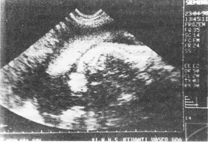
Foetal spine showing acute curvature
Fig. 2.
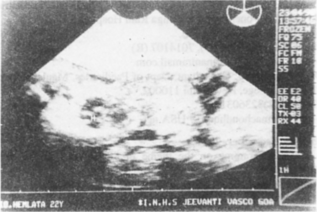
Liver lying outside the abdomen
Fig. 3.
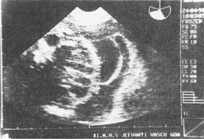
Amniotic band attaching to the side of the foetal head
Attempts to identify the umbilical cord insertion failed due to low amount of amniotie fluid and exteriorized bowel loops compromising the image acquisition. On the basis of US findings a diagnosis of ABS was made and MTP carried out.
Examination of foetus confirmed the antenatal US findings (Fig 4). The additional findings were a cleft palate, abnormal development of eyes and face, multiple digital amputations in hands and feet, large thoraco-abdominal cleft with normal umbilical cord insertion, proximal shortening of the left humerus and diagnosis of ABS was confirmed.
Fig. 4.
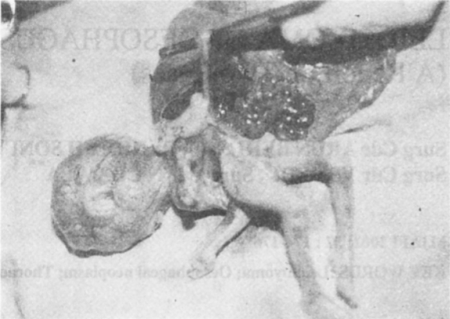
Foetus showing described abnormalities. Note cleft palate. abnormality of eyes and face and digital amputations in the hand
Discussion
The prevalence of ABS has been estimated to be around 1.16 per 10,000 live births [2]. Etiology of this syndrome is not precisely known. Some theories have been suggested such as teratogenic, multifactorial, and genetic factors causing a rupture of the amnion [3]. Rupture of the amnion in early pregnancy leads to entrapment of fetal structures by “sticky” mesodermic bands that originate from the chorionic side of the amnion, followed by disruption [4]. The syndrome results in structural anomalies that vary from minor to lethal forms. The most common findings are constriction rings around the digits, arms and legs. swelling of the extremities distal to the point of constriction, amputation of digits, arms and legs, craniofacial clefts, cephalocele, anencephaly, multiple joint contractures, clubfeet, simian crease, pseudosyndaetyly. microphthalmia, gastroschisis, omphalocoele, bladder exstrophy and ambiguous genitalia. The condition is diagnosed on antenatal US on the basis of association of multiple anomalies. Further visualization of amniotic bands attached to foetus with restriction of motion is diagnostic of the condition. The mere presence of amniotic bands is not considered sufficient to diagnose ABS as several types of membranes may be seen in normal pregnancies [1]. The case presented displays these features well.
Accurate antenatal diagnosis of this condition precludes the need for foetal karyotyping as this condition is not genetic or familial in nature [2]. Few familial recurrences have been reported in association with epidermolysis bullosa and Ehlers Danlos syndrome [5, 6].
The differential diagnosis considered antenatally included the limb-body wall complex or short umbilical cord syndrome, and pentology of Cantrell. The presence of a normally seen umbilical cord ruled out the first alternative. Pentology of Cantrell is a combination of an omphalocoele, ectopia cordis, and defects of the sternum, anterior diaphragm and diaphragmatic pericardium [1]. Foetal heart appeared to be entering the anterior cleft in the thoraco-abdominal wall antenatally and the possibility of a ruptured omphalocoele had to be entertained, as the umbilical cord insertion could not be identified antenatally. Postnatally, presence of a normal umbilical cord insertion confirmed the diagnosis of ABS.
The prognosis in case of ABS is dependent on the specific anomalies present. Minor digital amputations or constriction bands are unlikely to be diagnosed, they do not influence life expectancy and can be treated surgically. Severe and complex malformations as seen in this case are incompatible with extra uterine life and are an indication for termination of pregnancy [1].
References
- 1.Goncalves LF, Jeunty P. Ultrasound Evaluation of Foetal Abdominal Wall Defects. In: Callen PW, editor. Ultrasonography in Obstetrics and Gynaecology. 2nd ed. WB Saunders; Philadelphia: 1994. pp. 380–385. [Google Scholar]
- 2.Garza A, Cordero JF, Mulinare J. Epidemiology of the early amnion rupture spectrum of defects. Am J Dis Child. 1988;142(5):541–544. doi: 10.1001/archpedi.1988.02150050079037. [DOI] [PubMed] [Google Scholar]
- 3.Tadmor OP, Krcisberg GA, Achiron R, Porat S, Yagel S. Limb amputation in amniotic band syndrome: serial ultrasonographic and doppler observations. Ultrasound Obstet Gynecol. 1997;10:312–315. doi: 10.1046/j.1469-0705.1997.10050312.x. [DOI] [PubMed] [Google Scholar]
- 4.Topin R. Foetal Malformations Caused by Amnion Rupture During Gestation. Charles C Thomas; Springfield, IL: 1968. pp. 1–76. [Google Scholar]
- 5.Marras A, Dessi C, Macciotta A. Epidermolysis bullosa and amniotic bands. Am J Med Genet. 1984;19:815–817. doi: 10.1002/ajmg.1320190423. [DOI] [PubMed] [Google Scholar]
- 6.Young ID, Lindenbaum RH, Thompson EM, Pembrey ME. Amniotic band syndrome in in connective tissue disorders. Arch Dis Child. 1985;60:1061–1063. doi: 10.1136/adc.60.11.1061. [DOI] [PMC free article] [PubMed] [Google Scholar]