Abstract
Objectives:
This study sought to assess the effect of bracket base design on the shear bond strength (SBS) of the bracket to feldspathic porcelain.
Materials and Methods:
This in vitro, experimental study was conducted on 40 porcelain-fused-to-metal restorations and four different bracket base designs were bonded to these specimens. The porcelain surfaces were etched, silanized, and bonded to brackets. Specimens were thermocycler, incubated for 24 h and were subjected to SBS. Data were analyzed using Shapiro–Wilk test, Levene's test, one-way ANOVA, and Tukey's honest significant difference test. Adhesive remnant index was calculated and compared using Fisher's exact test.
Results:
One-way ANOVA showed that the SBS values were significantly different among the four groups (P < 0.001). Groups 1, 2, and 4 were not significantly different, but group 3 had significantly lower SBS (P < 0.001). Fractures mostly occurred at the porcelain-adhesive interface in Groups 1 and 2 while in Groups 3 and 4, bracket-adhesive and mixed failures were more common.
Conclusion:
The bracket base design significantly affects the SBS to feldspathic porcelain.
Keywords: Bond strength, dental porcelain, orthodontic brackets
INTRODUCTION
Demand for orthodontic treatment has greatly increased in adults in the recent years.[1] Among the types of brackets, metal brackets are the oldest and most commonly used type.[2] Porcelain-fused-to-metal (PFM) restorations are the most commonly used crowns due to high strength and accuracy of the cast metal and esthetic properties of the porcelain. Factors affecting the bracket bond strength to enamel have been extensively studied and include enamel surface preparation techniques, adhesive systems, and bracket-related factors such as the size and design of bracket base. Reportedly, 6–8 MPa bond strength is optimal for bracket bond to enamel.[3,4] Some studies confirm the effect of bracket base design on bond strength to enamel[5,6,7,8] while some others have rejected this theory.[9,10]
Type of porcelain surface treatment,[11] the adhesive system used and size and design of bracket base are believed to affect the bracket bond to porcelain.
There is a gap of information about the effect of bracket base design on the bond strength of bracket to porcelain. Thus, this in vitro study sought to assess the effect of bracket base design on the shear bond strength (SBS) of the bracket to feldspathic porcelain.
MATERIALS AND METHODS
This in vitro experimental study was conducted on PFM crowns. Bond strength of brackets with four different base designs to feldspathic porcelain was evaluated. Considering the Type I error of 0.05 and Type II error of 0.2 (power of 80%), the sample size was calculated to be 10 specimens in each group (a total of 40).
Fabrication of specimens
Mandibular premolars of both quadrants on a mandibular dentate model (Nissin, Tokyo, Japan) received the standard preparation for PFM crowns. Forty crowns were fabricated as such and randomly divided into four groups of 10. Each type of bracket was randomly allocated to one group, and brackets were bonded to PFM crowns under similar conditions.
The four types of brackets used were as follows:
Discovery stainless steel brackets with laser-structured base (Dentaurum, Ispringen, Germany)
Mini Master stainless steel brackets with photochemically micro-etched base (American Orthodontics, WI, USA)
Mini Twin stainless steel bracket with maximum injection molding base (Henry Schein Orthodontics, CA, USA)
4Radiance ceramic bracket with patented Radiance mechanical lock base (American Orthodontics, WI, USA).
Bracket bonding
The porcelain surface was first cleaned with a brush and then completely dried. Next, 9% hydrofluoric acid (Porcelain Etch®, Ultradent, UT, USA) was applied to the buccal surface of crowns using an applicator. After 2 min, the etchant was removed by rinsing with water, and the surface was air dried for 15 s using air spray to yield a porous, chalky porcelain surface. Using the sweeping motion of an applicator, silane (Ultradent Porcelain Repair Kit, USA) was applied to the porcelain surface. The bonding agent was applied. Self-cure composite resin (3M Unitek, USA) was applied to the posterior surface of the bracket in 1 mm thickness according to the manufacturer's instructions, and the bracket was placed on the porcelain surface using 150 N load measured by an orthodontic gauge (Dentaurum, Ispringen, Germany). Before composite polymerization, the excess composite resin was removed using the sharp tip of an explorer. Crowns on their respective dies were embedded in auto polymerizing acrylic resin (Meliodent, Germany) [Figure 1].
Figure 1.
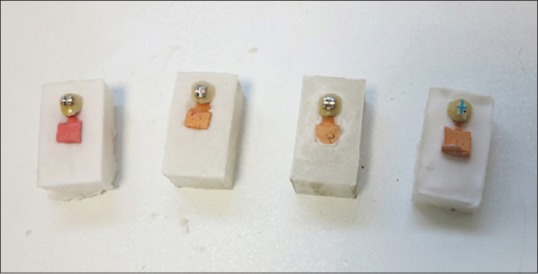
Crowns on their respective dies embedded in acrylic blocks
Thermocycling
All specimens were thermocycler (Dorsa, Tehran, Iran) for 5000 cycles at 5–55°C with 20 s of dwell time in each water bath and 20 s of transfer time. Next, the specimens were stored in an incubator (Pars Azma, Tehran, Iran) at 37°C and 100% humidity for 24 h.
Shear bond strength testing
The bracket-porcelain SBS was measured using UTM Instron machine (Zwick Roell Z020, Germany). The load was applied at a crosshead speed of 1 mm/min to the bracket-porcelain interface. To apply a load, each specimen was fixed by clamps to maintain the bonding interface parallel to the blade. Load was applied until fracture, and the load at fracture was recorded in Newton (N). To convert the SBS values to Megapascals (MPa), the load in N was divided by the cross-sectional area of the bracket base (m2) provided by the manufacturers (12.93 mm2 for Discovery, 11.04 mm2 for Mini Master, 9.42 mm2 for Mini Twin, and 13.94 mm2 for Radiance).
Adhesive remnant index
After separation of brackets from the porcelain, the bracket base and porcelain surface were examined under a stereomicroscope (Olympus SZX9 Research Stereomicroscope System, Japan) at × 10 magnification. To assess the adhesive remnant index (ARI) describing the amount of adhesive remained on the surface, the classification of Bordeaux et al.[12] was used.
Statistical analysis
The Shapiro–Wilk test was used to assess the normality of the SBS data and revealed that the data had a normal distribution. Levene's test was used to assess the equality of variances for SBS values and confirmed it. One-way ANOVA was applied for overall comparison of groups while the Tukey's honest significant difference (HSD) test was used for pairwise comparisons. ARI was calculated and compared among groups using Fisher's exact test. Type I error was considered as 0.05 and P < 0.05 was considered statistically significant [Figure 2].
Figure 2.
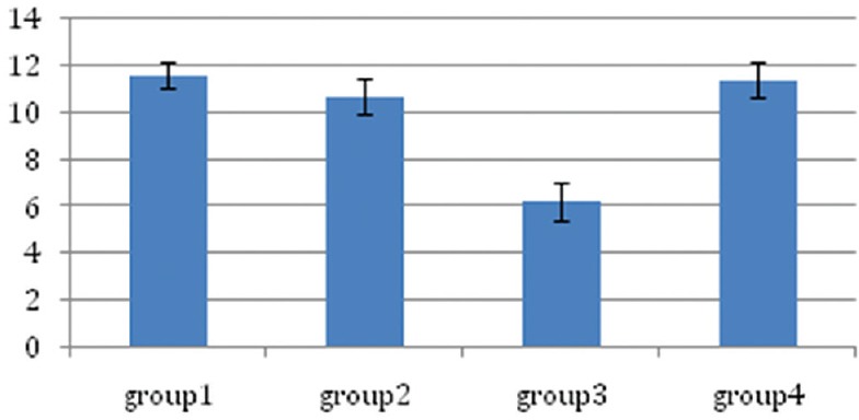
Error bar of the shear bond strength values
RESULTS
Shear bond strength
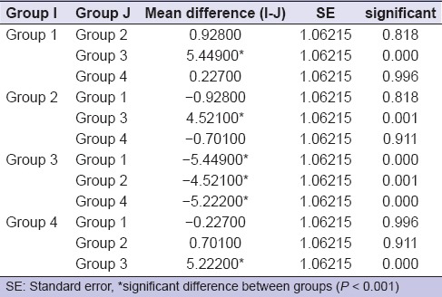
The mean SBS values for the four groups are demonstrated in Table 1. Comparison of the mean SBS values with one-way ANOVA revealed statistically significant differences among the four groups (P < 0.001). Pairwise comparison of groups with Tukey's HSD test revealed that groups one, two, and four were not significantly different regarding the mean SBS while the mentioned three groups had significantly higher mean SBS values than group 3 (P < 0.001 for all three). The highest mean SBS values belonged to groups 1 (11.61 MPa), 4 (11.38 MPa), and 2 (10.68 MPa), respectively, which were not significantly different. Group three had the lowest mean SBS (6.16 MPa), which was significantly lower than the corresponding values in the remaining three groups (P < 0.001) [Table 2].
Table 1.
The shear bond strength values in the four groups

Table 2.
Comparison of the shear bond strength values among the four groups using Tukey's honest significant difference test
Adhesive remnant index
The results of the ARI scores in the four groups based on the Fisher's exact test are shown in Table 3. The ARI scores were significantly different among the four groups (P < 0.001). As seen in Table 3, Type III ARI was the most frequent in groups one and two. However, in groups three and four, Types I and II indicative of bracket-adhesive and mixed failures, respectively were more common [Figure 3].
Table 3.
Adhesive remnant index scores in the four groups according to the Fisher's exact test
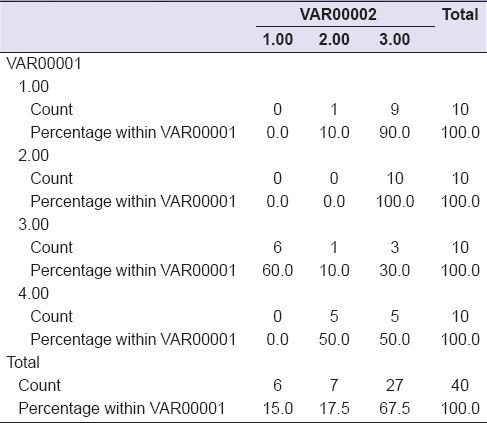
Figure 3.
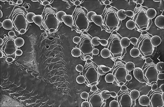
Electron micrograph of Discovery bracket base following laser irradiation
DISCUSSION
The purpose of this study was to compare the SBS of metal and ceramic brackets with different base designs to porcelain crowns and showed that the Discovery bracket had the highest mean SBS followed by the Radiance ceramic bracket and Mini Master bracket. Mini Twin bracket had the lowest mean SBS, showing significant differences with the values in other groups. This confirms the effect of bracket base design on SBS to porcelain.
In the fabrication process of Discovery brackets, the base surface is selectively melted in a controlled fashion by laser irradiation in the final step of fabrication causing droplets of variable sizes. The undercuts created by these droplets provide retention. These structures are probably responsible for high SBS of this bracket to porcelain demonstrated in this study. The manufacturing process of Radiance ceramic bracket is similar to that of other monocrystalline ceramic brackets. Radiance ceramic bracket is fabricated using Patented Quad Matte technology. For the fabrication of Mini Master brackets, 80-gauge mesh is placed over an etched foil base and is subjected to photochemical etching. The resultant porosities provide retention. Mini Twin brackets, with the lowest SBS in this study, are fabricated as a single mass using maximum injection molding technique and then the base is subjected to micro etching to provide retention. Such base design explains the low SBS obtained in this group. The undercuts created in this technique probably provide less mechanical retention. However, in all four groups, the mean SBS values were higher than the minimum required bond strength value of 6–8 MPa[3,4] and thus, they may be safely used in the clinical setting.
The ARI scores in this study confirmed our SBS results. In groups one, two, and four (almost) with the stronger bond between bracket and composite, the ARI score was mostly Type III. This indicates a strong bracket-adhesive bond. In contrast, in group three with the lowest SBS, fractures were mostly Type I and at the bracket-adhesive interface. As observed in this study, ceramic brackets had high-bond strength comparable to that of metal brackets, which is in line with the results of Reddy et al., and Stumpf Ade et al.[13,14] Samruajbenjakul andKukiattrakoon,[15] in 2009, evaluated the effect of the base design of ceramic and metal brackets on the SBS to feldspathic porcelain. They showed that bracket base design significantly affected the SBS, which confirms present findings despite some differences between their study and the present study.
Bishara et al.,[9,16] in 2004, assessed the SBS of two brackets with single and double mesh bases to enamel in vitro and revealed no significant difference in bond strength. They showed that the difference between the two base designs did not affect the bond strength. Cucu et al.[10] evaluated the effect of base size and mesh size on SBS in an in vitro study and reported no significant difference between brackets with different base sizes and similar mesh size. They demonstrated that base size had no effect on bond strength. Since this was an in vitro study, further clinical studies are required to better elucidate this subject.
CONCLUSION
The bracket base design significantly affects the SBS of brackets to feldspathic porcelain, and debonding characteristics are related to bracket base design.
Financial support and sponsorship
Nil.
Conflicts of interest
There are no conflicts of interest.
REFERENCES
- 1.Whitehouse JA. Everyday uses of adult orthodontics. Dent Today. 2004;23:116–118. 120. [PubMed] [Google Scholar]
- 2.Keim RG, Gottlieb EL, Nelson AH, Vogels DS., 3rd 2008 JCO study of orthodontic diagnosis and treatment procedures, part 1: Results and trends. J Clin Orthod. 2008;42:625–40. [PubMed] [Google Scholar]
- 3.Charles A, Senkutvan R, Ramya RS, Jacob S. Evaluation of shear bond strength with different enamel pretreatments: An in vitro study. Indian J Dent Res. 2014;25:470–4. doi: 10.4103/0970-9290.142537. [DOI] [PubMed] [Google Scholar]
- 4.Whitlock BO, 3rd, Eick JD, Ackerman RJ, Jr, Glaros AG, Chappell RP. Shear strength of ceramic brackets bonded to porcelain. Am J Orthod Dentofacial Orthop. 1994;106:358–64. doi: 10.1016/S0889-5406(94)70056-7. [DOI] [PubMed] [Google Scholar]
- 5.Algera TJ, Feilzer AJ, Prahl-Andersen B, Kleverlaan CJ. A comparison of finite element analysis with in vitro bond strength tests of the bracket-cement-enamel system. Eur J Orthod. 2011;33:608–12. doi: 10.1093/ejo/cjq112. [DOI] [PubMed] [Google Scholar]
- 6.Cozza P, Martucci L, De Toffol L, Penco SI. Shear bond strength of metal brackets on enamel. Angle Orthod. 2006;76:851–6. doi: 10.1043/0003-3219(2006)076[0851:SBSOMB]2.0.CO;2. [DOI] [PubMed] [Google Scholar]
- 7.Sorel O, El Alam R, Chagneau F, Cathelineau G. Comparison of bond strength between simple foil mesh and laser-structured base retention brackets. Am J Orthod Dentofacial Orthop. 2002;122:260–6. doi: 10.1067/mod.2002.125834. [DOI] [PubMed] [Google Scholar]
- 8.Wang WN, Li CH, Chou TH, Wang DD, Lin LH, Lin CT. Bond strength of various bracket base designs. Am J Orthod Dentofacial Orthop. 2004;125:65–70. doi: 10.1016/j.ajodo.2003.01.003. [DOI] [PubMed] [Google Scholar]
- 9.Bishara SE, Soliman MM, Oonsombat C, Laffoon JF, Ajlouni R. The effect of variation in mesh-base design on the shear bond strength of orthodontic brackets. Angle Orthod. 2004;74:400–4. doi: 10.1043/0003-3219(2004)074<0400:TEOVIM>2.0.CO;2. [DOI] [PubMed] [Google Scholar]
- 10.Cucu M, Driessen CH, Ferreira PD. The influence of orthodontic bracket base diameter and mesh size on bond strength. SADJ. 2002;57:16–20. [PubMed] [Google Scholar]
- 11.Yassaei S, Moradi F, Aghili H, Kamran MH. Shear bond strength of orthodontic brackets bonded to porcelain following etching with Er: YAG laser versus hydrofluoric acid. Orthodontics (Chic.) 2013;14:e82–7. doi: 10.11607/ortho.856. [DOI] [PubMed] [Google Scholar]
- 12.Bordeaux JM, Moore RN, Bagby MD. Comparative evaluation of ceramic bracket base designs. Am J Orthod Dentofacial Orthop. 1994;105:552–60. doi: 10.1016/S0889-5406(94)70139-3. [DOI] [PubMed] [Google Scholar]
- 13.Reddy YG, Sharma R, Singh A, Agrawal V, Agrawal V, Chaturvedi S. The shear bond strengths of metal and ceramic brackets: An in-vitro comparative study. J Clin Diagn Res. 2013;7:1495–7. doi: 10.7860/JCDR/2013/5435.3172. [DOI] [PMC free article] [PubMed] [Google Scholar]
- 14.Stumpf Ade S, Bergmann C, Prietsch JR, Vicenzi J. Shear bond strength of metallic and ceramic brackets using color change adhesives. Dental Press J Orthod. 2013;18:76–80. doi: 10.1590/s2176-94512013000200017. [DOI] [PubMed] [Google Scholar]
- 15.Samruajbenjakul B, Kukiattrakoon B. Shear bond strength of ceramic brackets with different base designs to feldspathic porcelains. Angle Orthod. 2009;79:571–6. doi: 10.2319/060308-290.1. [DOI] [PubMed] [Google Scholar]
- 16.Bishara SE, Ajlouni R, Oonsombat C, Laffoon J. Bonding orthodontic brackets to porcelain using different adhesives/enamel conditioners: A comparative study. World J Orthod. 2005;6:17–24. [PubMed] [Google Scholar]


