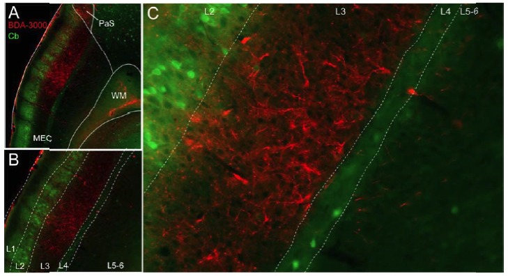
Author response image 1. Laminar organization of projections from the PreS to MEC.

(A) Parasagittal sectionthrough MEC showing anterogradely-labeled axons following injectionof the anterogradetracer BDA -10k (red) in the ipsilateral PreS. Entorhinal layers are outlined via calbindin staining (Cb, green). Note the massive arborization of PreS afferents within L3 of the MEC. (B and C) show high-magnifications viewof the section shown in A.
